Mario Roccuzzo - Peri‑Implant Soft‑Tissue Integration and Management
Здесь есть возможность читать онлайн «Mario Roccuzzo - Peri‑Implant Soft‑Tissue Integration and Management» — ознакомительный отрывок электронной книги совершенно бесплатно, а после прочтения отрывка купить полную версию. В некоторых случаях можно слушать аудио, скачать через торрент в формате fb2 и присутствует краткое содержание. Жанр: unrecognised, на английском языке. Описание произведения, (предисловие) а так же отзывы посетителей доступны на портале библиотеки ЛибКат.
- Название:Peri‑Implant Soft‑Tissue Integration and Management
- Автор:
- Жанр:
- Год:неизвестен
- ISBN:нет данных
- Рейтинг книги:3 / 5. Голосов: 1
-
Избранное:Добавить в избранное
- Отзывы:
-
Ваша оценка:
Peri‑Implant Soft‑Tissue Integration and Management: краткое содержание, описание и аннотация
Предлагаем к чтению аннотацию, описание, краткое содержание или предисловие (зависит от того, что написал сам автор книги «Peri‑Implant Soft‑Tissue Integration and Management»). Если вы не нашли необходимую информацию о книге — напишите в комментариях, мы постараемся отыскать её.
Peri‑Implant Soft‑Tissue Integration and Management — читать онлайн ознакомительный отрывок
Ниже представлен текст книги, разбитый по страницам. Система сохранения места последней прочитанной страницы, позволяет с удобством читать онлайн бесплатно книгу «Peri‑Implant Soft‑Tissue Integration and Management», без необходимости каждый раз заново искать на чём Вы остановились. Поставьте закладку, и сможете в любой момент перейти на страницу, на которой закончили чтение.
Интервал:
Закладка:
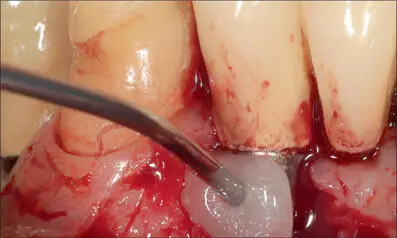
Fig 12d 24% EDTA applied for two minutes for decontamination.
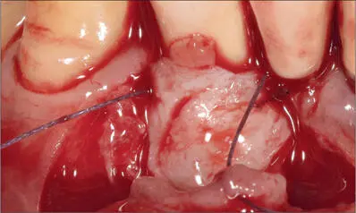
Fig 12e Connective-tissue graft, taken from the maxillary tuberosity and properly trimmed, adapted over the defect.
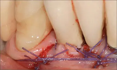
Fig 12f Flap sutured with 4-0 Vicryl to completely cover the connective-tissue graft.
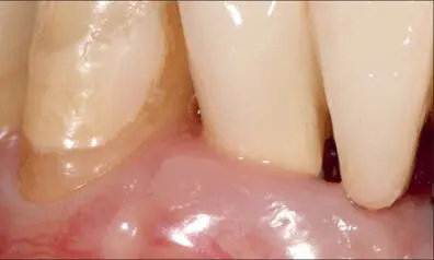
Fig 12g Seven-year follow-up. No signs of inflammation. The soft-tissue recession was reduced.
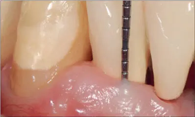
Fig 12h Minimal probing depth and no bleeding at the nine-year follow-up visit.
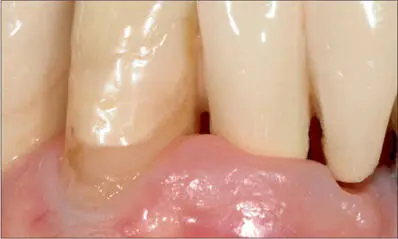
Fig 12i Twenty-five years after implant placement. The situation is still stable and free from signs of inflammation.
Recently, a reduced width of keratinized tissue around dental implants has been considered a risk indicator for the severity of peri-implant mucositis (Grischke and coworkers 2019) even in highly compliant, periodontally healthy patients. Based on this observation, in cases of peri-implant mucositis associated with insufficient keratinized tissue, a soft-tissue graft may be indicated to reduce the risk of recurrence. Figures 13a-yillustrate a case where the treatment of severe peri-implant mucositis was accompanied by the application of an FGG graft to improve the quality of the peri-implant soft tissue and to improve the chances of successful long-term maintenance.
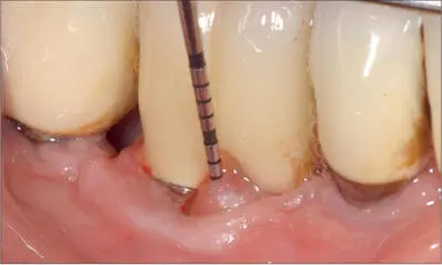
Fig 13a Mucositis around an implant (SLA S, diameter 4.1 mm, length 8 mm; Institut Straumann AG) placed more than ten years previously.
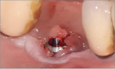
Fig 13b The crown and abutment were removed to provide access to the inflamed area.
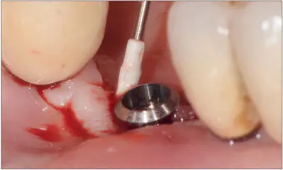
Fig 13c The treatment consisted of careful debridement and gentle cleaning of the area with titanium curettes and an ultrasonic device with a PTFE-coated tip.
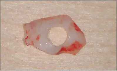
Fig 13d An FGG was harvested from the palate and perforated with a 4-mm biopsy punch.
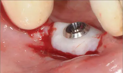
Fig 13e Precisely adapted FGG around the smooth collar of the implant.

Fig 13f Graft secured by means of 5-0 Vicryl sutures.
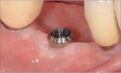
Fig 13g Six months after treatment.
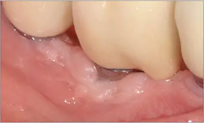
Fig 13h New screw-retained ceramic crown in situ.
Dentists should focus on more than just the implants and prosthetic restorations to achieve long-term clinical success.
Patients must be strongly motivated to adhere strictly to SPT and should be made to understand that supportive care is a key factor in enhancing the long-term outcome by controlling reinfection. Although the topic remains controversial due to a lack of proper scientific evidence, there is still enough clinical evidence that adequate keratinized tissue and vestibular depth may positively impact the health of the peri-implant mucosa. Furthermore, soft-tissue augmentation around implants in specific clinical situations may be of major help in ensuring long-term stability. Only if both the clinician and the patient understand the importance of a custom-planned SPT program—which may also include mucogingival surgery—can the risk of biological complications be reduced to a minimum.
Конец ознакомительного фрагмента.
Текст предоставлен ООО «ЛитРес».
Прочитайте эту книгу целиком, купив полную легальную версию на ЛитРес.
Безопасно оплатить книгу можно банковской картой Visa, MasterCard, Maestro, со счета мобильного телефона, с платежного терминала, в салоне МТС или Связной, через PayPal, WebMoney, Яндекс.Деньги, QIWI Кошелек, бонусными картами или другим удобным Вам способом.
Интервал:
Закладка:
Похожие книги на «Peri‑Implant Soft‑Tissue Integration and Management»
Представляем Вашему вниманию похожие книги на «Peri‑Implant Soft‑Tissue Integration and Management» списком для выбора. Мы отобрали схожую по названию и смыслу литературу в надежде предоставить читателям больше вариантов отыскать новые, интересные, ещё непрочитанные произведения.


Обсуждение, отзывы о книге «Peri‑Implant Soft‑Tissue Integration and Management» и просто собственные мнения читателей. Оставьте ваши комментарии, напишите, что Вы думаете о произведении, его смысле или главных героях. Укажите что конкретно понравилось, а что нет, и почему Вы так считаете.