Mario Roccuzzo - Peri‑Implant Soft‑Tissue Integration and Management
Здесь есть возможность читать онлайн «Mario Roccuzzo - Peri‑Implant Soft‑Tissue Integration and Management» — ознакомительный отрывок электронной книги совершенно бесплатно, а после прочтения отрывка купить полную версию. В некоторых случаях можно слушать аудио, скачать через торрент в формате fb2 и присутствует краткое содержание. Жанр: unrecognised, на английском языке. Описание произведения, (предисловие) а так же отзывы посетителей доступны на портале библиотеки ЛибКат.
- Название:Peri‑Implant Soft‑Tissue Integration and Management
- Автор:
- Жанр:
- Год:неизвестен
- ISBN:нет данных
- Рейтинг книги:3 / 5. Голосов: 1
-
Избранное:Добавить в избранное
- Отзывы:
-
Ваша оценка:
Peri‑Implant Soft‑Tissue Integration and Management: краткое содержание, описание и аннотация
Предлагаем к чтению аннотацию, описание, краткое содержание или предисловие (зависит от того, что написал сам автор книги «Peri‑Implant Soft‑Tissue Integration and Management»). Если вы не нашли необходимую информацию о книге — напишите в комментариях, мы постараемся отыскать её.
Peri‑Implant Soft‑Tissue Integration and Management — читать онлайн ознакомительный отрывок
Ниже представлен текст книги, разбитый по страницам. Система сохранения места последней прочитанной страницы, позволяет с удобством читать онлайн бесплатно книгу «Peri‑Implant Soft‑Tissue Integration and Management», без необходимости каждый раз заново искать на чём Вы остановились. Поставьте закладку, и сможете в любой момент перейти на страницу, на которой закончили чтение.
Интервал:
Закладка:
These findings were later confirmed in systematic reviews that concluded that the lack of an adequate zone of keratinized attached tissue may not be mandatory for maintaining soft-tissue health around dental implants, as long as an optimal level of oral hygiene is ensured (Wennström and Derks 2012; Gobbato and coworkers 2013; Lin and coworkers 2013). However, preclinical and clinical data indicate that in the absence of stable keratinized attached mucosa, plaque control is more difficult, which in turn may lead to peri-implant soft-tissue inflammation and, eventually, bone loss (Warrer and coworkers 1995; Wennström and Derks 2012; Gobbato and coworkers 2013; Lin and coworkers 2013).
Roccuzzo and coworkers (2016) evaluated the clinical conditions around dental implants placed in the posterior mandible of healthy or moderately periodontally compromised patients as a function of the presence or absence of keratinized attached mucosa (KAM). The results showed that the absence of KAM was associated with higher plaque accumulation, greater soft-tissue recession (REC), and a higher number of sites that required additional surgical or antibiotic treatment, indicating that implants not surrounded by KAM are more prone to plaque accumulation and to developing soft-tissue recessions despite adequate oral hygiene and supportive periodontal therapy. These findings are in line with the results of three recent reviews, which concluded that the presence of an adequate width of KAM around dental implants is associated with better soft and hard tissue stability, less plaque accumulation, soft-tissue recession, and a lower incidence of peri-implant mucositis (Sculean and coworkers 2017; Chackartchi and coworkers 2019).
Taken together, the by far greater part of the available evidence indicates that the lack of an adequate width of KAM around dental implants is associated with more plaque accumulation, inflammation, soft-tissue recession, and attachment loss (Warrer and coworkers 1995; Wennström and Derks 2012; Gobbato and coworkers 2013; Lin and coworkers 2013; Sculean and coworkers 2017; Chackartchi and coworkers 2019; Iorio-Siciliano and coworkers 2019).
A recent systematic review evaluated the effects of soft-tissue augmentation procedures on peri-implant health or disease in partially and fully edentulous patients (Thoma and coworkers 2018a), using soft-tissue grafting procedures to increase the width of the KAM or the thickness of the peri-implant mucosa. The findings indicated that soft-tissue grafting by means of autologous grafts may favor peri-implant health through a gain of KAM, improved bleeding scores, and less marginal bone loss.
In the esthetic zone, autologous connective-tissue grafts resulted in increased mucosal thickness around implants and were associated with statistically significantly less marginal bone loss over time. However, the data failed to reveal statistically significant changes in terms of bleeding on probing, probing depths, or plaque scores at grafted sites compared to sites without grafting. Nevertheless, the authors concluded that based on the available evidence, it is generally accepted that soft-tissue augmentation is beneficial to establishing and maintaining peri-implant health (Thoma and coworkers 2018a).
Regarding the thickness of the peri-implant mucosa, findings of preclinical and clinical studies suggest a threshold value of 2 mm for establishing a natural appearance of the peri-implant mucosa and minimal soft-tissue discoloration at implant-supported prosthetic reconstructions (Jung and coworkers 2007; Cosgarea and coworkers 2015; Ioannidis and coworkers 2017; Thoma and coworkers 2016). Moreover, an adequate mucosal thickness was associated with a decreased risk of mucosal recessions in immediate-placement protocols or in specific anatomic situations (e.g., minimal or no facial bony wall, orofacial implant malposition, various angles of the implant fixtures) (Buser and coworkers 2004; Evans and coworkers 2008; Sculean and coworkers 2017).
In summary, despite the fact that the available evidence is still inconclusive, there is reason to suggest that the presence of KAM favors peri-implant health through facilitating oral hygiene measures, with a consequent reduction in both inflammation (lower bleeding scores) and marginal bone loss. Furthermore, its presence or absence also plays a key role in ensuring esthetics.
Acknowledgments
Photography
Professor Dieter D. Bosshardt – Bern, Switzerland
3 Soft-Tissue Management Around Tissue-Level Implants
M. Roccuzzo
3.1 Soft-Tissue Management at Implant Placement
From the biological point of view, the apicocoronal positioning of an implant, particularly those of tissue-level design, should follow the principle of “as shallow as possible, as deep as necessary” (Buser and coworkers 2004) in order to avoid deep peri-implant probing depths, taking into account the prosthetic and esthetic factors in the area.
This concept has recently been confirmed in a case-control study on 19 patients that evaluated the modifying effect of a deep mucosal tunnel (DMT, ≥ 3 mm) on the induction and resolution phases of experimental peri-implant mucositis (Chan and coworkers 2019). All patients, each with a properly placed tissue-level implant, were assigned either to the test group (DMT, depth ≥ 3 mm) or to the control group (shallow mucosal tunnel, SMT, ≤ 1 mm). The subjects underwent a standard experimental peri-implant mucositis protocol, characterized by an oral-hygiene optimization phase, a three-week induction phase using an acrylic stent to prevent self-performed oral hygiene at the experimental implant, and a three-plus-two-week resolution phase.
The modified plaque index (mPI), gingival index (mGI), and IL-1β concentrations in the peri-implant sulcus fluid were determined over time. Both the mPI and the mGI increased during the induction phase. After normal oral hygiene had resumed, the mPI and mGI resolved towards baseline values in the SMT group, while they diverged in the DMT group. Although plaque accumulation was resolved in the DMT group, the resolution of inflammation was delayed and found to be of smaller magnitude during the first three weeks after resumption of oral hygiene. IL-1β Concentrations were significantly higher in the DMT group at the end of induction and during the resolution phase, corroborating the clinical findings. Removal of the crown and submucosal professional cleaning were needed to revert mGI to baseline values in the DMT group.
The fact that the depth of the peri-implant sulcus influenced the resolution of experimental mucositis raised doubts as to the efficacy of self-performed oral hygiene in scenarios where implants are placed too deeply. Therefore, since the risk of mucositis evolving into peri-implantitis appears to be higher in such clinical situations, clinicians should make every effort to place implants properly—not only for esthetic, but also for biological reasons (Berglundh and coworkers 2018).
It should be noted that, from a clinical point of view, this may be more easily achievable for implants without adjacent teeth, but more challenging if the implant has to be placed between two teeth, particularly if these teeth are periodontally compromised. Figures 1a-bshow examples of correct implant positioning. Figures 2a-bshow examples of incorrect implant positioning.
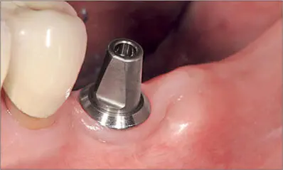
Fig 1a The implant was carefully selected and positioned in a periodontally compromised patient so as to present minimal probing depth (time of crown cementation).
Fig 1b Healthy interdental papilla between two tissue-level implants, after removing seven-year-old single ceramic crowns which had been kept in place with provisional cement.
Читать дальшеИнтервал:
Закладка:
Похожие книги на «Peri‑Implant Soft‑Tissue Integration and Management»
Представляем Вашему вниманию похожие книги на «Peri‑Implant Soft‑Tissue Integration and Management» списком для выбора. Мы отобрали схожую по названию и смыслу литературу в надежде предоставить читателям больше вариантов отыскать новые, интересные, ещё непрочитанные произведения.


Обсуждение, отзывы о книге «Peri‑Implant Soft‑Tissue Integration and Management» и просто собственные мнения читателей. Оставьте ваши комментарии, напишите, что Вы думаете о произведении, его смысле или главных героях. Укажите что конкретно понравилось, а что нет, и почему Вы так считаете.