Salivary Gland Pathology
Здесь есть возможность читать онлайн «Salivary Gland Pathology» — ознакомительный отрывок электронной книги совершенно бесплатно, а после прочтения отрывка купить полную версию. В некоторых случаях можно слушать аудио, скачать через торрент в формате fb2 и присутствует краткое содержание. Жанр: unrecognised, на английском языке. Описание произведения, (предисловие) а так же отзывы посетителей доступны на портале библиотеки ЛибКат.

- Название:Salivary Gland Pathology
- Автор:
- Жанр:
- Год:неизвестен
- ISBN:нет данных
- Рейтинг книги:3 / 5. Голосов: 1
-
Избранное:Добавить в избранное
- Отзывы:
-
Ваша оценка:
Salivary Gland Pathology: краткое содержание, описание и аннотация
Предлагаем к чтению аннотацию, описание, краткое содержание или предисловие (зависит от того, что написал сам автор книги «Salivary Gland Pathology»). Если вы не нашли необходимую информацию о книге — напишите в комментариях, мы постараемся отыскать её.
now features case presentations to place the information in context, as well as additional treatment algorithms in each chapter to assist in clinical decision making. Written by highly respected clinicians, educators, and researchers with extensive expertise in oral and maxillofacial surgery, this authoritative resource:
Reviews the etiology, diagnosis, and treatment of all salivary gland pathologies, with detailed explanations and hundreds of full-color clinical images Incorporates new information on the taxonomy of salivary gland tumors, neoplastic and non-neoplastic entities, image guided biopsies of salivary gland lesions, and complications in traditional and non-traditional forms of salivary gland surgery Offers expanded coverage of histopathology, including classification, grading, and staging of salivary gland tumors Features up-to-date chapters on anatomy and physiology, imaging, cysts, systemic diseases, tumors, trauma, and innovative salivary gland surgical techniques Includes a discussion of meta-analyses and systematic reviews to support evidence-based practice remains the definitive resource for oral and maxillofacial surgeons, otolaryngologists, head and neck surgeons, and general surgeons as well as their residents providing care for patients with salivary gland pathology.
Salivary Gland Pathology — читать онлайн ознакомительный отрывок
Ниже представлен текст книги, разбитый по страницам. Система сохранения места последней прочитанной страницы, позволяет с удобством читать онлайн бесплатно книгу «Salivary Gland Pathology», без необходимости каждый раз заново искать на чём Вы остановились. Поставьте закладку, и сможете в любой момент перейти на страницу, на которой закончили чтение.
Интервал:
Закладка:
3 Repeat CT scanning in staged parotid surgeries permits the assessment of interval growth of masses prior to the second parotid surgery. The increased growth of the mass in this case increased the pretest probability of a neoplastic process, and specifically a Warthin tumor.
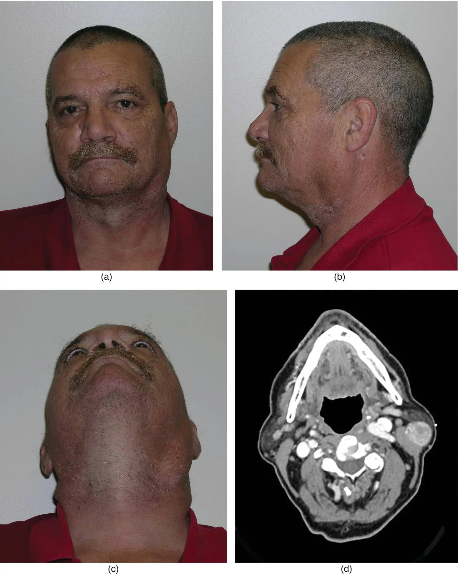
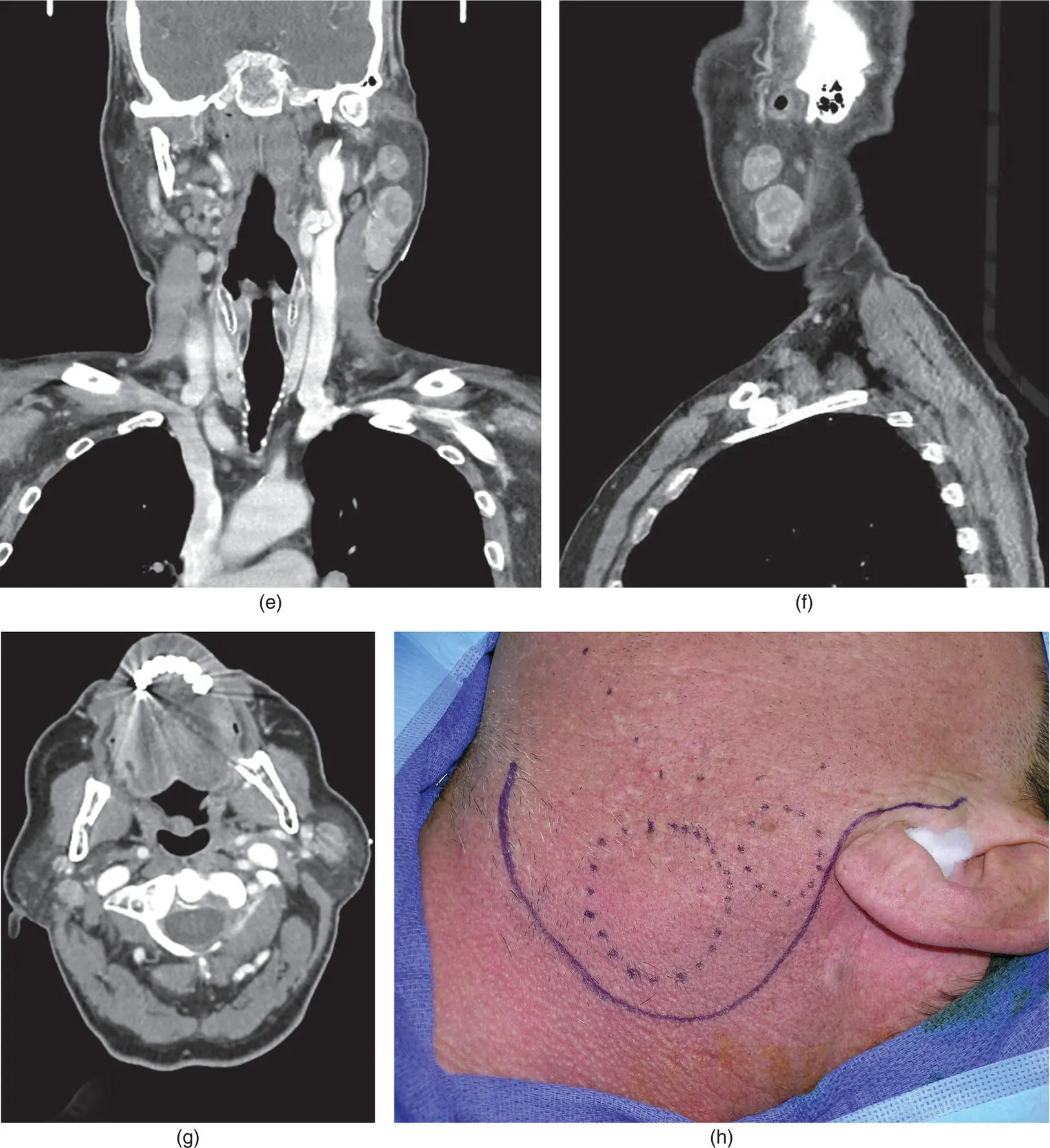
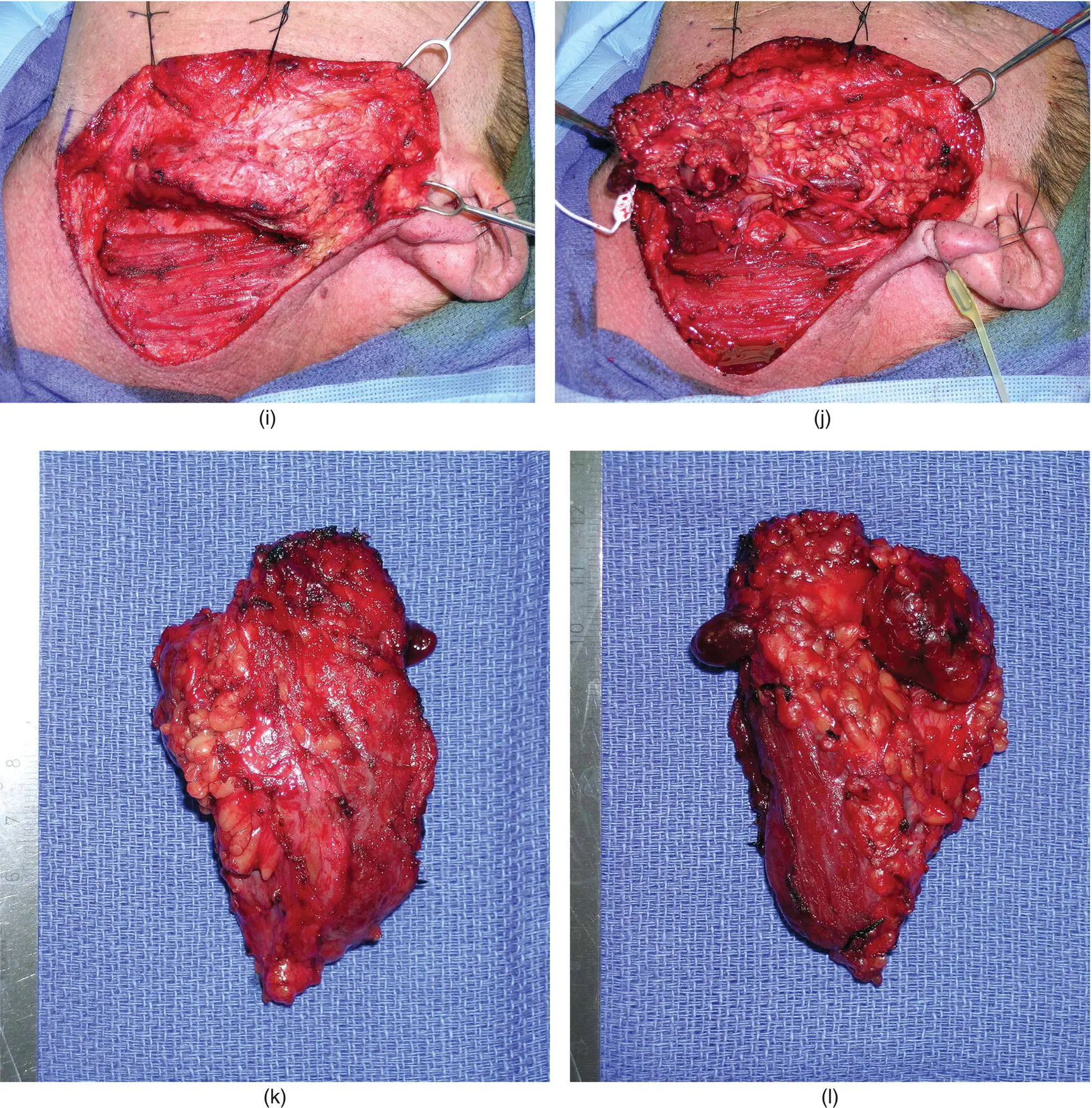
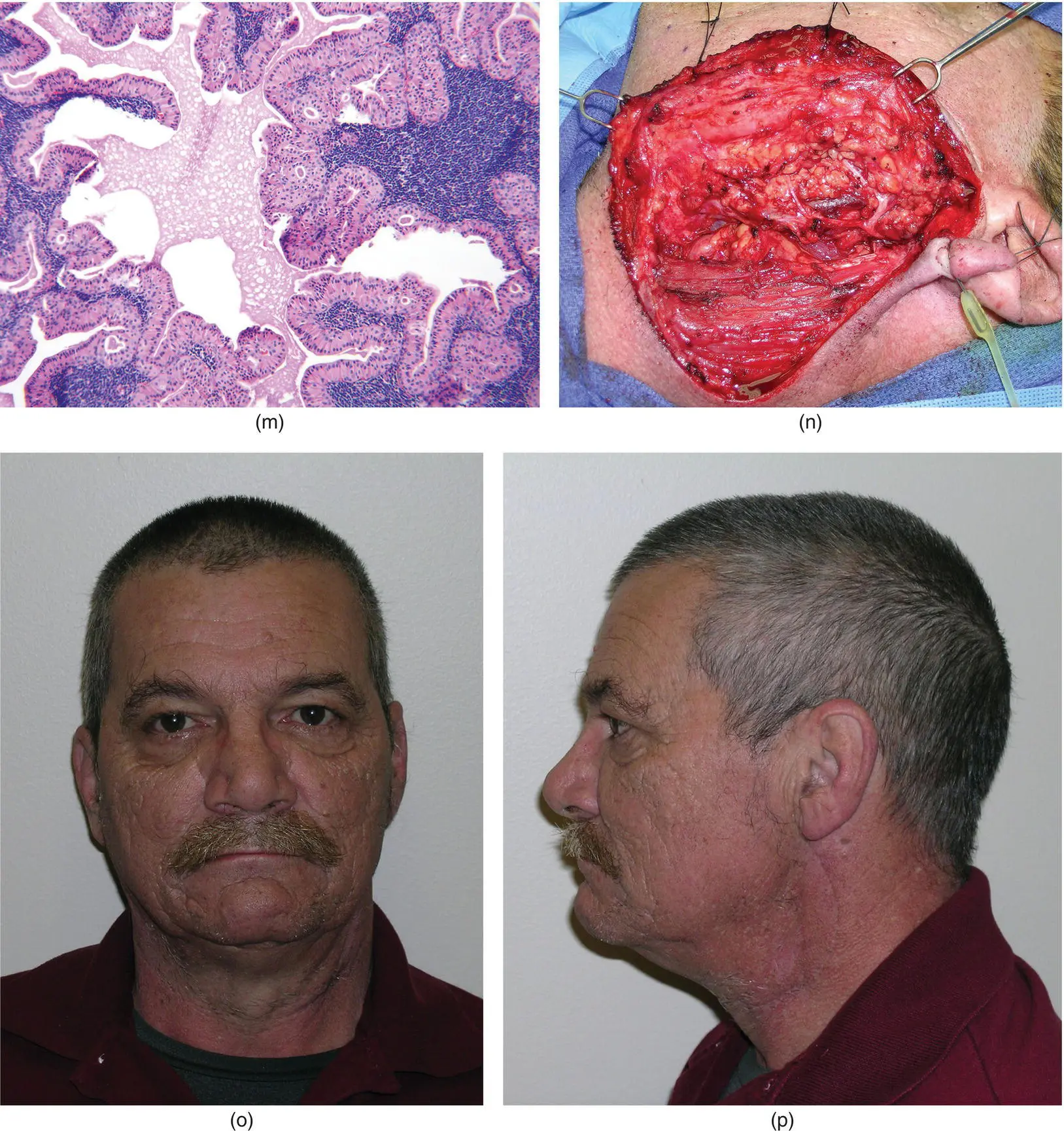
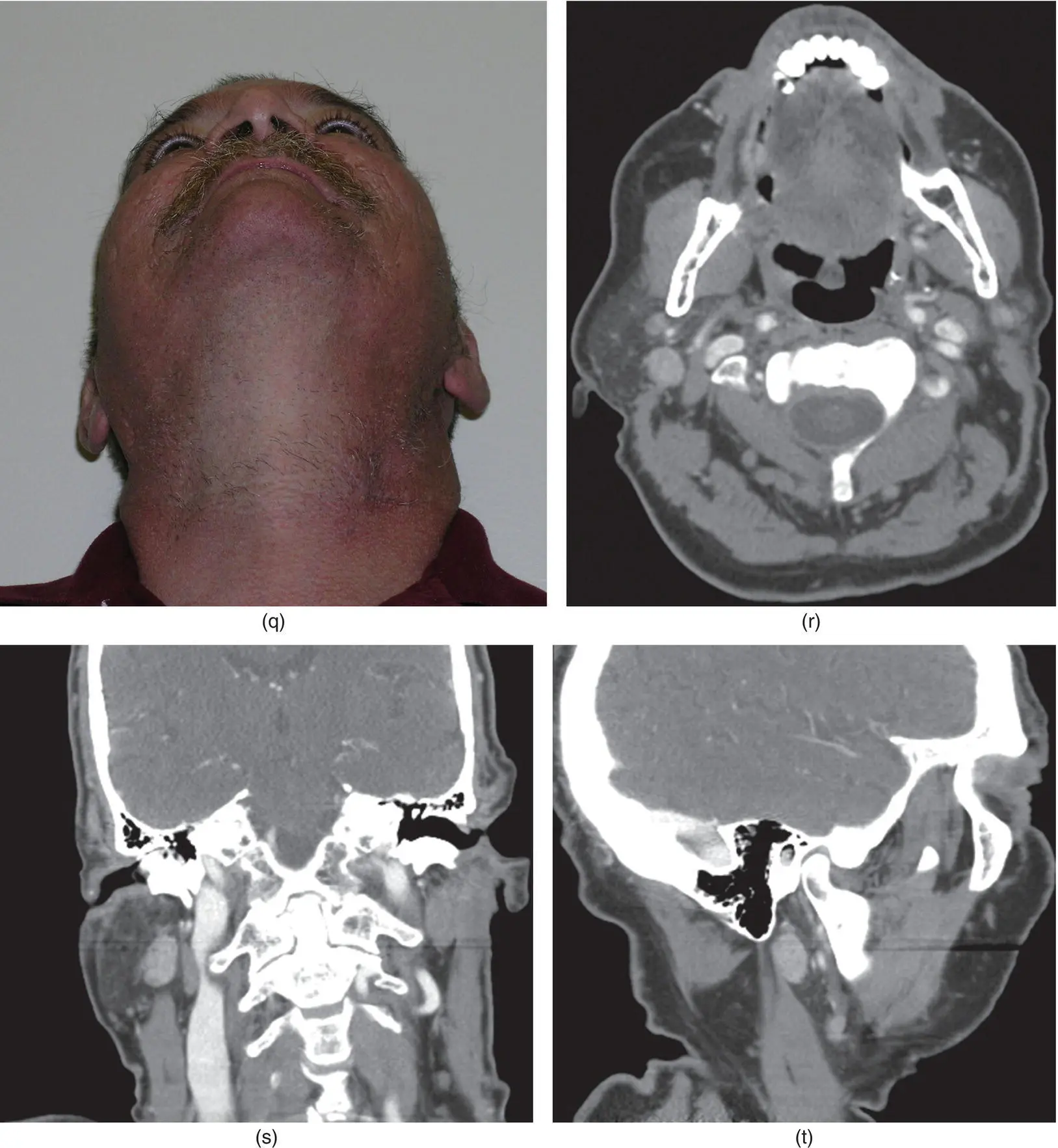
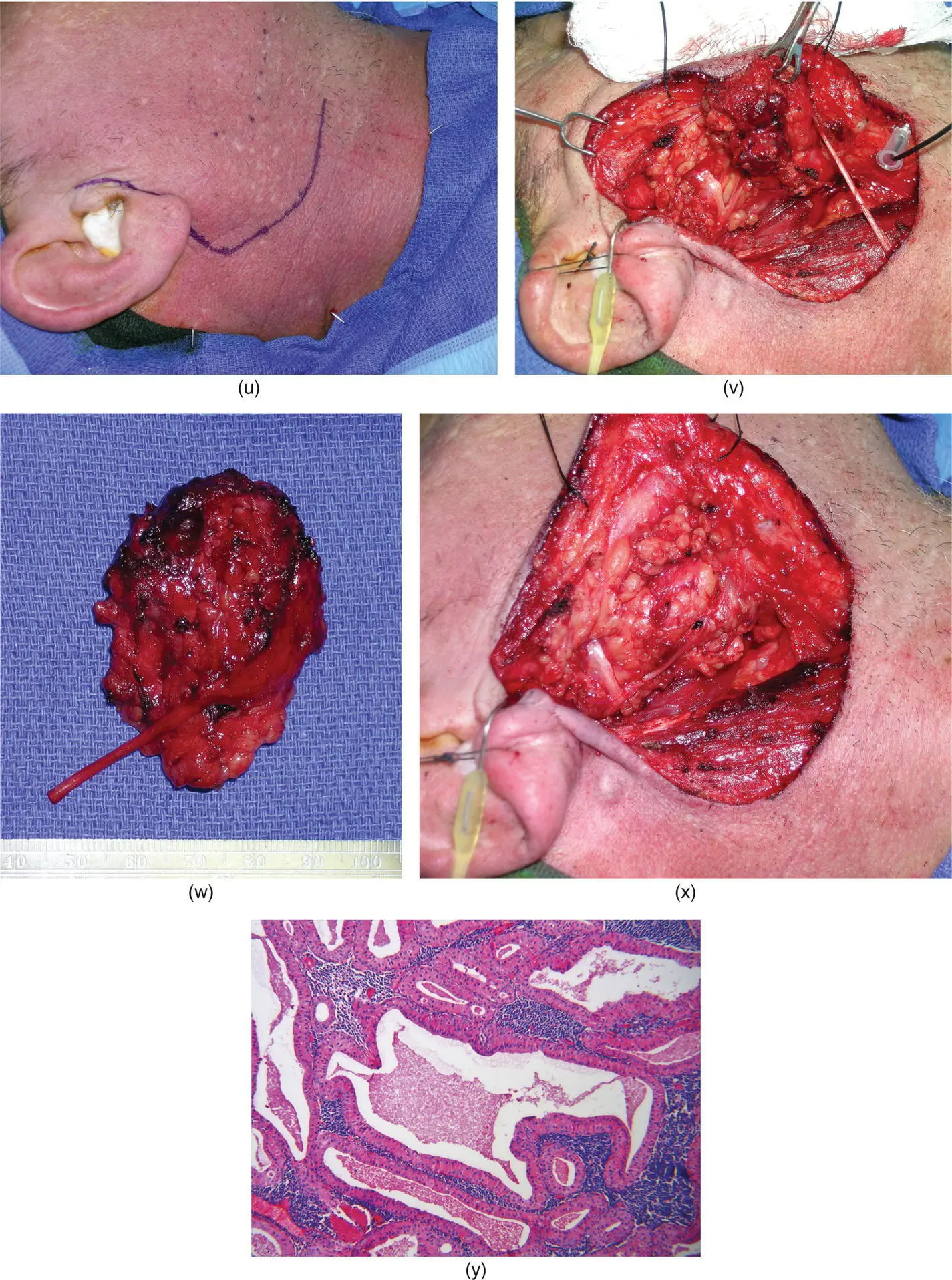
Figure 2.85. The patient demonstrates obvious left facial swelling in the region of the parotid gland (a–c). Axial (d), coronal (e), and sagittal (f) views demonstrate two tumors in the superficial lobe of the left parotid gland. Additionally, an enhancing but smaller tumor is noted in the right parotid gland (g). Synchronous, bilateral Warthin tumors are the suspected diagnoses. The patient underwent left superficial parotidectomy via a modified Blair incision (h). The skin flap is elevated anteriorly, superficial to the parotid capsule (i). The main trunk of the facial nerve and its peripheral branches are identified as the specimen is elevated (j). The specimen is delivered and inspected on its lateral (k) and medial surfaces (l). Final pathology identifies Warthin tumors of the left parotid gland (m). The resultant tissue bed and dissected facial nerve are noted (n). The patient is noted at five months postoperatively (o–q). A repeat CT scan is obtained and axial (r), coronal (s), and sagittal (t) images identify a larger tumor of the right parotid gland than was noted at the time of the patient's original presentation. He underwent a right superficial parotidectomy via a modified Blair incision (u). The specimen is elevated off the identified and preserved facial nerve (v). The specimen is delivered (w) and the resultant tissue bed is noted (x). Final pathology confirmed the clinical suspicion of Warthin tumor of the right parotid gland (y). (m = Hematoxylin and eosin, original magnification × 100; y = Hematoxylin and eosin, original magnification × 100).
References
1 Abdel‐Razek A, Kandeel A, Soliman N et al. 2007. Role of diffusion‐weighted echo‐planar MR imaging in differentiation of residual or recurrent head and neck tumors and post‐treatment changes. Am J Neuroradiol 28:1146–1152.
2 Alexander de Ru J, Van Leeuwen M, Van Benthem P et al. 2007. Do magnetic resonance imaging and ultrasound add anything to the workup of parotid gland tumors? J Oral Maxillofac Surg 65:945–952.
3 Alibek S, Zenk J, Bozzato A et al. 2007. The value of dynamic MRI studies in parotid tumors. Acad Radiol 14:701–710.
4 Ando M, Matsuzaki M, Murofushi T. 2005. Mucosa‐associated lymphoid tissue lymphoma presents as diffuse swelling of the parotid gland. Am J Otolaryngol 26:285–288.
5 Andre’ R, Becker M, Lombardi T, Buchholzer S, Marchal F, Seebach JD. 2021. Comparison of clinical characteristics and magnetic resonance imaging of salivary glands with magnetic resonance sialography in Sjogren’s syndrome. Laryngoscope 131 (1):E83–E89.
6 Aribandi M, Wood W, Elston D, Weiss D. 2006. CT features of plexiform neurofibroma of the submandibular gland. Am J Neuroradiol 27:126–128.
7 Baba S, Engles J, Huso D et al. 2007. Comparison of uptake of multiple clinical radiotracers into brown adipose tissue under cold‐stimulated and nonstimulated conditions. J Nucl Med 48:1715–1723.
8 Beale T, Madani G. 2006. Anatomy of the salivary glands. Semin Ultrasound CT MRI 27:436–439.
9 Beaulier S, Kinaha P, Tseng J et al. 2003. SUV varies with time after injection in 18F‐FDG PET of breast cancer: Characterization and method to adjust for time differences. J Nucl Med 44:1044–1050.
10 Bialek E, Jakubowski W, Zajkowski P et al. 2006. US of the major salivary glands: Anatomy and spatial relationships, pathologic conditions and pitfalls. Radiographics 26:745–763.
11 Bron L, Traynor S, McNeil E et al. 2003. Primary and metastatic cancer of the parotid: Comparison of clinical behavior in 232 cases. Laryngoscope 113(6):1070–1075.
12 Bui C, Ching A, Carlos R et al. 2003. Diagnostic accuracy of 2‐[fluorine‐18]‐fluro‐2‐deoxy‐D‐Glucose Positron Emission Tomography imaging in non‐squamous tumors of the head and neck. Invest Radiol 38:593–601.
13 Burke CJ, Thomas RH, Howlett D. 2011. Imaging the major salivary glands. Br J Oral Maxillofac Surg 49:261–269.
14 Burrell S, Van den Abbeele A. 2005. 2‐Deoxy‐2‐(F‐18)‐Fluoro‐D‐glucose‐‐Positron Emission tomography of the head and neck: An atlas of normal uptake and variants. Mol Imaging Biol 7:244–256.
15 Cermik T, Mavi A, Acikgoz G et al. 2007. FDG PET in detecting primary and recurrent malignant salivary gland tumors. Clin Nucl Med 32(4):286–291.
16 Chang P, Fischbein N, McCalmont T et al. 2004. Perineural spread of malignant melanoma of the head and neck: Clinical and imaging features. Am J Neuroradiol 25:5–11.
17 Ching A, Ahuja A, King A et al. 2001. Comparison of the sonographic features of acalculous and calculous submandibular sialadenitis. J Clin Ultrasound 29(6):332–338.
18 Cholankeril J, Scioscia P. 1993. Post‐traumatic sialoceles and mucoceles of the salivary glands. Clin Imaging 17(1):41–45.
19 Cohade C, Mourtzikos K, Wahl R. 2003a. USA‐fat: Prevalence is related to ambient outdoor temperature‐evaluation with 18F‐FDG PET/CT J Nucl Med 44:1267–1270.
20 Cohade C, Osman M, Pannu H, Wahl R. 2003b. Uptake in supraclavicular area fat (USA‐Fat): Description on 18F‐FDG PET/CT J Nucl Med 44:170–176.
21 Delbeke D, Coleman R, Guiberteau M et al. 2006. Procedure guidelines for tumor imaging with 18F‐FDG PET/CT J Nucl Med 47:887–895.
22 Drumond J. 1995. Tomographic measurements of age changes in the human parotid gland. Gerodontology 12(1):26–30.
23 Eichhorn K, Iakovos A, Ridder G. 2002. Malignant non‐Hodgkin’s lymphoma mimicking a benign parotid tumor: Sonographic findings. J Clin Ultrasound 30(1):42–44.
24 Eida S, Sumi M, Sakihama N et al. 2007. Apparent diffusion coefficient mapping of salivary gland tumors: Prediction of the benignancy and malignancy. Am J Neuroradiol 28:116–121.
25 Gerstle R, Aylward S, Kromhout‐Schiro S et al. 2000. The role of neural networks in improving the accuracy of MR spectroscopy for the diagnosis of head and neck squamous cell carcinoma. Am J Neuroradiol 21:1133–1138.
26 Golder W, Stiller M. 2014. Distribution pattern of Sjogren’s syndrome: A sialographical study. Z Rheumatol 10:1–6.
27 Habermann C, Gossrau P, Kooijman H et al. 2007. Monitoring of gustatory stimulation of salivary glands by diffusion weighted MR imaging: Comparison of 1.5T and 3T. Am J Neuroradiol 28:1547–1551.
28 Hadi M, Chen C, Millie W et al. 2007. PET/CT, and 123I‐MIBG SPECT: A study of patients being evaluated for pheochromocytoma. J Nucl Med 48:1077–1083.
29 Haldar S, Sinnott JD, Tekeli KM, Turner SS, Howlett DC. 2016. Biopsy of parotid masses: Review of current techniques. World J Radiol 8(5):501–505.
Читать дальшеИнтервал:
Закладка:
Похожие книги на «Salivary Gland Pathology»
Представляем Вашему вниманию похожие книги на «Salivary Gland Pathology» списком для выбора. Мы отобрали схожую по названию и смыслу литературу в надежде предоставить читателям больше вариантов отыскать новые, интересные, ещё непрочитанные произведения.


Обсуждение, отзывы о книге «Salivary Gland Pathology» и просто собственные мнения читателей. Оставьте ваши комментарии, напишите, что Вы думаете о произведении, его смысле или главных героях. Укажите что конкретно понравилось, а что нет, и почему Вы так считаете.