Salivary Gland Pathology
Здесь есть возможность читать онлайн «Salivary Gland Pathology» — ознакомительный отрывок электронной книги совершенно бесплатно, а после прочтения отрывка купить полную версию. В некоторых случаях можно слушать аудио, скачать через торрент в формате fb2 и присутствует краткое содержание. Жанр: unrecognised, на английском языке. Описание произведения, (предисловие) а так же отзывы посетителей доступны на портале библиотеки ЛибКат.

- Название:Salivary Gland Pathology
- Автор:
- Жанр:
- Год:неизвестен
- ISBN:нет данных
- Рейтинг книги:3 / 5. Голосов: 1
-
Избранное:Добавить в избранное
- Отзывы:
-
Ваша оценка:
Salivary Gland Pathology: краткое содержание, описание и аннотация
Предлагаем к чтению аннотацию, описание, краткое содержание или предисловие (зависит от того, что написал сам автор книги «Salivary Gland Pathology»). Если вы не нашли необходимую информацию о книге — напишите в комментариях, мы постараемся отыскать её.
now features case presentations to place the information in context, as well as additional treatment algorithms in each chapter to assist in clinical decision making. Written by highly respected clinicians, educators, and researchers with extensive expertise in oral and maxillofacial surgery, this authoritative resource:
Reviews the etiology, diagnosis, and treatment of all salivary gland pathologies, with detailed explanations and hundreds of full-color clinical images Incorporates new information on the taxonomy of salivary gland tumors, neoplastic and non-neoplastic entities, image guided biopsies of salivary gland lesions, and complications in traditional and non-traditional forms of salivary gland surgery Offers expanded coverage of histopathology, including classification, grading, and staging of salivary gland tumors Features up-to-date chapters on anatomy and physiology, imaging, cysts, systemic diseases, tumors, trauma, and innovative salivary gland surgical techniques Includes a discussion of meta-analyses and systematic reviews to support evidence-based practice remains the definitive resource for oral and maxillofacial surgeons, otolaryngologists, head and neck surgeons, and general surgeons as well as their residents providing care for patients with salivary gland pathology.
Salivary Gland Pathology — читать онлайн ознакомительный отрывок
Ниже представлен текст книги, разбитый по страницам. Система сохранения места последней прочитанной страницы, позволяет с удобством читать онлайн бесплатно книгу «Salivary Gland Pathology», без необходимости каждый раз заново искать на чём Вы остановились. Поставьте закладку, и сможете в любой момент перейти на страницу, на которой закончили чтение.
Интервал:
Закладка:
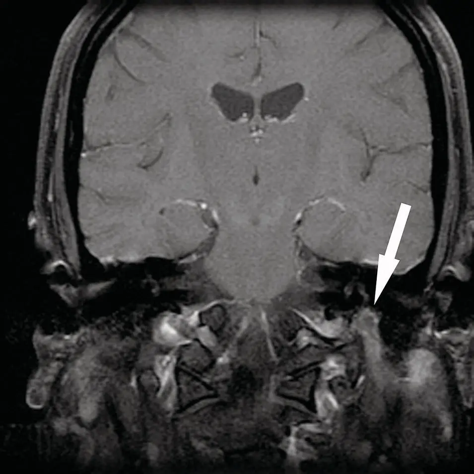
Figure 2.74. Coronal T1 contrast‐enhanced MRI demonstrating a mass in the left parotid gland with smooth margins. The mass extends superiorly into the skull base at the stylomastoid foramen (arrow). A benign schwannoma was diagnosed.
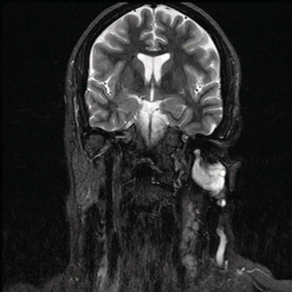
Figure 2.75.Coronal T2 MRI corresponding to the case illustrated in Figure 2.74.
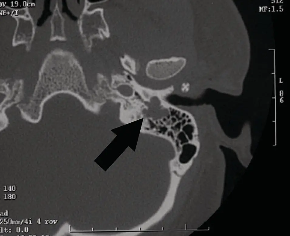
Figure 2.76. Axial CT at the skull base displayed in bone window showing dilatation of the stylomastoid foramen with soft tissue mass (arrow). A benign schwannoma was diagnosed.
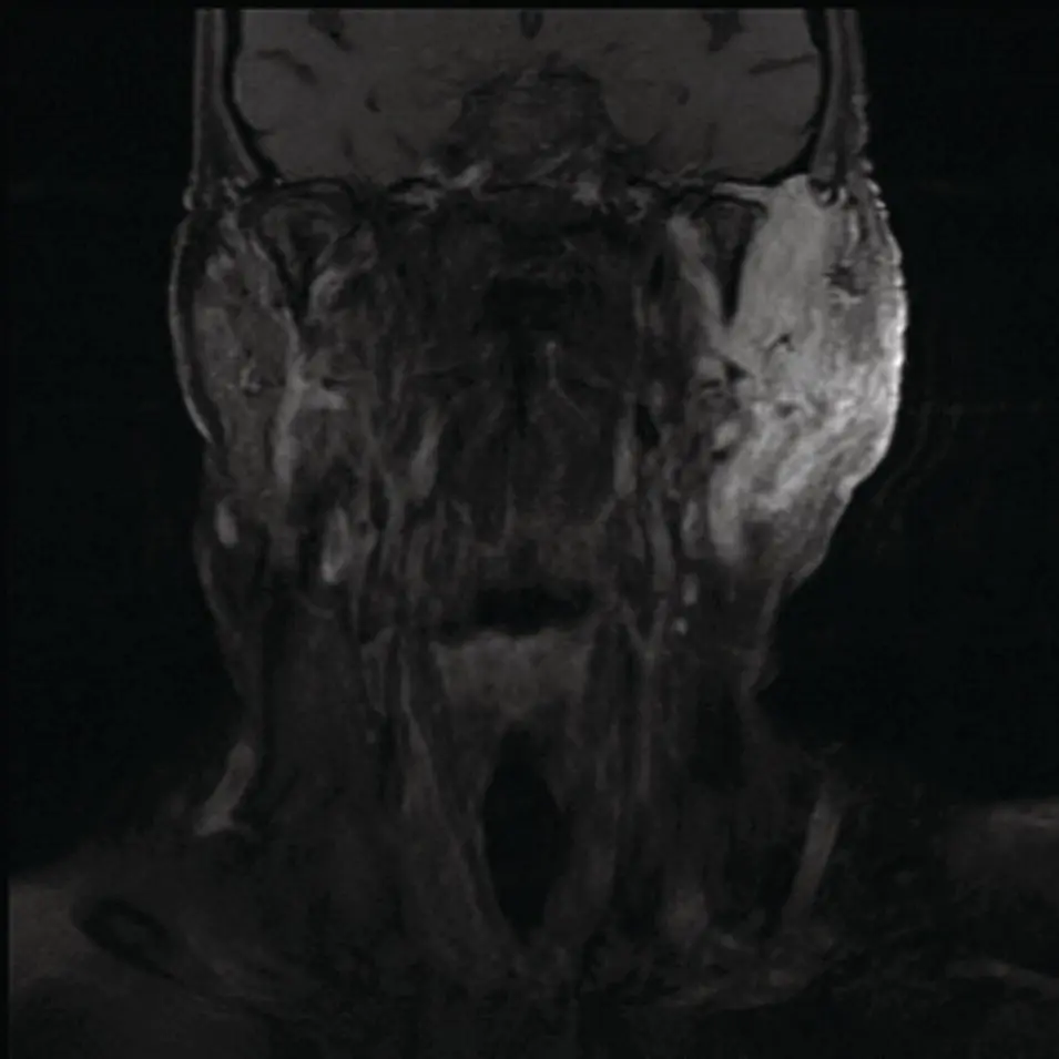
Figure 2.77. Coronal T1 contrast image showing a very ill‐defined mass with heterogenous enhancement in the parotid gland with skull base extension via the stylomastoid foramen. A malignant schwannoma was diagnosed.
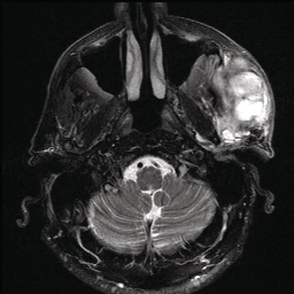
Figure 2.78. Axial T2 MRI image corresponding to the case illustrated in Figure 2.77.
Malignant
Lymphoma
Both primary and secondary lymphomas of the salivary glands are rare. Primary lymphoma of the salivary glands is the mucosa‐associated lymphoid tissue subtype (MALT). MALT lymphomas constitute about 5% of non‐Hodgkin lymphomas (Jhanvar and Straus 2006). These lymphomas are seen in the gastrointestinal tract and are associated with chronic inflammatory or autoimmune diseases. The salivary glands do not typically contain MALT but may in the setting of chronic inflammation (Ando et al. 2005). The MALT lymphomas found in the gastrointestinal tract are not typically associated with Sjögren syndrome. The MALT lymphoma, a low‐grade B‐cell type, tends to be a slow‐growing neoplasm. Metastases tend to occur at other mucosal sites, a demonstration of tissue tropism. The MALT lymphomas are amenable to radiotherapy but can relapse in the contralateral gland, demonstrating tropism for the glandular tissue (MacManus et al. 2007). In Sjögren syndrome, there is an approximately 40‐fold increased incidence of developing lymphoma compared to age‐controlled populations. Of the various subtypes of lymphoma that are seen associated with Sjögren syndrome (follicular, diffuse large B‐cell, large cell, and immunoblastic), the MALT subtype is the most common, at around 50% (Tonami et al. 2002). The parotid gland is the most commonly affected (80%). Less commonly, the submandibular and rarely the sublingual gland may be involved. Clinically, it may present with a focal mass or diffuse unilateral or bilateral glandular swelling.
67Ga‐citrate scintigraphy had been the standard imaging modality used to assess staging and post‐therapy follow‐up for lymphomas (Hodgkin and non‐Hodgkin) for many decades. PET/CT with FDG is quickly becoming the standard for staging and follow‐up for many lymphoma subtypes (Jhanvar and Straus 2006).
The imaging findings in salivary lymphomas, however, are not specific. CT may demonstrate focal or diffuse low‐ to intermediate‐density mass with cystic areas and calcifications. MRI shows the soft‐tissue areas to be isointense to skeletal muscle on T1 images and hypointense relative to fat on T2 images along with diffuse enhancement post‐contrast (Tonami et al. 2002). Although, there may be cystic changes demonstrated by CT, MRI, or US, they are thought to be dilated ducts resulting from compression of terminal ducts (Ando et al. 2005). The US characteristics of MALT lymphoma may demonstrate multifocal hypoechoic intraparotid nodules and cysts (which may be dilated ducts) and calcification as well. Large B‐cell intraparotid lymphoma has been described as hypoechoic, homogenous, well‐marginated mass exhibiting increased thru transmission (a characteristic of cysts) and hypervascularity (Eichhorn et al. 2002). Although there are reports of hypermetabolism in MALT lymphomas, PET imaging findings are also not conclusive (MacManus et al. 2007). Uptake in the tumor and a background of chronic inflammatory changes of chronic sialadenitis may result in variably elevated uptake of FDG.
Secondary lymphomas (Hodgkin and non‐Hodgkin) are also quite rare, with the histology most commonly encountered being of the large cell type. There is typically extra‐glandular lymphadenopathy associated. The imaging features are also non‐specific, although there is usually no associated chronic sialadenitis ( Figures 2.79through 2.81).
Metastases
Intraglandular lymph nodes are found in the parotid gland due to its early encapsulation during development. The sublingual gland and submandibular gland do not contain lymph nodes. The parotid and periparotid lymph nodes are the first order nodal site for lesions that affect the scalp, skin of the upper face, and external ear (Ollila et al. 1999). The most common malignancy to metastasize to the parotid nodes is squamous cell carcinoma followed by melanoma and, less commonly, Merkel cell carcinoma (Bron et al. 2003) ( Figures 2.82through 2.84).
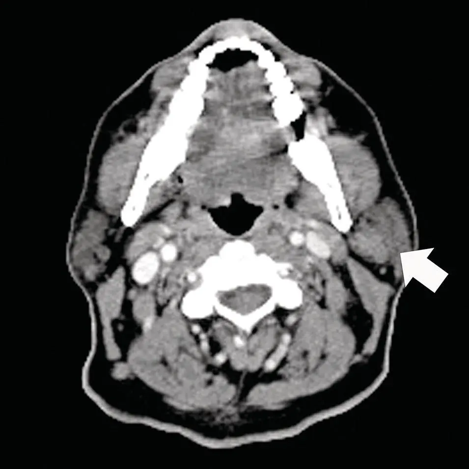
Figure 2.79. Axial CT scan with contrast at the level of the parotid tail demonstrating an ill‐defined heterogeneously enhancing mass adjacent to or exophytic from the parotid tail medially (arrow). Lymphoma in cervical lymphadenopathy was diagnosed at surgery.
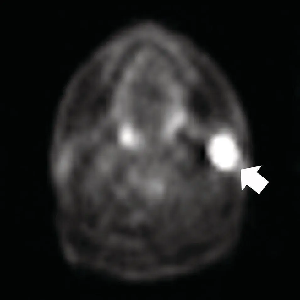
Figure 2.80.Axial PET scan image corresponding to the case in Figure 2.79. A large mass of the left parotid gland (arrow) is noted.
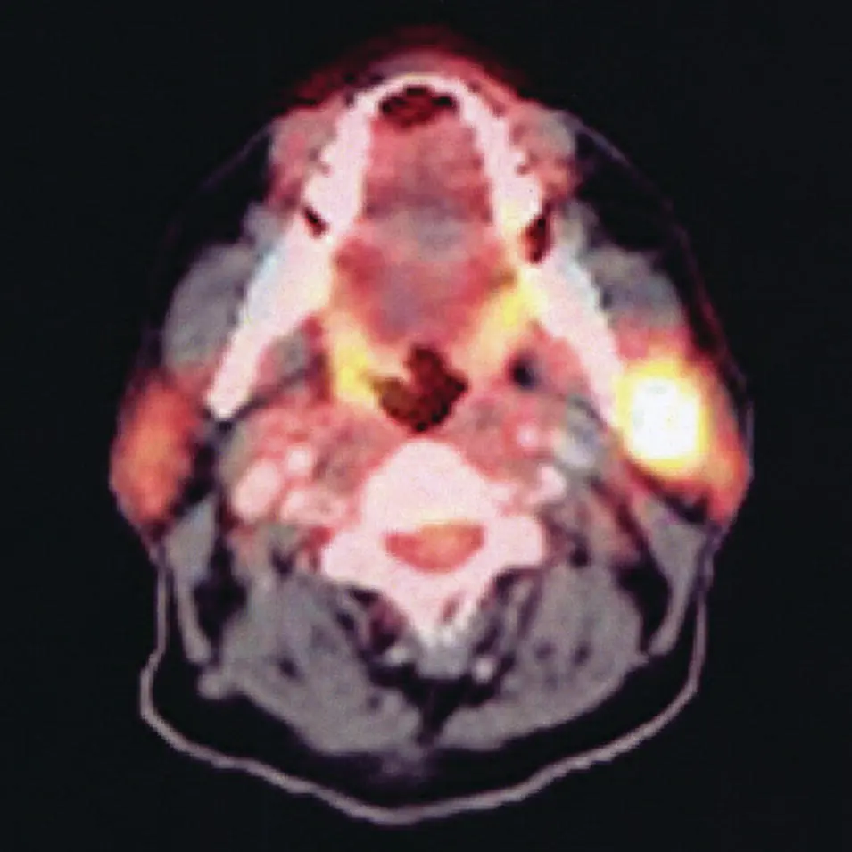
Figure 2.81. Fused axial PET/CT image corresponding to the case illustrated in Figure 2.79.
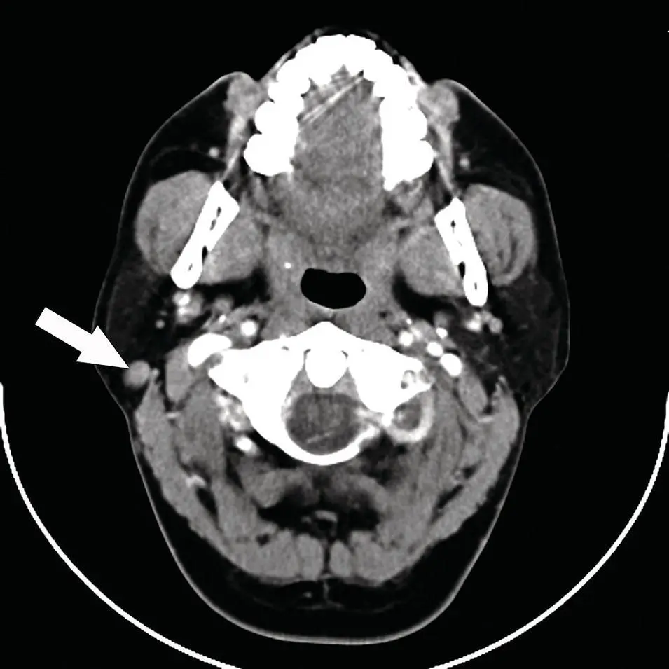
Figure 2.82. Axial CT of a mass in the right parotid gland with homogenous enhancement. The patient had a history of right facial melanoma. Metastatic melanoma was diagnosed at surgery.
The imaging findings are not specific. CT in early stages demonstrates the nodes to have sharp margins, round or ovoid architecture but without a fatty hilum. Late in the disease, mass can mimic infected or inflammatory nodes with heterogenous borders, enhancement, and necrosis. Late in the disease with extranodal spread, the margins blur and are ill‐defined. Contrast enhancement is heterogenous. Similar findings are seen on MRI with T1 showing low to intermediate signal pre‐contrast and homogenous to heterogenous signal post‐contrast depending on intranodal versus extranodal disease. PET with FDG is abnormal in infectious, inflammatory, and neoplastic etiology and is not typically helpful within the parotid, but can aid in localizing the site of the primary lesion as well as other sites of metastases. This can be significant since the incidence of clinically occult neck disease is high in skin cancer metastatic to the parotid gland (Bron et al. 2003). Local failure was highest with metastatic squamous cell carcinoma and distant metastases were higher in melanoma (Bron et al. 2003).
Читать дальшеИнтервал:
Закладка:
Похожие книги на «Salivary Gland Pathology»
Представляем Вашему вниманию похожие книги на «Salivary Gland Pathology» списком для выбора. Мы отобрали схожую по названию и смыслу литературу в надежде предоставить читателям больше вариантов отыскать новые, интересные, ещё непрочитанные произведения.


Обсуждение, отзывы о книге «Salivary Gland Pathology» и просто собственные мнения читателей. Оставьте ваши комментарии, напишите, что Вы думаете о произведении, его смысле или главных героях. Укажите что конкретно понравилось, а что нет, и почему Вы так считаете.