Salivary Gland Pathology
Здесь есть возможность читать онлайн «Salivary Gland Pathology» — ознакомительный отрывок электронной книги совершенно бесплатно, а после прочтения отрывка купить полную версию. В некоторых случаях можно слушать аудио, скачать через торрент в формате fb2 и присутствует краткое содержание. Жанр: unrecognised, на английском языке. Описание произведения, (предисловие) а так же отзывы посетителей доступны на портале библиотеки ЛибКат.

- Название:Salivary Gland Pathology
- Автор:
- Жанр:
- Год:неизвестен
- ISBN:нет данных
- Рейтинг книги:3 / 5. Голосов: 1
-
Избранное:Добавить в избранное
- Отзывы:
-
Ваша оценка:
Salivary Gland Pathology: краткое содержание, описание и аннотация
Предлагаем к чтению аннотацию, описание, краткое содержание или предисловие (зависит от того, что написал сам автор книги «Salivary Gland Pathology»). Если вы не нашли необходимую информацию о книге — напишите в комментариях, мы постараемся отыскать её.
now features case presentations to place the information in context, as well as additional treatment algorithms in each chapter to assist in clinical decision making. Written by highly respected clinicians, educators, and researchers with extensive expertise in oral and maxillofacial surgery, this authoritative resource:
Reviews the etiology, diagnosis, and treatment of all salivary gland pathologies, with detailed explanations and hundreds of full-color clinical images Incorporates new information on the taxonomy of salivary gland tumors, neoplastic and non-neoplastic entities, image guided biopsies of salivary gland lesions, and complications in traditional and non-traditional forms of salivary gland surgery Offers expanded coverage of histopathology, including classification, grading, and staging of salivary gland tumors Features up-to-date chapters on anatomy and physiology, imaging, cysts, systemic diseases, tumors, trauma, and innovative salivary gland surgical techniques Includes a discussion of meta-analyses and systematic reviews to support evidence-based practice remains the definitive resource for oral and maxillofacial surgeons, otolaryngologists, head and neck surgeons, and general surgeons as well as their residents providing care for patients with salivary gland pathology.
Salivary Gland Pathology — читать онлайн ознакомительный отрывок
Ниже представлен текст книги, разбитый по страницам. Система сохранения места последней прочитанной страницы, позволяет с удобством читать онлайн бесплатно книгу «Salivary Gland Pathology», без необходимости каждый раз заново искать на чём Вы остановились. Поставьте закладку, и сможете в любой момент перейти на страницу, на которой закончили чтение.
Интервал:
Закладка:
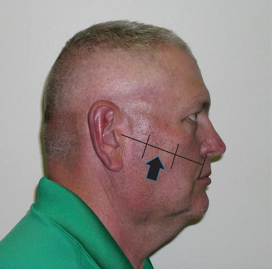
Figure 1.8. The surface markings for the location of the parotid duct.
Nerve Supply to the Parotid
The parasympathetic secretomotor nerve supply comes from the inferior salivatory nucleus in the brain stem ( Figure 1.9). From there, the fibers run in the tympanic branch of the glossopharyngeal nerve contributing to the tympanic plexus in the middle ear. The lesser petrosal nerve arises from the tympanic plexus leaving the middle ear and running in a groove on the petrous temporal bone in the middle cranial fossa. From here, it exits through the foramen ovale to the otic ganglion which lies on the medial aspect of the mandibular branch of the trigeminal nerve. Postsynaptic postganglionic fibers leave the ganglion to join the auriculotemporal nerve which distributes the parasympathetic secretomotor fibers throughout the parotid gland. Some authorities suggest that there are also some parasympathetic innervations to the parotid from the chorda tympani branch of the facial nerve.
The sympathetic nerve supply to the parotid arises from the superior cervical sympathetic ganglion. The sympathetic fibers reach the gland via the plexus around the middle meningeal artery. They then pass through the otic ganglion without synapsing and innervate the gland through the auriculotemporal nerve. There is also sympathetic innervation to the gland arising from the plexuses that accompany the blood vessels supplying the gland.
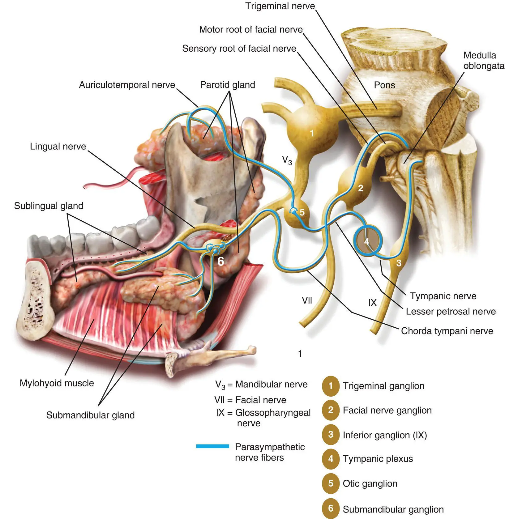
Figure 1.9. The parasympathetic innervations of the salivary glands. The parasympathetic fibers are shown as blue lines.
Sensory fibers arising from the connective tissue within the parotid gland merge into the auriculotemporal nerve and pass proximally through the otic ganglion without synapsing. From there, the fibers join the mandibular division of the trigeminal nerve. The sensory innervation of the parotid capsule is via the great auricular nerve.
Submandibular Gland
EMBRYOLOGY
The submandibular gland begins to form at the 13 mm stage in the seventh week of intrauterine life (Zhang et al. 2010; Berta et al. 2013; Chadi et al. 2017) as an epithelial outgrowth into the mesenchyme forming the floor of the mouth in the linguogingival groove. This proliferates rapidly giving off numerous branching processes which eventually develop lumina. Initially, the developing gland opens into the floor of the mouth posteriorly, lateral to the tongue. The walls of the groove into which it drains come together to form the submandibular duct. This process commences posteriorly and moves forwards so that ultimately the orifice of the duct comes to lie anteriorly below the tip of the tongue close to the midline.
ANATOMY
The submandibular gland consists of a larger superficial lobe lying within the digastric triangle in the neck and a smaller deep lobe lying within the floor of the mouth posteriorly ( Figure 1.10). The two lobes are continuous with each other around the posterior border of the mylohyoid muscle. As in the parotid gland, the two “lobes” are not true lobes embryologically as the gland arises as a single epithelial outgrowth (Langdon 1998a). However, surgically it consists of the two lobes as described above. It is a mixed seromucinous gland.
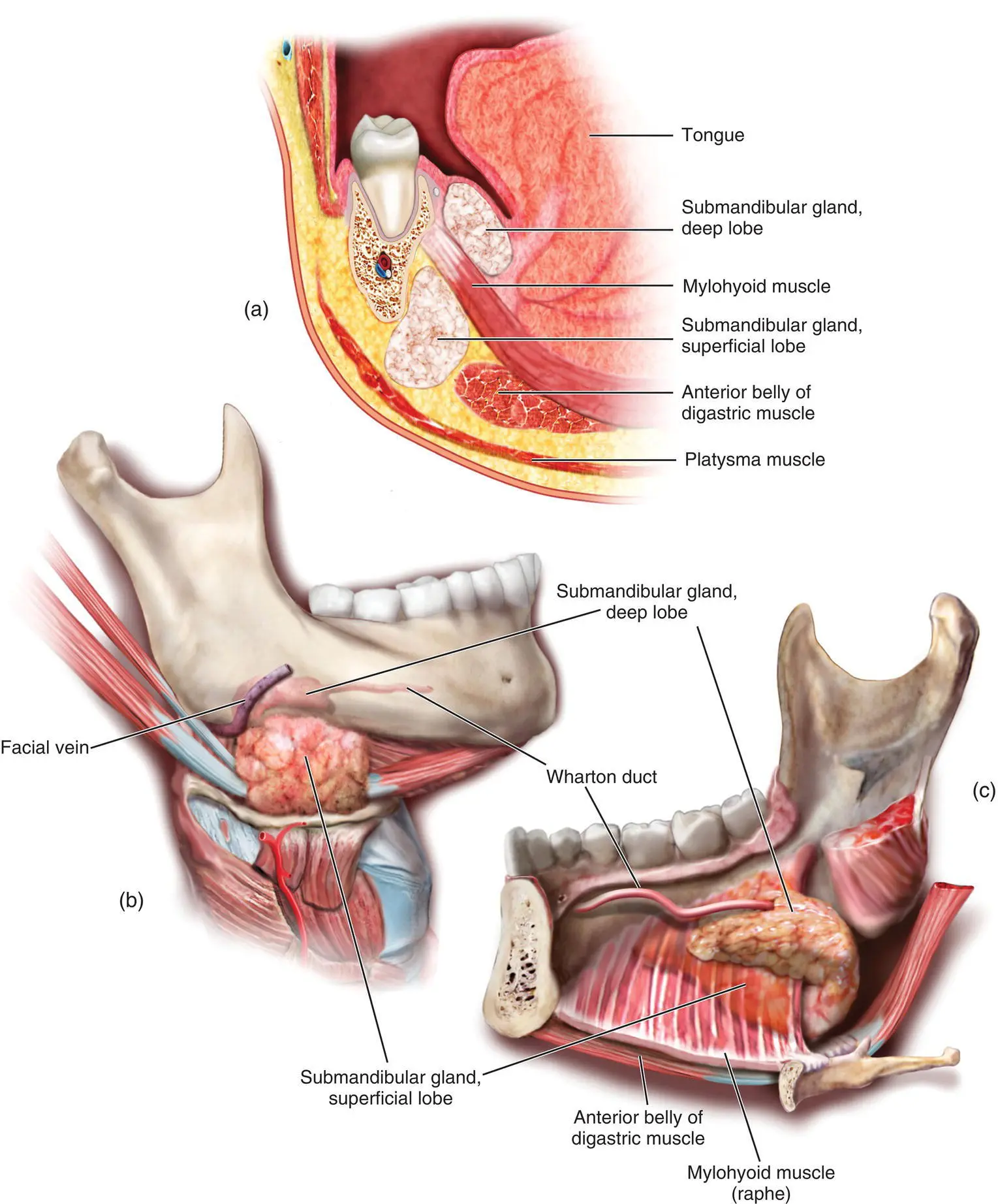
Figure 1.10. The relationship of the superficial and deep lobes of the submandibular gland. (a) cross‐sectional anatomy. (b) The superficial lobe from outside. (c) The relationship of the deep and superficial lobes to the mylohyoid muscle.
Superficial Lobe
The superficial lobe lies within the digastric triangle. Its anterior pole reaches the anterior belly of the digastric muscle and the posterior pole reaches the stylomandibular ligament. This structure is all that separates the superficial lobe of the submandibular gland from the parotid gland. It is important to realize just how close the lower pole of the parotid is to the posterior pole of the submandibular gland as confusion can arise if a mass in the region is incorrectly ascribed to the wrong anatomical structure ( Figure 1.2). Superiorly, the superficial lobe lies medial to the body of the mandible. Inferiorly, it often overlaps the intermediate tendon of the digastric muscles and the insertion of the stylohyoid muscle. The lobe is partially enclosed between the two layers of the deep cervical fascia that arise from the greater cornu of the hyoid bone and is in intimate proximity of the facial vein and artery ( Figure 1.11). The superficial layer of the fascia is attached to the lower border of the mandible and covers the inferior surface of the superficial lobe. The deep layer of fascia is attached to the mylohyoid line on the inner aspect of the mandible and therefore covers the medial surface of the lobe.
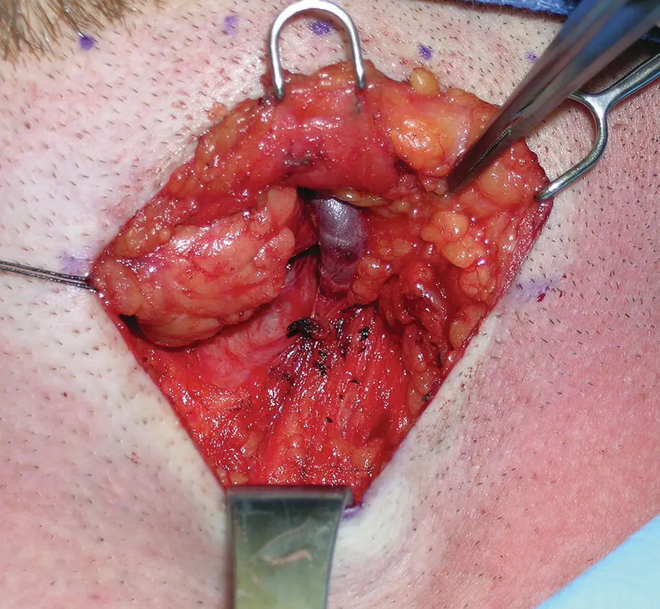
Figure 1.11 Superficial dissection of the left submandibular gland. The investing layer of the deep cervical fascia is elevated off the submandibular gland and the facial vein is identified.
The inferior surface, which is covered by skin, subcutaneous fat, platysma, and the deep fascia, is crossed by the facial vein and the cervical branch of the facial nerve which loops down from the angle of the mandible and subsequently innervates the lower lip. The submandibular lymph nodes lie between the salivary gland and the mandible. Sometimes one or more lymph nodes may be embedded within the salivary gland.
The lateral surface of the superficial lobe is related to the submandibular fossa, a concavity on the medial surface of the mandible, and the attachment of the medial pterygoid muscle. The facial artery grooves its posterior part lying at first deep to the lobe and then emerging between its lateral surface and the mandibular attachment of the medial pterygoid muscle from which it reaches the lower border of the mandible.
The medial surface is related anteriorly to the mylohyoid from which it is separated by the mylohyoid nerve and submental vessels. Posteriorly, it is related to the styloglossus muscle, the stylohyoid ligament, and the glossopharyngeal nerve separating it from the pharynx. Between these, the medial aspect of the lobe is related to the hyoglossus muscle from which it is separated by the styloglossus muscle, the lingual nerve, the submandibular ganglion, the hypoglossal nerve, and the deep lingual vein. More inferiorly, the medial surface is related to the stylohyoid muscle and the posterior belly of digastric.
Deep Lobe
The deep lobe of the gland arises from the superficial lobe at the posterior free edge of the mylohyoid muscle and extends forward to the back of the sublingual gland ( Figure 1.12). It lies between the mylohyoid inferolaterally, the hyoglossus, and the styloglossus muscles medially, the lingual nerve superiorly and the hypoglossal nerve and deep lingual vein inferiorly.
Submandibular Duct
The submandibular duct is about 62 mm long and 3 mm in diameter in the adult. The wall of the submandibular duct is thinner than that of the parotid duct. It arises from numerous tributaries in the superficial lobe and emerges from the medial surface of this lobe just behind the posterior border of the mylohyoid. It crosses the deep lobe, passing upwards, and slightly backward for 5 mm before running forwards between the mylohyoid and hyoglossus muscles. As it passes forward, it runs between the sublingual gland and genioglossus to open into the floor of the mouth on the summit of the sublingual papilla at the side of the lingual frenum just below the tip of the tongue. It lies between the lingual and hypoglossal nerves on the hyoglossus. At the anterior border of the hyoglossus muscle, it is crossed by the lingual nerve. As the duct traverses the deep lobe of the gland, it receives tributaries draining that lobe.
Читать дальшеИнтервал:
Закладка:
Похожие книги на «Salivary Gland Pathology»
Представляем Вашему вниманию похожие книги на «Salivary Gland Pathology» списком для выбора. Мы отобрали схожую по названию и смыслу литературу в надежде предоставить читателям больше вариантов отыскать новые, интересные, ещё непрочитанные произведения.


Обсуждение, отзывы о книге «Salivary Gland Pathology» и просто собственные мнения читателей. Оставьте ваши комментарии, напишите, что Вы думаете о произведении, его смысле или главных героях. Укажите что конкретно понравилось, а что нет, и почему Вы так считаете.