Salivary Gland Pathology
Здесь есть возможность читать онлайн «Salivary Gland Pathology» — ознакомительный отрывок электронной книги совершенно бесплатно, а после прочтения отрывка купить полную версию. В некоторых случаях можно слушать аудио, скачать через торрент в формате fb2 и присутствует краткое содержание. Жанр: unrecognised, на английском языке. Описание произведения, (предисловие) а так же отзывы посетителей доступны на портале библиотеки ЛибКат.

- Название:Salivary Gland Pathology
- Автор:
- Жанр:
- Год:неизвестен
- ISBN:нет данных
- Рейтинг книги:3 / 5. Голосов: 1
-
Избранное:Добавить в избранное
- Отзывы:
-
Ваша оценка:
Salivary Gland Pathology: краткое содержание, описание и аннотация
Предлагаем к чтению аннотацию, описание, краткое содержание или предисловие (зависит от того, что написал сам автор книги «Salivary Gland Pathology»). Если вы не нашли необходимую информацию о книге — напишите в комментариях, мы постараемся отыскать её.
now features case presentations to place the information in context, as well as additional treatment algorithms in each chapter to assist in clinical decision making. Written by highly respected clinicians, educators, and researchers with extensive expertise in oral and maxillofacial surgery, this authoritative resource:
Reviews the etiology, diagnosis, and treatment of all salivary gland pathologies, with detailed explanations and hundreds of full-color clinical images Incorporates new information on the taxonomy of salivary gland tumors, neoplastic and non-neoplastic entities, image guided biopsies of salivary gland lesions, and complications in traditional and non-traditional forms of salivary gland surgery Offers expanded coverage of histopathology, including classification, grading, and staging of salivary gland tumors Features up-to-date chapters on anatomy and physiology, imaging, cysts, systemic diseases, tumors, trauma, and innovative salivary gland surgical techniques Includes a discussion of meta-analyses and systematic reviews to support evidence-based practice remains the definitive resource for oral and maxillofacial surgeons, otolaryngologists, head and neck surgeons, and general surgeons as well as their residents providing care for patients with salivary gland pathology.
Salivary Gland Pathology — читать онлайн ознакомительный отрывок
Ниже представлен текст книги, разбитый по страницам. Система сохранения места последней прочитанной страницы, позволяет с удобством читать онлайн бесплатно книгу «Salivary Gland Pathology», без необходимости каждый раз заново искать на чём Вы остановились. Поставьте закладку, и сможете в любой момент перейти на страницу, на которой закончили чтение.
Интервал:
Закладка:
On entering the parotid gland, the facial nerve divides into two divisions, temporofacial and cervicofacial the former being the larger. The division of the facial nerve is sometimes called the pes anserinus due to its resemblance to the foot of a goose. From the temporofacial and cervicofacial divisions, the facial nerve gives rise to five named branches – temporal, zygomatic, buccal, mandibular, and cervical ( Figure 1.5). The peripheral branches of the facial nerve form variable anastomotic arcades between adjacent branches to form the parotid plexus. These anastomoses are important during facial nerve dissection as accidental damage to a small branch often fails to result in any facial weakness due to dual innervation from adjacent branches. Davis et al. (1956) studied these patterns following the dissection of 350 facial nerves in cadavers. The anastomotic relationships between adjacent branches fell into six patterns ( Figure 1.6). They showed that in only 6% of cases (type VI) is there any anastomosis between the mandibular branch and adjacent branches. This explains why, when transient facial weakness follows facial nerve dissection, it is usually the mandibular branch that is affected.
Auriculotemporal Nerve
The auriculotemporal nerve arises from the posterior division of the mandibular division of the trigeminal nerve in the infratemporal fossa. It runs backward beneath the lateral pterygoid muscle between the medial aspect of the condylar neck and the sphenomandibular ligament. It enters the anteromedial surface of the parotid gland passing upwards and outwards to emerge at the superior border of the gland between the temporomandibular joint and the external acoustic meatus. This nerve communicates widely with the temporofacial division of the facial nerve and limits the mobility of the facial nerve during surgery (Flatau and Mills 1995). Further communications with the temporal and zygomatic branches loop around the transverse facial and superficial temporal vessels (Bernstein and Nelson 1984).
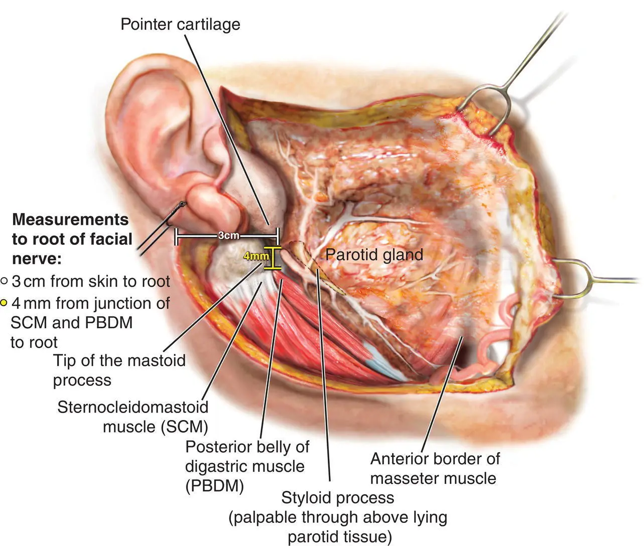
Figure 1.4. Anatomical landmarks of the extratemporal facial nerve.
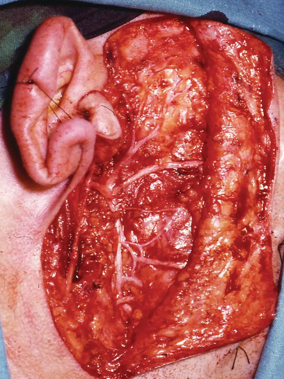
Figure 1.5. Clinical photograph of dissected facial nerve following superficial parotidectomy.
Source: Published with permission, Martin Dunitz, London, Langdon JD, Berkowitz BKB, Moxham BJ, editors, Surgical Anatomy of the Infratemporal Fossa. DOI: 10.1002/9781118949139.ch1.
Retromandibular Vein
The vein is formed within the parotid gland by the union of the superficial temporal vein and the maxillary vein. The retromandibular vein passes downwards and close to the lower pole of the parotid where it often divides into two branches passing out of the gland. The posterior branch passes backward to unite with the posterior auricular vein on the surface of the sternocleidomastoid muscle to form the external jugular vein. The anterior branch passes forward to join the facial vein.
The retromandibular vein is an important landmark during parotid gland surgery. The division of the facial nerve into its temporofacial and cervicofacial divisions occurs just behind the retromandibular vein ( Figure 1.7). The two divisions lie just superficial to the vein in contact with it. It is all too easy to tear the vein while exposing the division of the facial nerve!

Figure 1.6. The branching patterns of the facial nerve.
Source: Berkovitz et al. 2003/Taylor & Francis.
| IType I, 13% | VType V, 9% | 3 Buccal branch |
| IIType II, 20% | VIType VI, 6% | 4 Mandibular branch |
| IIIType III, 28% | Temporal branch | 5 Cervical branch |
| IVType IV, 24% | 2 Zygomatic branch |
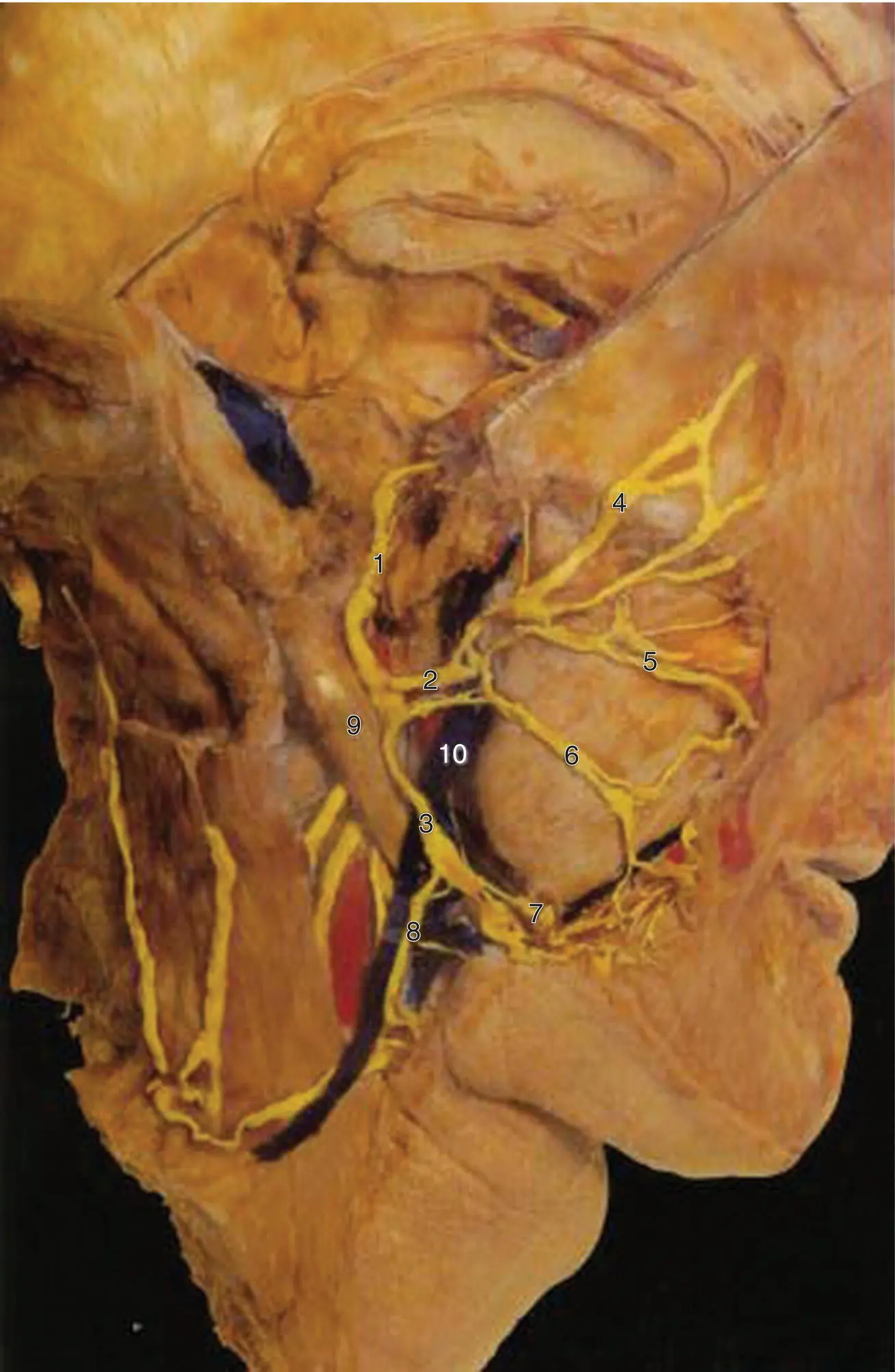
Figure 1.7. The facial nerve and its relationship to the retromandibular vein within the parotid gland.
Source: Published with permission, Martin Dunitz, London, Langdon JD, Berkowitz BKB, Moxham BJ, editors, Surgical Anatomy of the Infratemporal Fossa. DOI: 10.1002/9781118949139.ch1.
1 Facial nerve at stylomastoid foramen
2 Temporofacial branch of facial nerve
3 Cervicofacial branch of facial nerve
4 Temporal branch of facial nerve
5 Zygomatic branch of facial nerve
6 Buccal branch of facial nerve
7 Mandibular branch of facial nerve
8 Cervical branch of facial nerve
9 Posterior belly of the digastric muscle
10 Retromandibular vein and external carotid artery
External Carotid Artery
The external carotid artery runs deeply within the parotid gland. It appears from behind the posterior belly of the digastric muscle and grooves the parotid before entering it. It gives off the posterior auricular artery before ascending and dividing into its terminal branches, the superficial temporal and maxillary arteries at the level of the condyle. The superficial temporal artery continues vertically to emerge at the superior border of the gland and crosses the zygomatic arch. Within the substance of the parotid, it gives off the transverse facial artery which emerges at the anterior border of the gland to run across the face above the parotid duct. The maxillary artery emerges from the deep aspect of the gland anteriorly to enter the infratemporal fossa. The maxillary artery gives off the deep auricular artery and the anterior tympanic artery within the substance of the parotid. All these branches from the external carotid also give off numerous small branches within the parotid to supply the gland itself.
Parotid Lymph Nodes
Lymph nodes are found within the subcutaneous tissues overlying the parotid to form the preauricular nodes and also within the substance of the gland (Goldenberg et al. 2000). There are typically 10 nodes within the substance of the gland, the majority being within the superficial lobe and therefore superficial to the plane of the facial nerve. Only one or two nodes lie within the deep lobe (Marks 1984; McKean et al. 1984; Garatea‐Crelgo et al. 1993). All the parotid nodes drain into the upper deep cervical chain.
Parotid Duct
The parotid duct emerges from the anterior border of the parotid gland and passes horizontally across the masseter muscle. The surface markings of the duct are obtained by drawing a line from the mid‐point of the tragal cartilage to the middle of a straight line from the ipsilateral ala to the commissure ( Figure 1.8). This line is divided into three equal parts and the middle section corresponds to the position of the parotid duct. The duct lies approximately 1 cm below the transverse facial vessels. The accessory lobe of the parotid gland, when present, drains into its upper border via one or two tributaries (Kulkarni et al. 2011). Anastomosing branches between the buccal and zygomatic branches of the facial nerve cross the duct. At the anterior border of the masseter, the duct bends sharply to perforate the buccal pad of fat and the buccinator muscle at the level of the upper molar teeth. The duct then bends again to pass forward for a short distance before entering the oral cavity at the parotid papilla.
Читать дальшеИнтервал:
Закладка:
Похожие книги на «Salivary Gland Pathology»
Представляем Вашему вниманию похожие книги на «Salivary Gland Pathology» списком для выбора. Мы отобрали схожую по названию и смыслу литературу в надежде предоставить читателям больше вариантов отыскать новые, интересные, ещё непрочитанные произведения.


Обсуждение, отзывы о книге «Salivary Gland Pathology» и просто собственные мнения читателей. Оставьте ваши комментарии, напишите, что Вы думаете о произведении, его смысле или главных героях. Укажите что конкретно понравилось, а что нет, и почему Вы так считаете.