Salivary Gland Pathology
Здесь есть возможность читать онлайн «Salivary Gland Pathology» — ознакомительный отрывок электронной книги совершенно бесплатно, а после прочтения отрывка купить полную версию. В некоторых случаях можно слушать аудио, скачать через торрент в формате fb2 и присутствует краткое содержание. Жанр: unrecognised, на английском языке. Описание произведения, (предисловие) а так же отзывы посетителей доступны на портале библиотеки ЛибКат.

- Название:Salivary Gland Pathology
- Автор:
- Жанр:
- Год:неизвестен
- ISBN:нет данных
- Рейтинг книги:3 / 5. Голосов: 1
-
Избранное:Добавить в избранное
- Отзывы:
-
Ваша оценка:
Salivary Gland Pathology: краткое содержание, описание и аннотация
Предлагаем к чтению аннотацию, описание, краткое содержание или предисловие (зависит от того, что написал сам автор книги «Salivary Gland Pathology»). Если вы не нашли необходимую информацию о книге — напишите в комментариях, мы постараемся отыскать её.
now features case presentations to place the information in context, as well as additional treatment algorithms in each chapter to assist in clinical decision making. Written by highly respected clinicians, educators, and researchers with extensive expertise in oral and maxillofacial surgery, this authoritative resource:
Reviews the etiology, diagnosis, and treatment of all salivary gland pathologies, with detailed explanations and hundreds of full-color clinical images Incorporates new information on the taxonomy of salivary gland tumors, neoplastic and non-neoplastic entities, image guided biopsies of salivary gland lesions, and complications in traditional and non-traditional forms of salivary gland surgery Offers expanded coverage of histopathology, including classification, grading, and staging of salivary gland tumors Features up-to-date chapters on anatomy and physiology, imaging, cysts, systemic diseases, tumors, trauma, and innovative salivary gland surgical techniques Includes a discussion of meta-analyses and systematic reviews to support evidence-based practice remains the definitive resource for oral and maxillofacial surgeons, otolaryngologists, head and neck surgeons, and general surgeons as well as their residents providing care for patients with salivary gland pathology.
Salivary Gland Pathology — читать онлайн ознакомительный отрывок
Ниже представлен текст книги, разбитый по страницам. Система сохранения места последней прочитанной страницы, позволяет с удобством читать онлайн бесплатно книгу «Salivary Gland Pathology», без необходимости каждый раз заново искать на чём Вы остановились. Поставьте закладку, и сможете в любой момент перейти на страницу, на которой закончили чтение.
Интервал:
Закладка:
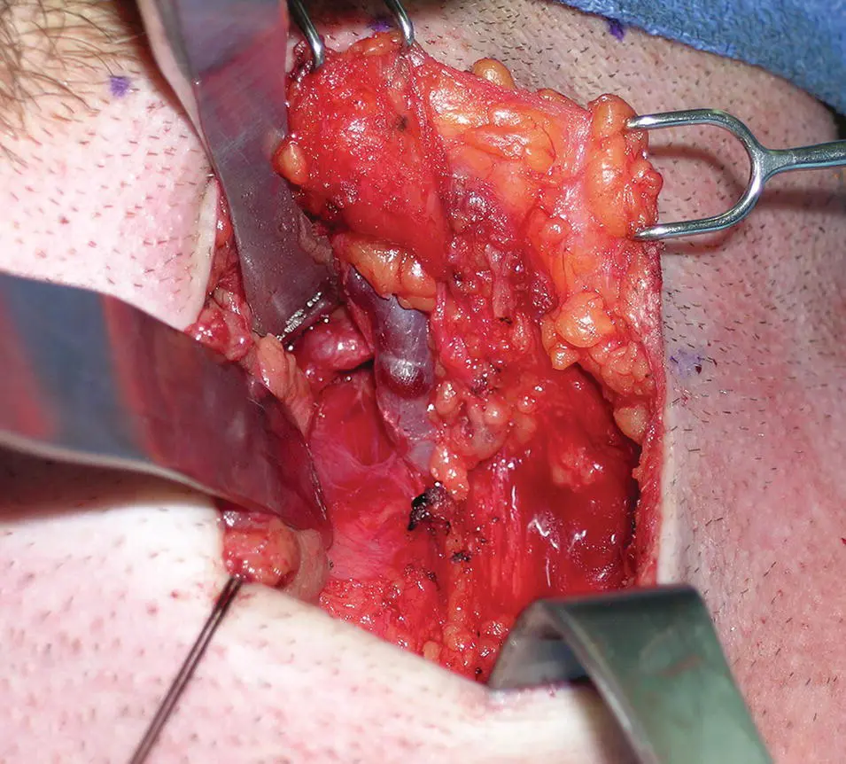
Figure 1.12. Deep dissection of the left submandibular gland. With the submandibular gland retracted, the facial artery is identified in proximity to the facial vein.
Blood Supply and Lymphatic Drainage
The arterial blood supply arises from multiple branches of the facial and lingual arteries. Venous blood drains predominantly into the deep lingual vein. The lymphatics drain into the deep cervical group of nodes, mostly into the jugulo‐omohyoid node, via the submandibular nodes.
Nerve Supply to the Submandibular Gland
Parasympathetic innervation
The secretomotor supply to the submandibular gland arises from the submandibular (sublingual) ganglion. This is a small ganglion lying on the upper part of the hyoglossus muscle. There are additional ganglion cells at the hilum of the gland. The submandibular ganglion is suspended from the lingual nerve by anterior and posterior filaments ( Figure 1.13).
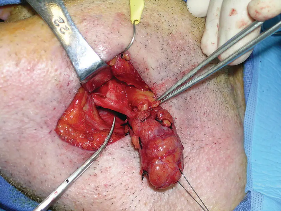
Figure 1.13. Clinical photograph showing the relationship of the lingual nerve to the submandibular gland.
The parasympathetic secretomotor fibers originate in the superior salivatory nucleus and the preganglionic fibers then travel via the facial nerve, chorda tympani, and lingual nerve to the ganglion via the posterior filaments connecting the ganglion to the lingual nerve. They synapse within the ganglion and the postganglionic fibers innervate the submandibular and sublingual glands ( Figure 1.9). Some fibers are thought to reach the lower pole of the parotid gland.
Sympathetic innervation
The sympathetic root is derived from the plexus on the facial artery. The postganglionic fibers arise from the superior cervical ganglion and pass through the submandibular ganglion without synapsing. They are vasomotor to the vessels supplying the submandibular and sublingual glands. Five or six branches from the ganglion supply the submandibular gland and its duct. Others pass back into the lingual nerve via the anterior filament to innervate the sublingual and other minor salivary glands in the region.
Sensory innervation
Sensory fibers arising from the submandibular and sublingual glands pass through the ganglion without synapsing and join the lingual nerve, itself a branch of the trigeminal nerve.
Sublingual Gland
EMBRYOLOGY
The sublingual gland arises in 20 mm embryos in the eighth week of intrauterine life as numerous small epithelial thickenings in the linguogingival groove and on the outer side of the groove. Each thickening forms its own canal and so many of the sublingual ducts open directly onto the summit of the sublingual fold. Those that arise within the linguogingival grove end up draining into the submandibular duct.
ANATOMY
The sublingual gland is the smallest of the major salivary glands. It is almond shaped and weighs approximately 4 g. It is predominantly a mucous gland. The gland lies on the mylohyoid and is covered by the mucosa of the floor of the mouth which is raised as it overlies the gland to form the sublingual fold. Posteriorly, the sublingual gland is in contact with the deep lobe of the submandibular gland. The sublingual fossa of the mandible is located laterally and the genioglossus muscle is located medially. The lingual nerve and the submandibular duct lie medial to the sublingual gland between it and the genioglossus.
Sublingual Ducts
The gland has a variable number of excretory ducts ranging from 8 to 20. The majority drain into the floor of the mouth at the crest of the sublingual fold. A few drain into the submandibular duct. Sometimes, a collection of draining ducts coalesce anteriorly to form a major duct (Bartholin's duct) which opens with the orifice of the submandibular duct at the sublingual papilla (Zhang et al. 2010).
Blood Supply, Innervation, and Lymphatic Drainage
The arterial supply is from the sublingual branch of the lingual artery and also the submental branch of the facial artery. Innervation is via the sublingual ganglion as described above. The lymphatics drain to the submental nodes.
Minor Salivary Glands
Minor salivary glands are distributed widely in the oral cavity and oropharynx. They are grouped as labial, buccal, palatoglossal, palatal, and lingual glands. The labial and buccal glands contain both mucous and serous acini whereas the palatoglossal glands are mucous secreting. The palatal glands which are also mucous secreting occur in both the hard and soft palates. The anterior and posterior lingual glands are mainly mucous secreting. The anterior glands are embedded within the muscle ventrally and they drain via four or five ducts near the lingual frenum. The posterior lingual glands are located at the root of the tongue. The deep posterior lingual glands are predominantly serous secreting. Additional serous glands (glands of von Ebner) occur around the circumvallate papillae on the dorsum of the tongue. Their watery secretion is thought to be important in spreading taste stimuli over the taste buds.
Tubarial Salivary Glands
In 2020, Valstar et al reported on the serendipitous presence of bilateral macroscopic salivary gland structures in the nasopharynx of humans. Their existence was visualized by positron emission tomography/computed tomography with prostate‐specific membrane antigen ligands (PSMA PET/CT). The presence of the PSMA‐positive nasopharyngeal regions was elucidated in a retrospective cohort of 100 consecutive patients with prostate or urethral gland cancer. The designated area of the posterior nasopharynx was also studied with hematoxylin and eosin (H&E) and PSMA and alpha‐amylase) immunohistochemistry in two human cadavers. All 100 patients (99 males, one female; median age 69.5, range 53–84) demonstrated a well‐demarcated bilateral PSMA‐positive region on PSMA PET/CT. This 3.9 cm cranio‐caudal structure (range 1.0–5.7 cm) extended from the skull base along the posterolateral pharyngeal wall on the pharyngeal aspect of the superior pharyngeal constrictor muscle with a PSMA‐positive structure located predominantly over the torus tubarius. The tracer uptake in these structures was similar to the uptake of the sublingual glands. The dissected structures from the two human cadavers demonstrated a large aggregate of mucous salivary gland tissue with multiple visible draining duct openings in the dorsolateral pharyngeal wall. The authors concluded that the human body contains a pair of once overlooked and clinically significant salivary glands in the posterior nasopharynx, and the authors proposed the name tubarial glands. Their particular interest in these structures was in gland sparing radiation protocols for head and neck cancer in the best interests of maintaining salivary function and the quality of life of patients.
Histology of the Salivary Glands
The salivary glands are composed of large numbers of secretory acini that may be tubular or globular in shape. Each acinus drains into a duct. These microscopic ducts coalesce to form lobular ducts. Each lobule has its own duct and these then merge to form the main ducts. The individual lobes and lobules are separated by dense connective tissue that is continuous with the gland capsule. The ducts, blood vessels, lymphatics, and nerves run through and are supported by this connective tissue.
Читать дальшеИнтервал:
Закладка:
Похожие книги на «Salivary Gland Pathology»
Представляем Вашему вниманию похожие книги на «Salivary Gland Pathology» списком для выбора. Мы отобрали схожую по названию и смыслу литературу в надежде предоставить читателям больше вариантов отыскать новые, интересные, ещё непрочитанные произведения.


Обсуждение, отзывы о книге «Salivary Gland Pathology» и просто собственные мнения читателей. Оставьте ваши комментарии, напишите, что Вы думаете о произведении, его смысле или главных героях. Укажите что конкретно понравилось, а что нет, и почему Вы так считаете.