Diatom Microscopy
Здесь есть возможность читать онлайн «Diatom Microscopy» — ознакомительный отрывок электронной книги совершенно бесплатно, а после прочтения отрывка купить полную версию. В некоторых случаях можно слушать аудио, скачать через торрент в формате fb2 и присутствует краткое содержание. Жанр: unrecognised, на английском языке. Описание произведения, (предисловие) а так же отзывы посетителей доступны на портале библиотеки ЛибКат.
- Название:Diatom Microscopy
- Автор:
- Жанр:
- Год:неизвестен
- ISBN:нет данных
- Рейтинг книги:5 / 5. Голосов: 1
-
Избранное:Добавить в избранное
- Отзывы:
-
Ваша оценка:
Diatom Microscopy: краткое содержание, описание и аннотация
Предлагаем к чтению аннотацию, описание, краткое содержание или предисловие (зависит от того, что написал сам автор книги «Diatom Microscopy»). Если вы не нашли необходимую информацию о книге — напишите в комментариях, мы постараемся отыскать её.
The main goal of the book is to demonstrate the wide variety of microscopy methods being used to investigate natural and altered diatom structures. Diatom Microscopy
Diatom Microscopy — читать онлайн ознакомительный отрывок
Ниже представлен текст книги, разбитый по страницам. Система сохранения места последней прочитанной страницы, позволяет с удобством читать онлайн бесплатно книгу «Diatom Microscopy», без необходимости каждый раз заново искать на чём Вы остановились. Поставьте закладку, и сможете в любой момент перейти на страницу, на которой закончили чтение.
Интервал:
Закладка:
The rate of diatom silicification depends on environmental factors, such as water temperature, pH and depth. Znachor and Nedoma [1.78] used fluorescence microscopy for imaging and measuring single-cell PDMPO fluorescence to observe relative differences in silicification among diatom species. The acquired images showed that the fluorescence signals from diatoms and chloroplasts are easily discerned, which can be used to confirm an entire community in a silicified state. Onesto et al . [1.45] cultivated diatom frustules with Au NPs, which is abbreviation as D24 metallic NPs, at a ratio of 1:10 ratio for 10 minutes in order to use the diatoms to detect chemical pollutants (mineral oil). Under a fluorescence microscope, the diatoms absorbed light at a wavelength of 441 nm and emitted a fluorescence signal at 485 nm, as shown in Figure 1.8c. This system proved highly selective, specific and sensitive. Bovine serum albumin (BSA) can be detected in simple diatom frustules, and Au nanoparticles in D24 systems highlight the presence of the aromatic components of BSA at 1392 cm -1and in the 1556–1576 cm -1band, which are confirmed by surface-enhanced Raman spectroscopy. This method can be used to detect BSA at low concentrations, for use in bioengineering, medicine and pollution monitoring. Furthermore, Delalat et al . used genetic engineering to promote the expression of the IgG-binding domain of G protein on the surface of diatom frustules (Thalassiosira pseudonana) that was treated as drug carriers for the selective killing of neuroblastoma and B lymphoma cells. Thus, diatoms can be used as a target for drug delivery and absorption in the body, where the therapeutic effects can be examined and monitored under a fluorescence microscope [1.10]. The polyunsaturated aldehydes (PUAs) secreted by diatoms also have anti-cancer effects. Clementina et al . [1.61] demonstrated that PUA substances killed lung cancer cells and colorectal cancer cells without adversely affecting normal cells.
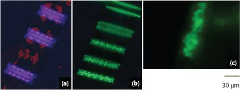
Figure 1.8 Images of new silicon deposits and chlorophyll a (Chl a) of coastal and subarctic diatoms under fluorescence microscopy. (a) PDMPO-stained diatom valves in the blue emission spectrum images using a DAPI filter. (b) Newly deposited valves exhibited an intense yellow-green fluorescence, and Chl a may in some cases be visible simultaneously in red. From [1.35] with permission of John Wiley and Sons. (c) Fluorescence microscopic image showing D24 systems after incubation with fluorescent 50 nm yellow microspheres. Subsequent fluorescence analysis with background-free images revealed device localization, selectivity, and specificity. From [1.45] with permission of Springer Nature.
1.4 Confocal Laser Scanning Microscopy
Confocal laser scanning microscopy (CLSM) has been used with hyperspectral analysis to characterize the optical properties of diatom frustules [1.55]. Enhanced light transmission through the diatom frustule was observed at roughly 636 and 663 nm, which respectively match the maximum light absorption of chlorophyll a and c , as shown in Figure 1.9. This is a clear demonstration that the efficient light trapping of the frustule can be attributed to strong asymmetry between cribrum and foramen pseudoperiodic structures, which prevents the backscattering of transmitted light resulting in a corresponding increase in light absorption.
In terms of diatom ecology, Buhmann et al . [1.4] established a system for the incubation of pure phototrophic biofilms in the laboratory, with the aim of elucidating microbial population dynamics and community interactions associated with biofilm development. That system proved effective in the growth and monitoring of three strains of Planothidium sp . strain 8c isolated from epilithic (epilithic: growing on rock surfaces) biofilms of an oligotrophic freshwater lake for co-cultivation with axenic (axenic: the state of a culture where only a single species is presented that is free from other cultivatable organisms) diatom phototrophic biofilms. Automated photometric monitoring of biofilm density under controlled illumination revealed a 3x increase in biofilm turbidity (turbidity: degree of light attenuation) when co-cultured with Planothidium sp . strain 8c, indicating the co-existence the chlorophyll autofluorescence and stained-bacteria. This type of system can be used to evaluate diatom biofilms and study the interactions in diatom-bacteria biofilms. Diatom diazotroph (diazotroph: mostly bacteria and archaea that can transform nitrogen gas into a usable form such as ammonia) association (DDA) is among the oldest planktons, which lives on the surface of the sea at elevated temperatures under high CO 2levels, such as those predicted by most models of global warming. DDAs are marine plankton that combine with bacteria with fixed nitrogen (N 2) from a variety of diatom genera. The development and distribution of DDA can be observed using molecular genetics methods to assess cells collected in the field via single-cell CLSM. The distribution and evolution of DDAs and their phylogenetic diversity can be characterized using confocal analysis with 3-D imaging re-evaluation, as shown in Figures 1.10a-1.10c[1.5].
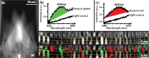
Figure 1.9 Hyperspectral analysis of a single valve of Coscinodiscus centralis oriented with the foramen side upwards and the direction of the incident light indicated by the white arrow (a). The wavelength regions (green and red) where transmission spectra (b, c) differing from the light source are detected. Close view of hyper-map in a (d). Close-up view of composite image in a (e). The yellow lines in (d, e) indicate the transition between the foramen and cribrum layers of the valve, with the honeycombed structure in the middle. From [1.55] with permission of Springer Nature.
McNair et al . [1.39] proposed a method for quantifying biogenic silica (bSiO 2) production, in which the fluorescent dye PDMPO is used to dissolve diatom frustules. PDMPO is incorporated in SiO 2deposition vesicles (SDV) and newly formed diatom frustules. The use of PDMPO to measure bSiO 2production by a single diatom cells requires quantification of single-cell fluorescence and accurate calibration of the relationship between the incorporated PDMPO and the amount of newly polymerized silica. CLSM is used to detect single diatom cells. Improvements in PDMPO labeling have made it possible to progress from qualitative assessment to quantitative measurement of bSiO 2in newly deposited diatom communities and single diatom cells. On the other hand, diatoms are commonly divided into two breeding methods of which the silicification process start from the SDV inside the cell membrane. However, there is few discussions on the SDV membrane protein of diatoms or other silica-forming organisms. For this, Kotzsch et al . [1.32] discovered a unique SiMat7-like protein, as the organic component of diatom silica, which is significantly different from other proteins in the secondary structure. It was also proved that SiMat7 is the original member of a new type of silica biomineralization protein family, named as Siliconnin-1 (Sin1). After protein analysis, Sin1 sequence was divided the into two regions, an NQ (asparagine and glutamine)-rich domain and a cytosolic domain. By connecting GFP to different positions, it formed into Sin1-GFP Nand Sin1-GFP Cshown in different parts of living cells, which can be observed under CLSM (see Figure 1.11). The NQ-rich domain is flipped out of the membrane, and the cytosolic domain remained between the cells, manifesting that Sin1 is a transmembrane protein.
Читать дальшеИнтервал:
Закладка:
Похожие книги на «Diatom Microscopy»
Представляем Вашему вниманию похожие книги на «Diatom Microscopy» списком для выбора. Мы отобрали схожую по названию и смыслу литературу в надежде предоставить читателям больше вариантов отыскать новые, интересные, ещё непрочитанные произведения.


Обсуждение, отзывы о книге «Diatom Microscopy» и просто собственные мнения читателей. Оставьте ваши комментарии, напишите, что Вы думаете о произведении, его смысле или главных героях. Укажите что конкретно понравилось, а что нет, и почему Вы так считаете.