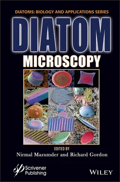
Diatom Microscopy
Здесь есть возможность читать онлайн «Diatom Microscopy» — ознакомительный отрывок электронной книги совершенно бесплатно, а после прочтения отрывка купить полную версию. В некоторых случаях можно слушать аудио, скачать через торрент в формате fb2 и присутствует краткое содержание. Жанр: unrecognised, на английском языке. Описание произведения, (предисловие) а так же отзывы посетителей доступны на портале библиотеки ЛибКат.
- Название:Diatom Microscopy
- Автор:
- Жанр:
- Год:неизвестен
- ISBN:нет данных
- Рейтинг книги:5 / 5. Голосов: 1
-
Избранное:Добавить в избранное
- Отзывы:
-
Ваша оценка:
- 100
- 1
- 2
- 3
- 4
- 5
Diatom Microscopy: краткое содержание, описание и аннотация
Предлагаем к чтению аннотацию, описание, краткое содержание или предисловие (зависит от того, что написал сам автор книги «Diatom Microscopy»). Если вы не нашли необходимую информацию о книге — напишите в комментариях, мы постараемся отыскать её.
The main goal of the book is to demonstrate the wide variety of microscopy methods being used to investigate natural and altered diatom structures. Diatom Microscopy
Diatom Microscopy — читать онлайн ознакомительный отрывок
Ниже представлен текст книги, разбитый по страницам. Система сохранения места последней прочитанной страницы, позволяет с удобством читать онлайн бесплатно книгу «Diatom Microscopy», без необходимости каждый раз заново искать на чём Вы остановились. Поставьте закладку, и сможете в любой момент перейти на страницу, на которой закончили чтение.
Интервал:
Закладка:
Table of Contents
1 Cover
2 Title Page
3 Copyright
4 Preface References
5 1 Investigation of Diatoms with Optical Microscopy 1.1 Introduction 1.2 Light Microscopy 1.3 Fluorescence Microscopy 1.4 Confocal Laser Scanning Microscopy 1.5 Multiphoton Microscopy 1.6 Super-Resolution Optical Microscopy 1.7 Conclusion Acknowledgement References
6 2 Nanobioscience Studies of Living Diatoms Using Unique Optical Microscopy Systems Abbreviations 2.1 Trajectory Analysis of Gliding Among Individual Diatom Cells Using Microchamber Systems 2.2 Direct Observation of Floating Phenomena of Individual Diatoms Using a “Tumbled” Microscope System 2.3 Three-Dimensional Physical Imaging of Living Diatom Cells Using a Holographic Microscope System Acknowledgements References
7 3 Recent Insights Into the Ultrastructure of Diatoms Using Scanning and Transmission Electron-Microscopy 3.1 Introduction 3.2 Scanning Electron Microscopy (SEM) of Diatoms 3.3 Transmission Electron Microscopy (TEM) of Diatoms 3.4 Conclusion References
8 4 Atomic Force Microscopy Study of Diatoms 4.1 Introduction 4.2 Types of AFM Modes 4.3 Sample Preparation and Methods 4.4 Study of Diatom Ultrastructure Under AFM 4.5 Conclusion Glossary Acknowledgement References
9 5 Refractive Index Tomography for Diatom Analysis 5.1 Introduction 5.2 Fundamentals of PC-ODT 5.3 Experimental Setup for PC-ODT 5.4 Diatom RI Reconstructions with Bright-Field Illumination 5.5 Illumination Impact on PC-ODT Performance 5.6 Concluding Remarks Acknowledgement References
10 6 Luminescent Diatom Frustules: A Review on the Key Research Applications 6.1 Introduction 6.2 Key Research Applications of Luminescence Properties of Diatom Frustules 6.3 Future Perspectives 6.4 Conclusion Acknowledgement References
11 7 Micro to Nano Ornateness of Diatoms from Geographically Distant Origins of the Globe 7.1 Introduction 7.2 Materials and Methods 7.3 Diatoms from Different Geographical Origins of the World 7.4 Conclusion 7.5 Acknowledgements References
12 8 Types of X-Ray Techniques for Diatom Research 8.1 Introduction 8.2 Applications 8.3 Conclusions Glossary References
13 9 Diatom Assisted SERS 9.1 Introduction 9.2 Diatom 9.3 Raman Scattering 9.4 SERS Through Diatom: Fundamentals and Application Overview 9.5 Conclusion and Future Outlook References
14 10 Diatoms as Sensors and Their Applications 10.1 Introduction 10.2 Diatoms as Biosensors 10.3 Conclusion Acknowledgments References
15 11 Diatom Frustules: A Transducer Platform for Optical Detection of Molecules 11.1 Introduction 11.2 Optical Properties of Diatom Frustules 11.3 Methods Involved in Thin Film Deposition of Diatom Frustules 11.4 Diatom as an Optical Transducer for Biosensors 11.5 Diatom as an Optical Transducer for Gas/Chemical Sensors 11.6 Conclusion References
16 12 Effects of Light on Physico-Chemical Properties of Diatoms 12.1 Introduction 12.2 Effect of Light on Diatom Function and Morphology 12.3 Conclusion Acknowledgment References
17 Index
18 Also of Interest
19 End User License Agreement
List of Illustrations
1 Chapter 1 Figure 1.1 Selected diatoms taxa in the Baryczka stream obtained using phase con... Figure 1.2 Comparison of diatom defects and man-made photonic crystal fiber. The... Figure 1.3 Quantitative phase image of diatom cell recorded using 20 x/0.45 micr... Figure 1.4 QPIs results showing a variety of diatom samples; (a) diatom recorded... Figure 1.5 Images illustrating the relationship between the bacterium L. monocyt... Figure 1.6 Dark field micrographs of control cells and cells exposed to Ag NPs. ... Figure 1.7 (a) Dark field image of single valve of C. wailesii diatom and (b) co... Figure 1.8 Images of new silicon deposits and chlorophyll a (Chl a) of coastal a... Figure 1.9 Hyperspectral analysis of a single valve of Coscinodiscus centralis o... Figure 1.10 Laser confocal microscope images of H. hauckii-R . intracellularis sy... Figure 1.11 Live cells, biosilica and biosilica-associated organic matrix from t... Figure 1.12 Quantification of carbolines in healthy and oomycete-infected diatom... Figure 1.13 Evaluation of siRNA uptake and cellular internalization using confoc... Figure 1.14 Specific particle endocytosis of FITC/PEI/FA-functionalized silica n... Figure 1.15 Left: Multi-photon image of living centric diatoms Coscinodiscus wai... Figure 1.16 (a) Asterionellopsis glacialis and (b) Proboscia alata captured usin... Figure 1.17 (a) Average fluorescence lifetime images of T. weissflogii exposed t... Figure 1.18 PALM analysis of tpSil3. (a) Comparison of epifluorescence image and...
2 Chapter 2 Figure 2.1 Diatom studies from the viewpoint of nanobiological physics using opt... Figure 2.2 Microscopy images of (a) PDMS microchamber, (b) spiral grooves, and (... Figure 2.3 Trajectories (red lines) of movements of the same diatom cell (a) bef... Figure 2.4 Overview of a “tumbled” microscope system with a microchamber. A phot... Figure 2.5 A typical example of snapshots and trajectories of the settlement of ... Figure 2.6 Typical examples of snapshots and trajectories of floating diatom cel... Figure 2.7 A DHM image of a living Cylindrotheca spp. cell. Ranges of refractive...
3 Chapter 3Figure 3.1 SEM images of dehydrated mucilage, (a) 5 μm, (b) 2 μm [3.9]. From Top...Figure 3.2 (a) Overall view of Karayevia amoena , (b) valve without raphe (c) val...Figure 3.3 (a–c) Scanning electron micrographs of Pd3Co-D(100)-G [3.55], and bef...Figure 3.4 TEM micrographs of (a–c) Pd3Co-D (100)-G [3.55], (d) T. pseudonana, (...Figure 3.5 TEM micrographs of Pseudo-nitzschia species (a–c) P. calliantha , (d—f...Figure 3.6 CCMP470 structures obtained using (a) bright field LM, (b) phase cont...
4 Chapter 4Figure 4.1 Schematic setup of an Atomic Force Microscope. Reproduced with permis...Figure 4.2 Comparison of the cell jackets fibrillar network obtained with (a) SE...Figure 4.3 Mesoscale structures of diatomaceous silica studied under AFM, (a) Li...Figure 4.4 The hypothetical path insertion of TiO 2in the diatom frustule by a t...Figure 4.5 Ultrastructure of Cyclotella cryptica observed under AFM: (a) Wide an...Figure 4.6 The centric Coscinodiscus sp. diatom frustule with mesh-like porous s...Figure 4.7 (a, b) showing the effect of stretching of polysaccharide network ove...
5 Chapter 5Figure 5.1 (p x-p z) slices of the normalized OTFs corresponding to a microscope e...Figure 5.2 Experimental setup for PC-ODT, consisting of a bright-field microscop...Figure 5.3 (a1–a2) Volumetric representation of the reconstructed 3D RI contrast...Figure 5.4 Intensity images and the corresponding RI for a Cocconeis placentula ...Figure 5.5 Reconstructed RI for (a) Cymbella subturgidula and (b) Diploneis elli...Figure 5.6 Intensity distributions in the condenser plane (xc, yc) along with th...Figure 5.7 Normalized p x-p zsection of AOTFs for (a) BFI with NA c=0.48, (b) BFI ...Figure 5.8 |POTF| sections calculated for ideal (theoretical) illumination: (a) ...Figure 5.9 RI contrast slices of a diatom immersed in oil and reconstructed with...
6 Chapter 6Figure 6.1 Fluorescence images of the single valve of C. wailesii frustule with ...Figure 6.2 Schematic representation of the steps in the synthesis of the composi...Figure 6.3 (a) Photoluminescence quenching of diatoms in the presence of several...Figure 6.4 (a) Photoluminescence spectra of silicon diatoms after each functiona...
7 Chapter 7Figure 7.1 (a) Diatoms and Desmids from biofilm at rocks of Rajghat, Sagar, Madh...Figure 7.2.I (a) Hyalodiscus sp.; (b) (Calcareous nannofossil, Chiasmolithus sp....Figure 7.2.II (a) Unknown
Читать дальшеИнтервал:
Закладка:
Похожие книги на «Diatom Microscopy»
Представляем Вашему вниманию похожие книги на «Diatom Microscopy» списком для выбора. Мы отобрали схожую по названию и смыслу литературу в надежде предоставить читателям больше вариантов отыскать новые, интересные, ещё непрочитанные произведения.
Обсуждение, отзывы о книге «Diatom Microscopy» и просто собственные мнения читателей. Оставьте ваши комментарии, напишите, что Вы думаете о произведении, его смысле или главных героях. Укажите что конкретно понравилось, а что нет, и почему Вы так считаете.


