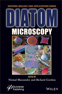
Diatom Microscopy
Здесь есть возможность читать онлайн «Diatom Microscopy» — ознакомительный отрывок электронной книги совершенно бесплатно, а после прочтения отрывка купить полную версию. В некоторых случаях можно слушать аудио, скачать через торрент в формате fb2 и присутствует краткое содержание. Жанр: unrecognised, на английском языке. Описание произведения, (предисловие) а так же отзывы посетителей доступны на портале библиотеки ЛибКат.
- Название:Diatom Microscopy
- Автор:
- Жанр:
- Год:неизвестен
- ISBN:нет данных
- Рейтинг книги:5 / 5. Голосов: 1
-
Избранное:Добавить в избранное
- Отзывы:
-
Ваша оценка:
- 100
- 1
- 2
- 3
- 4
- 5
Diatom Microscopy: краткое содержание, описание и аннотация
Предлагаем к чтению аннотацию, описание, краткое содержание или предисловие (зависит от того, что написал сам автор книги «Diatom Microscopy»). Если вы не нашли необходимую информацию о книге — напишите в комментариях, мы постараемся отыскать её.
The main goal of the book is to demonstrate the wide variety of microscopy methods being used to investigate natural and altered diatom structures. Diatom Microscopy
Diatom Microscopy — читать онлайн ознакомительный отрывок
Ниже представлен текст книги, разбитый по страницам. Система сохранения места последней прочитанной страницы, позволяет с удобством читать онлайн бесплатно книгу «Diatom Microscopy», без необходимости каждый раз заново искать на чём Вы остановились. Поставьте закладку, и сможете в любой момент перейти на страницу, на которой закончили чтение.
Интервал:
Закладка:
8 Chapter 8Figure 8.1 X-ray microscopy of diatoms. (A) X-PEEM images of a diatom frustule d...Figure 8.2 X-ray spectroscopy of diatoms. (A) Percentage of adsorbed zinc as a f...
9 Chapter 9Figure 9.1 Representation of diatoms (adapted from [9.33]).Figure 9.2 Schematics of SERS (a) normal raman scattering (b) SERS.Figure 9.3 Schematic illustration of diatom assisted SERS.
10 Chapter 10Figure 10.1 SEM images of the diatom valves of Coscinodiscus wailesii at differe...Figure 10.2 SEM images of diatoms fabricated by in situ growth and self-assembly...Figure 10.3 Diatom-based immunoassay sensors made by a covalent bond between the...Figure 10.4 Schematic representation of the preparation of diatom-based immunose...Figure 10.5 Schematic representation of SERS-based diatom immunosensor. (a) Modi...Figure 10.6 Photoluminescence emission from diatom-based optical sensors upon (a...Figure 10.7 Schematic representation of dissolved ammonia sensing mechanism in b...Figure 10.8 Schematic representation of ribose-induced conformational change in ...
11 Chapter 11Figure 11.1 PL spectra of diatom frustules with multiple emission peaks when exc...Figure 11.2 Various types of optical properties present in diatom frustules.Figure 11.3 Methods involved in thin film deposition.Figure 11.4 Monolayer formation of diatom frustules.Figure 11.5 Steps involved in the coating antibody over diatom and bonding proce...Figure 11.6 Schematic representation of functionalization of diatom frustules.Figure 11.7 (a) Electron micrograph of amine functionalized diatom (AFD) and (b)...Figure 11.8 Electron micrographs of (a) Nitzschia sp . (b) its pore arrangement.Figure 11.9 Mechanism of Meisenheimer complex formation.Figure 11.10 Photoluminescent spectra of AFD with different concentration of 4-N...
12 Chapter 12Figure 12.1 Eukaryotic phylogenetic tree showing major groups. Diatoms belong to...Figure 12.2 Single valves of centric diatoms (a) C. wailessi (b) Actinoptychus s...Figure 12.3 Effect of monochromatic LED lights (red, green and blue) on the cell...Figure 12.4 (a) Effect of monochromatic lights on fatty acid composition in Gole...Figure 12.5 Growth and photosynthetic ability of T. pseudonana and P. tricornutu...Figure 12.6 (a) The association between growth rates and absorption coefficient ...Figure 12.7 Light absorption spectra of various characteristic pigments of Nitzs...Figure 12.8 Depending on the depth, the diatoms are subjected to different wavel...Figure 12.9 (a) Fucoxanthin content and productivities of N. laevis in different...Figure 12.10 Differences in the photosynthetic apparatus organization, thylakoid...Figure 12.11 Growth curves of P. tricornutum under (a) mFL after high light of 1...Figure 12.12 Incorporation of molecular antennae, Cy5 antennae dye in vivo in T....
List of Tables
1 Chapter 3Table 3.1 SEM and TEM analysis of diatoms.
2 Chapter 4Table 4.1 Comparison of characteristics of various microscopic techniques [4.45]...Table 4.2 Difference between pennate and centric diatoms based on AFM studies.
3 Chapter 5Table 5.1 Main works which have contribute to PC-ODT development.
4 Chapter 6Table 6.1 Some representative examples in which luminescence properties of diato...
5 Chapter 7Table 7.1 Twenty one water bodies of Haryana selected for sampling for the year ...Table 7.2 Diatom Map (D-Map) from different water bodies of Haryana during year ...Table 7.3 Diatom Map (D-Map) from different water bodies of Haryana during year ...
6 Chapter 8Table 8.1 Various X-ray techniques for diatom studies.
7 Chapter 10Table 10.1 Various diatoms used in the bio-derived sensors and their application...
8 Chapter 12Table 12.1 Role of chlorophylls and carotenoids presents in diatoms.Table 12.2 Changes in the content of pigments in response to light intensity and...
Guide
1 Cover
2 Table of Contents
3 Title Page
4 Copyright
5 Preface
6 Begin Reading
7 Index
8 Also of Interest
9 End User License Agreement
Pages
1 v
2 ii
3 iii
4 iv
5 xi
6 xii
7 xiii
8 xiv
9 xv
10 1
11 2
12 3
13 4
14 5
15 6
16 7
17 8
18 9
19 10
20 11
21 12
22 13
23 14
24 15
25 16
26 17
27 18
28 19
29 20
30 21
31 22
32 23
33 24
34 25
35 26
36 27
37 28
38 29
39 30
40 31
41 33
42 34
43 35
44 36
45 37
46 38
47 39
48 40
49 41
50 42
51 43
52 44
53 45
54 46
55 47
56 48
57 49
58 50
59 51
60 52
61 53
62 54
63 55
64 56
65 57
66 58
67 59
68 60
69 61
70 62
71 63
72 64
73 65
74 66
75 67
76 68
77 69
78 70
79 71
80 72
81 73
82 74
83 75
84 76
85 77
86 78
87 79
88 81
89 82
90 83
91 84
92 85
93 86
94 87
95 88
96 89
97 90
98 91
99 92
100 93
101 94
102 95
103 96
104 97
105 98
106 99
107 100
108 101
109 102
110 103
111 104
112 105
113 106
114 107
115 108
116 109
117 111
118 112
119 113
120 114
121 115
122 116
123 117
124 118
125 119
126 120
127 121
128 122
129 123
130 124
131 125
132 126
133 127
134 128
135 129
136 130
137 131
138 132
139 133
Читать дальшеИнтервал:
Закладка:
Похожие книги на «Diatom Microscopy»
Представляем Вашему вниманию похожие книги на «Diatom Microscopy» списком для выбора. Мы отобрали схожую по названию и смыслу литературу в надежде предоставить читателям больше вариантов отыскать новые, интересные, ещё непрочитанные произведения.
Обсуждение, отзывы о книге «Diatom Microscopy» и просто собственные мнения читателей. Оставьте ваши комментарии, напишите, что Вы думаете о произведении, его смысле или главных героях. Укажите что конкретно понравилось, а что нет, и почему Вы так считаете.


