Diatom Microscopy
Здесь есть возможность читать онлайн «Diatom Microscopy» — ознакомительный отрывок электронной книги совершенно бесплатно, а после прочтения отрывка купить полную версию. В некоторых случаях можно слушать аудио, скачать через торрент в формате fb2 и присутствует краткое содержание. Жанр: unrecognised, на английском языке. Описание произведения, (предисловие) а так же отзывы посетителей доступны на портале библиотеки ЛибКат.
- Название:Diatom Microscopy
- Автор:
- Жанр:
- Год:неизвестен
- ISBN:нет данных
- Рейтинг книги:5 / 5. Голосов: 1
-
Избранное:Добавить в избранное
- Отзывы:
-
Ваша оценка:
Diatom Microscopy: краткое содержание, описание и аннотация
Предлагаем к чтению аннотацию, описание, краткое содержание или предисловие (зависит от того, что написал сам автор книги «Diatom Microscopy»). Если вы не нашли необходимую информацию о книге — напишите в комментариях, мы постараемся отыскать её.
The main goal of the book is to demonstrate the wide variety of microscopy methods being used to investigate natural and altered diatom structures. Diatom Microscopy
Diatom Microscopy — читать онлайн ознакомительный отрывок
Ниже представлен текст книги, разбитый по страницам. Система сохранения места последней прочитанной страницы, позволяет с удобством читать онлайн бесплатно книгу «Diatom Microscopy», без необходимости каждый раз заново искать на чём Вы остановились. Поставьте закладку, и сможете в любой момент перейти на страницу, на которой закончили чтение.
Интервал:
Закладка:
1.2.2 Differential Interference Contrast (DIC) Microscopy
In differential interference contrast (DIC) microscopy, two cross-polarized light beams pass through a specimen and then combine to form interference fringes indicated by the thickness and birefringence of features of interest. DIC microscopy has been used in forensic medicine to characterize the morphology of diatoms, reproductive biology to characterize sperm cells and hematology for the early discovery of cancer cells [1.46]. This method provides high-resolution images with good contrast without halos around structures, even when imaging thick samples, in contrast to phase contrast microscopy. However, tiny changes or fine structures, which are below the resolution limit, might be invisible when imaged by conventional photography. Until 1980s, electronic contrast enhancement capabilities of video cameras were found to visualize 25-nm diameter micro-tubules by DIC microscopy with the resolution nearly an order of magnitude smaller than the diffraction limit [1.1, 1.59]. In the natural environment, phytoplankton and bacteria live in close proximity. Highresolution video-enhanced differential interference contrast (VEDIC) microscopy has been used to characterize the relationship between marine diatoms and the bacterium L. monocytogenes . As shown in Figure 1.5, the relationship between listeria and the benthic diatom Navicula sp . is parasitic in nature, whereas the relationship between the listeria and the planktonic diatom Phaeodactylum tricornutum is protocooperative (protocooperative: the co-cultivation of planktonic diatom with any of the L. monocytogenes strains can enhance the growth of both the diatom and the listeria cells.) [1.65].
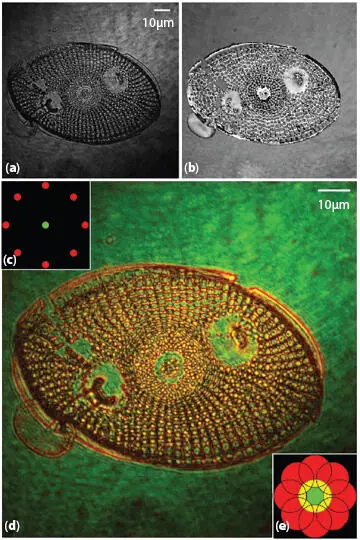
Figure 1.3 Quantitative phase image of diatom cell recorded using 20 x/0.45 microscope objective. Shown here are the (a) intensity and (b) phase images of the complex transmittance. (c) Illustration of simulated Rheinberg filter located at the rear focal plane of a microscope condenser lens. The filter comprises eight red point sources located around a center green point source. (d) Resulting image from simulated Rheinberg illumination. (e) Filtering implemented in the DFT domain, for each of the independent point sources in the filter. From [1.17] with permission of OSA.
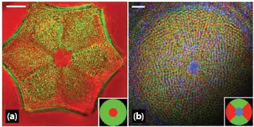
Figure 1.4 QPIs results showing a variety of diatom samples; (a) diatom recorded using a 63 × 1.3 NA microscope objective (scale bar = 5 μm); and (b) diatom cell recorded using a 20 × 0.4 NA microscope objective (scale bar = 10 μm). The color filters used in each case are illustrated in the bottom corner of the image. From [1.18] with permission of OSA.
Figure 1.5 Images illustrating the relationship between the bacterium L. monocytogenes strain P and the benthic diatom Navicula sp.: (a) first stage, (b) second stage, (c) third stage, and (d) control. The scale bars = 10 μm. From [1.65] with permission of Springer Nature.
1.2.3 Darkfield Microscopy
The widespread use of silver nanoparticles (Ag NPs) in industrial applications has prompted concerns about their effects on the environment [1.16]. Researchers have used dark field images to investigate the uptake of Ag NPs by the marine diatoms Cylindrotheca fusiformis and Cylindrotheca closterium and the biological effects on the organisms. As shown in Figure 1.6, Ag NPs detected throughout the diatom cells caused dramatic morphological changes within the cells but had little effect on the cell walls [1.49].
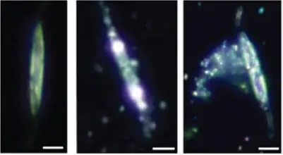
Figure 1.6 Dark field micrographs of control cells and cells exposed to Ag NPs. Ag NPs appear as bright, luminescent spots due to surface plasmon resonance. Scale bar = 10 μm. From [1.49] with permission of John Wiley and Sons.
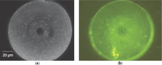
Figure 1.7 (a) Dark field image of single valve of C. wailesii diatom and (b) corresponding fluorescence macroscopic image. From [1.9] with permission of Hindawi.
At the macroscale, the wide bandgap of amorphous silica does not produce photoluminescence; however, dark field and photoluminescence imaging have shown that nanoscale silica particles exhibit photoluminescence at appropriate excitation wavelengths [1.52]. In addition to photoluminescence from nanostructured silica and diatom frustules, there is also a contribution of organic residuals incorporated in the porous silica matrix. Figure 1.7presents dark field and green fluorescence images at excitation wavelengths of 450-490 nm [1.9]. It is noted that the photoluminescence process converts the DNA-harmful UV radiation in blue radiation. Due to the maximal efficiency of photosynthesis is in the red spectral region, the green fluorescence appeared in the macroscopy images. These results indicate that there are three mechanisms by which the frustule protects the living diatoms from UV radiation: light absorption, light-collection inhibition and wavelength conversion.
1.3 Fluorescence Microscopy
Living diatoms present fluorescence from the frustule (frustule photoluminescence), chloroplasts, and lipid layers (autofluorescence). Both forms can be observed under a fluorescence microscope under an excitation wavelength of 450-490 nm, as exemplified by the C. wailesii diatom in Figure 1.7[1.9]. The enormous number of diatom species distributed throughout the world makes them an ideal vehicle by which to monitor water quality [1.29] and changes in streams and rivers over extended timespans [1.39]. In the last decade, researchers began using transgenic technology to alter diatoms with the aim of extending this analysis. For example [1.36], in a poor growth environment, diatoms will produce alkaline phosphatase, of which the promoter of alkaline phosphatase of P. tricornutum is very active. It can be used to connect the green fluorescent enzyme behind the promoters, so that in the absence of phosphate, the red autofluorescence from chloroplasts in diatoms illuminated by blue light would be changed into green autofluorescence. On the other hand, the fluorescent dye, PDMPO (2-(4-pyridyl)-5-((4-(2-dimethylaminoethylaminocarbamoyl)methoxy) phenyl)oxazole) combines with the silicic acid used by diatoms in the formation of silicified cell walls and the incorporation ratio of PDMPO and biosilica remain nearly constant for an extended duration to 2 years [1.35, 1.39]. The images in Figure 1.8were taken between 1 and 2 years after the sample was mounted on glass slides. They clearly demonstrate the persistence of PDMPO fluorescence in illustrating the fine structure of valve areolae and striae using a fluorescence microscope under UV light excitation. With this method, the silicification of an entire diatom community can be quantified by converting PDMPO incorporation to silica production using diatom bSiO2:PDMPO incorporation ratios [1.39].
Читать дальшеИнтервал:
Закладка:
Похожие книги на «Diatom Microscopy»
Представляем Вашему вниманию похожие книги на «Diatom Microscopy» списком для выбора. Мы отобрали схожую по названию и смыслу литературу в надежде предоставить читателям больше вариантов отыскать новые, интересные, ещё непрочитанные произведения.


Обсуждение, отзывы о книге «Diatom Microscopy» и просто собственные мнения читателей. Оставьте ваши комментарии, напишите, что Вы думаете о произведении, его смысле или главных героях. Укажите что конкретно понравилось, а что нет, и почему Вы так считаете.