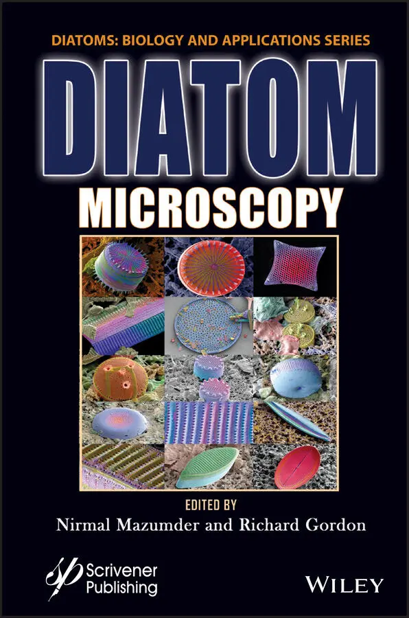
Diatom Microscopy
Здесь есть возможность читать онлайн «Diatom Microscopy» — ознакомительный отрывок электронной книги совершенно бесплатно, а после прочтения отрывка купить полную версию. В некоторых случаях можно слушать аудио, скачать через торрент в формате fb2 и присутствует краткое содержание. Жанр: unrecognised, на английском языке. Описание произведения, (предисловие) а так же отзывы посетителей доступны на портале библиотеки ЛибКат.
- Название:Diatom Microscopy
- Автор:
- Жанр:
- Год:неизвестен
- ISBN:нет данных
- Рейтинг книги:5 / 5. Голосов: 1
-
Избранное:Добавить в избранное
- Отзывы:
-
Ваша оценка:
Diatom Microscopy: краткое содержание, описание и аннотация
Предлагаем к чтению аннотацию, описание, краткое содержание или предисловие (зависит от того, что написал сам автор книги «Diatom Microscopy»). Если вы не нашли необходимую информацию о книге — напишите в комментариях, мы постараемся отыскать её.
The main goal of the book is to demonstrate the wide variety of microscopy methods being used to investigate natural and altered diatom structures. Diatom Microscopy
Diatom Microscopy — читать онлайн ознакомительный отрывок
Ниже представлен текст книги, разбитый по страницам. Система сохранения места последней прочитанной страницы, позволяет с удобством читать онлайн бесплатно книгу «Diatom Microscopy», без необходимости каждый раз заново искать на чём Вы остановились. Поставьте закладку, и сможете в любой момент перейти на страницу, на которой закончили чтение.
Интервал:
Закладка:
[1.2] Chakraborty, I., Chakrabarti, S., Managuli, V. and Mazumder, N. (2022) Chapter 4: Atomic force microscopy study of diatoms. In: Diatom Microscopy [DIMI, Volume in the series: Diatoms: Biology & Applications, series editors: Richard Gordon & Joseph Seckbach] . N. Mazumder and R. Gordon, (eds.) Wiley-Scrivener, Beverly, MA, USA: 81-110.
[1.3] De, P. and Mazumder, N. (2022) Chapter 10: Diatoms as sensors and their applications. In: Diatom Microscopy [DIMI, Volume in the series: Diatoms: Biology & Applications, series editors: Richard Gordon & Joseph Seckbach] . N. Mazumder and R. Gordon, (eds.) Wiley-Scrivener, Beverly, MA, USA: 251-282.
[1.4] Gopal, D., Chakrabarti, S., Venkata, D., Keshav, S., Gordon, R. and Mazumder, N. (2022) Chapter 3: Recent insights into the ultrastructure of diatoms using scanning and transmission electron-microscopy. In: Diatom Microscopy [DIMI, Volume in the series: Diatoms: Biology & Applications, series editors: Richard Gordon & Joseph Seckbach] . N. Mazumder and R. Gordon, (eds.) Wiley-Scrivener, Beverly, MA, USA: 57-80.
[1.5] Khan, M.J., Mathy, D. and Vinayak, V. (2022) Chapter 7: Micro to nano ornateness of diatoms from geographically distant origins of the globe. In: Diatom Microscopy [DIMI, Volume in the series: Diatoms: Biology & Applications, series editors: Richard Gordon & Joseph Seckbach] . N. Mazumder and R. Gordon, (eds.) Wiley-Scrivener, Beverly, MA, USA: 179-220.
[1.6] Lin, S.-T., Lee, M.-X. and Zhuo, G.-Y. (2022) Chapter 1: Investigation of diatoms with optical microscopy. In: Diatom Microscopy [DIMI, Volume in the series: Diatoms: Biology & Applications, series editors: Richard Gordon & Joseph Seckbach] . N. Mazumder and R. Gordon, (eds.) Wiley-Scrivener, Beverly, MA, USA: 1-32.
[1.7] Sen, J., Dhawan, P., De, P. and Mazumder, N. (2022) Chapter 12: Effects of light on physico-chemical properties of diatoms. In: Diatom Microscopy [DIMI, Volume in the series: Diatoms: Biology & Applications, series editors: Richard Gordon & Joseph Seckbach] . N. Mazumder and R. Gordon, (eds.) Wiley-Scrivener, Beverly, MA, USA: 307-334.
[1.8] Soto, J.M., Rodrigo, J.A. and Alieva, T. (2022) Chapter 5: Refractive index tomography for diatom analysis. In: Diatom Microscopy [DIMI, Volume in the series: Diatoms: Biology & Applications, series editors: Richard Gordon & Joseph Seckbach] . N. Mazumder and R. Gordon, (eds.) Wiley-Scrivener, Beverly, MA, USA: 111-138.
[1.9] Sunder, M., Acharya, N., Nayak, S., Gordon, R. and Mazumder, N. (2022) Chapter 8: Types of x-ray techniques for diatom research. In: Diatom Microscopy [DIMI, Volume in the series: Diatoms: Biology & Applications, series editors: Richard Gordon & Joseph Seckbach] . N. Mazumder and R. Gordon, (eds.) Wiley-Scrivener, Beverly, MA, USA: 221-236.
[1.10] Tisso, J., Shetty, S., Mazumder, N., Gogoi, A. and Ahmed, G.A. (2022) Chapter 6: Luminescent diatom frustules: A review on the key research applications. In: Diatom Microscopy [DIMI, Volume in the series: Diatoms: Biology & Applications, series editors: Richard Gordon & Joseph Seckbach] . N. Mazumder and R. Gordon, (eds.) Wiley-Scrivener, Beverly, MA, USA: 139-178.
[1.11] Umemura, K. (2022) Chapter 2: Nanobioscience studies of living diatoms using unique optical microscopy systems. In: Diatom Microscopy [DIMI, Volume in the series: Diatoms: Biology & Applications, series editors: Richard Gordon & Joseph Seckbach] . N. Mazumder and R. Gordon, (eds.) Wiley-Scrivener, Beverly, MA, USA: 33-56.
[1.12] Viji S., Ponpandian N. and Viswanathan C. (2022) Chapter 11: Diatom frustules: A transducer platform for optical detection of molecules. In: Diatom Microscopy [DIMI, Volume in the series: Diatoms: Biology & Applications, series editors: Richard Gordon & Joseph Seckbach] . N. Mazumder and R. Gordon, (eds.) Wiley-Scrivener, Beverly, MA, USA: 283-306.
1
Investigation of Diatoms with Optical Microscopy
Shih-Ting Lin1, Ming-Xin Lee2 and Guan-Yu Zhuo1,2*
1Integrative Stem Cell Center, China Medical University Hospital, Taichung, Taiwan
2Institute of New Drug Development, China Medical University, Taichung, Taiwan
Abstract
Diatoms are eukaryotic microalgae occurring in the water column as phytoplankton and on the ocean bed as benthic microalgae. Diatoms are similar to plants in the fact that they use photosynthesis, but differ in terms of evolutionary classification. More than 10,000 diatom species have been formally described, based primarily on the unique patterning of their silica shells. Over the last two decades, diatoms have attracted considerable attention for their potential use in synthesizing bio-functional materials due to the advantages of low production cost, prevention of using toxic chemicals and producing hazardous waste and intricate 3D hierarchical structures applicable for photonic applications, biosensors, catalysis, adsorption, and drug delivery systems. All such applications require a comprehensive understanding of the diatom structure, from the microscale to the nanoscale. Optical imaging provides spatial resolution at the sub-micrometer scale without harming the specimens. Image post-processing and reconstruction also make it possible to render the structure of samples in 3D via optical sectioning. In this chapter, we explore the various facets of optical microscopy within the context of diatom research and the applicability of this work to eco-environmental science and biomedicine. In the following sections, we respectively address light microscopy, fluorescence microscopy, confocal laser scanning microscopy, multiphoton microscopy, and super-resolution optical microscopy.
Keywords:Light microscopy, phase contrast microscopy, differential inteference contrast microscopy, darkfield microscopy, fluorescence microscopy, confocal microscopy, multiphoton microscopy, super-resolution optical microscopy
1.1 Introduction
Diatoms present a three-dimensional (3D) hierarchical structure featuring a nano-patterned porous silica shell (i.e., frustule), which is surprisingly similar to artificial photonic crystals in its ability to manipulate light [1.21, 1.67, 1.73, 1.77]. Diatom frustules comprise two parts, an upper cover (epitheca) and a lower cover (hypotheca), both of which possess a shell surface (valve) and several silicon segments referred to as a girdle band. The upper and lower covers overlap around the shell ring in a manner similar to a soap dish protecting the cell content within. There has been considerable research into the synthesis of porous silica nanoparticles (NP S), due to their large surface area and biocompatibility, which makes them ideally suited as sensors, optical devices and drug delivery systems [1.57]. The diatom frustule is a nano-patterned cell encasement made of amorphous biosilica, which occurs in an astonishing range of structures. This has prompted research into the fabrication of functional materials using diatom frustules cultivated under artificial conditions. The formation of silica shells involves highly phosphorylated proteins, long-chain polyamines and carbohydrates [1.33] in a series of biomineralization processes, many of which can be replicated in test tubes. The accumulation of diatoms on the ocean floor results in the formation of a sedimentary mineral referred to as diatomite, a highly porous silicate with low density, high absorption capacity and high surface roughness. Researchers are developing methods by which to modify the surface properties of diatomite particles with specific functional groups or combine diatomite with fluorescent particles for use in microscopic imaging applications [1.45, 1.69].
A number of microscopy techniques are used in the study of diatoms. Electron microscopy provides outstanding resolution, whereas optical microscopy permits the real-time dynamic observation required for biological research [1.23, 1.31]. In the last century, the historical development of optical microscopes has focused primarily on improving image contrast, leading to the development of phase contrast microscopy, differential interference contrast (DIC) microscopy, darkfield microscopy, and polarized light microscopy. Since the advent of dye-labeling technology [1.34], fluorescence microscopy has become the most common imaging tool in biological research; however, fluorescence microscopy is prone to interference from out-of-focal plane scattering, which prevents 3D imaging. Fluorescent molecules are also prone to photobleaching under irradiation by strong light [1.51].
Читать дальшеИнтервал:
Закладка:
Похожие книги на «Diatom Microscopy»
Представляем Вашему вниманию похожие книги на «Diatom Microscopy» списком для выбора. Мы отобрали схожую по названию и смыслу литературу в надежде предоставить читателям больше вариантов отыскать новые, интересные, ещё непрочитанные произведения.


Обсуждение, отзывы о книге «Diatom Microscopy» и просто собственные мнения читателей. Оставьте ваши комментарии, напишите, что Вы думаете о произведении, его смысле или главных героях. Укажите что конкретно понравилось, а что нет, и почему Вы так считаете.