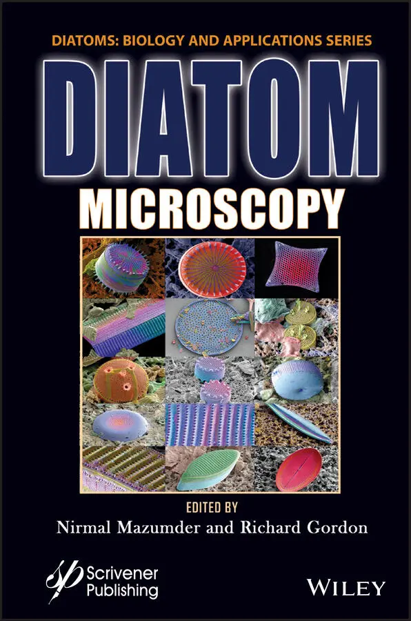
Diatom Microscopy
Здесь есть возможность читать онлайн «Diatom Microscopy» — ознакомительный отрывок электронной книги совершенно бесплатно, а после прочтения отрывка купить полную версию. В некоторых случаях можно слушать аудио, скачать через торрент в формате fb2 и присутствует краткое содержание. Жанр: unrecognised, на английском языке. Описание произведения, (предисловие) а так же отзывы посетителей доступны на портале библиотеки ЛибКат.
- Название:Diatom Microscopy
- Автор:
- Жанр:
- Год:неизвестен
- ISBN:нет данных
- Рейтинг книги:5 / 5. Голосов: 1
-
Избранное:Добавить в избранное
- Отзывы:
-
Ваша оценка:
Diatom Microscopy: краткое содержание, описание и аннотация
Предлагаем к чтению аннотацию, описание, краткое содержание или предисловие (зависит от того, что написал сам автор книги «Diatom Microscopy»). Если вы не нашли необходимую информацию о книге — напишите в комментариях, мы постараемся отыскать её.
The main goal of the book is to demonstrate the wide variety of microscopy methods being used to investigate natural and altered diatom structures. Diatom Microscopy
Diatom Microscopy — читать онлайн ознакомительный отрывок
Ниже представлен текст книги, разбитый по страницам. Система сохранения места последней прочитанной страницы, позволяет с удобством читать онлайн бесплатно книгу «Diatom Microscopy», без необходимости каждый раз заново искать на чём Вы остановились. Поставьте закладку, и сможете в любой момент перейти на страницу, на которой закончили чтение.
Интервал:
Закладка:
[1.9] De Tommasi, E. (2016) Light Manipulation by Single Cells: The Case of Diatoms. J Spectrosc 2016, 2490128.
[1.10] Delalat, B., Sheppard, V.C., Rasi Ghaemi, S., Rao, S., Prestidge, C.A., McPhee, G., Rogers, M.-L., Donoghue, J.F., Pillay, V., Johns, T.G., Kröger, N. and Voelcker, N.H. (2015) Targeted drug delivery using genetically engineered diatom biosilica. Nat Commun 6(1), 8791.
[1.11] Delasoie, J., Rossier, J., Haeni, L., Rothen-Rutishauser, B. and Zobi, F. (2018) Slow-targeted release of a ruthenium anticancer agent from vitamin B12 functionalized marine diatom microalgae. Dalton Trans 47(48), 17221–17232.
[1.12] Denk, W., Strickler, J.H. and Webb, W.W. (1990) Two-photon laser scanning fluorescence microscopy. Science 248(4951), 73–76.
[1.13] Dertinger, T., Colyer, R., Iyer, G., Weiss, S. and Enderlein, J. (2009) Fast, background-free, 3D super-resolution optical fluctuation imaging (SOFI). Proc Natl Acad Sci USA 106(52), 22287–22292.
[1.14] Drum, R.W. and Gordon, R. (2003) Star Trek replicators and diatom nanotechnology. Trends Biotechnol 21(8), 325–328.
[1.15] Ewelina, S. and Boguslaw, S. (2009) The use of benthic diatoms in estimating water quality of variously polluted rivers. Oceanol Hydrobiol Stud 38(1), 17–26.
[1.16] Fabrega, J., Luoma, S.N., Tyler, C.R., Galloway, T.S. and Lead, J.R. (2011) Silver nanoparticles: Behaviour and effects in the aquatic environment. Environ Int 37(2), 517–531.
[1.17] Fan, X., Healy, J.J., O’Dwyer, K. and Hennelly, B.M. (2019) Label-free color staining of quantitative phase images of biological cells by simulated Rheinberg illumination. Appl Opt 58(12), 3104–3114.
[1.18] Fan, X., Healy, J.J., O’Dwyer, K. and Hennelly, B.M. (2020) Label-free color staining of quantitative phase images. Opt Lasers Eng 129, 106049.
[1.19] Fernández-Suárez, M. and Ting, A.Y. (2008) Fluorescent probes for super-resolution imaging in living cells. Nat Rev Mol Cell Biol 9(12), 929–943.
[1.20] Fu, W., Chaiboonchoe, A., Khraiwesh, B., Sultana, M., Jaiswal, A., Jijakli, K., Nelson, D.R., Al-Hrout, A.a., Baig, B., Amin, A. and Salehi-Ashtiani, K. (2017) Intracellular spectral recompositioning of light enhances algal photosynthetic efficiency. Sci Adv 3(9), e1603096.
[1.21] Ghobara, M.M., Ghobara, M.M., Mazumder, N., Vinayak, V., Reissig, L., Gebeshuber, I.C., Tiffany, M.A., Gordon, R. and Gordon, R. (2019) On Light and Diatoms: A Photonics and Photobiology Review. In: Diatoms: Fundamentals and Applications . 129–189.
[1.22] Gröger, P., Poulsen, N., Klemm, J., Kröger, N. and Schlierf, M. (2016) Establishing super-resolution imaging for proteins in diatom biosilica. Sci Rep 6(1), 36824.
[1.23] Hasle, G.R. and Fryxell, G.A. (1970) Diatoms: Cleaning and Mounting for Light and Electron Microscopy. Trans Am Microsc Soc 89(4), 469–474.
[1.24] Hell, S.W., Dyba, M. and Jakobs, S. (2004) Concepts for nanoscale resolution in fluorescence microscopy. Curr Opin Neurobiol 14(5), 599–609.
[1.25] Hell, S.W. and Kroug, M. (1995) Ground-state-depletion fluorscence microscopy: A concept for breaking the diffraction resolution limit. Appl Phys B 60(5), 495–497.
[1.26] Hell, S.W. and Wichmann, J. (1994) Breaking the diffraction resolution limit by stimulated emission: stimulated-emission-depletion fluorescence microscopy. Opt Lett 19(11), 780–782.
[1.27] Horton, N.G., Wang, K., Kobat, D., Clark, C.G., Wise, F.W., Schaffer, C.B. and Xu, C. (2013) In vivo three-photon microscopy of subcortical structures within an intact mouse brain. Nat photonics 7(3), 205–209.
[1.28] Huang, B., Babcock, H. and Zhuang, X. (2010) Breaking the diffraction barrier: super-resolution imaging of cells. Cell 143(7), 1047–1058.
[1.29] Köhler, J. (1997) Use of algae for monitoring rivers II. - Innsbruck. Int Revue ges Hydrobiol 82(3), 340.
[1.30] Kieu, K., Mehravar, S., Gowda, R., Norwood, R.A. and Peyghambarian, N. (2013) Label-free multi-photon imaging using a compact femtosecond fiber laser mode-locked by carbon nanotube saturable absorber. Biomed Opt Express 4(10), 2187–2195.
[1.31] Korte, V.L. and Blinn, D.W. (1983) Diatom Colonization on Artificial Substrata in Pool and Riffle Zones Studied by Light and Scanning Electron Microscopy. J Phycol 19(3), 332–341.
[1.32] Kotzsch, A., Gröger, P., Pawolski, D., Bomans, P.H.H., Sommerdijk, N.A.J.M., Schlierf, M. and Kröger, N. (2017) Silicanin-1 is a conserved diatom membrane protein involved in silica biomineralization. BMC Biol 15(1), 65–65.
[1.33] Kröger, N. and Poulsen, N. (2008) Diatoms-from cell wall biogenesis to nanotechnology. Annu Rev Genet 42, 83–107.
[1.34] Kremers, G.-J., Gilbert, S.G., Cranfill, P.J., Davidson, M.W. and Piston, D.W. (2011) Fluorescent proteins at a glance. J Cell Sci 124, 157–160.
[1.35] Leblanc, K. and Hutchins, D.A. (2005) New applications of a biogenic silica deposition fluorophore in the study of oceanic diatoms. Limnology and Oceanography: Methods 3(10), 462–476.
[1.36] Lin, H.-Y., Yen, S.-C., Kuo, P.-C., Chung, C.-Y., Yeh, K.-L., Huang, C.-H., Chang, J. and Lin, H.-J. (2017) Alkaline phosphatase promoter as an efficient driving element for exogenic recombinant in the marine diatom Phaeodactylum tricornutum. Algal Res 23, 58–65.
[1.37] Marter, P., Schmidt, S., Kiontke, S. and Moog, D. (2020) Optimized mRuby3 is a Suitable Fluorescent Protein for in vivo Co-localization Studies with GFP in the Diatom Phaeodactylum tricornutum. Protist 171(1), 125715.
[1.38] Mazumder, N., Balla, N.K., Zhuo, G.-Y., Kistenev, Y.V., Kumar, R., Kao, F.-J., Brasselet, S., Nikolaev, V.V. and Krivova, N.A. (2019) Label-Free Non-linear Multimodal Optical Microscopy—Basics, Development, and Applications. Frontiers in Physics 7, 170.
[1.39] McNair, H.M., Brzezinski, M.A. and Krause, J.W. (2015) Quantifying diatom silicification with the fluorescent dye, PDMPO. Limnology and Oceanography: Methods 13(10), 587–599.
[1.40] Minsky, M. (1988) Memoir on inventing the confocal scanning microscope. Scanning 10(4), 128–138.
[1.41] Moerner, W.E. (2006) Single-molecule mountains yield nanoscale cell images. Nat Methods 3(10), 781–782.
[1.42] Montazer, Z., Habibi Najafi, M.B. and Levin, D.B. (2019) Microbial degradation of low-density polyethylene and synthesis of polyhydroxyalkanoate polymers. Can J Microbiol 65(3), 224–234.
[1.43] Nieuwenhuizen, R.P.J., Lidke, K.A., Bates, M., Puig, D.L., Grünwald, D., Stallinga, S. and Rieger, B. (2013) Measuring image resolution in optical nanoscopy. Nat Methods 10(6), 557–562.
[1.44] Noga, T., Stanek-Tarkowska, J., Kochman, N., Peszek, Ł., Pajączek, A. and Woźniak, K. (2013) Application of diatoms to assess the quality of the waters of the baryczka stream, left-side tributary of the river san. J Ecol Eng 14(3), 8–23.
[1.45] Onesto, V., Villani, M., Coluccio, M.L., Majewska, R., Alabastri, A., Battista, E., Schirato, A., Calestani, D., Coppedé, N., Cesarelli, M., Amato, F., Di Fabrizio, E. and Gentile, F. (2018) Silica diatom shells tailored with Au nanoparticles enable sensitive analysis of molecules for biological, safety and environment applications. Nanoscale Res Lett 13(1), 94.
[1.46] Patrascu, E., Melinte, P. and Dragoi, G. (2014) Optimized morphologic evaluation of biostructures by examination in polarized light and differential interference contrast microscopy. Romanian J Leg Med 22, 275–282.
[1.47] Patterson, G.H. (2009) Fluorescence microscopy below the diffraction limit. Semin Cell Dev Biol 20(8), 886–893.
Читать дальшеИнтервал:
Закладка:
Похожие книги на «Diatom Microscopy»
Представляем Вашему вниманию похожие книги на «Diatom Microscopy» списком для выбора. Мы отобрали схожую по названию и смыслу литературу в надежде предоставить читателям больше вариантов отыскать новые, интересные, ещё непрочитанные произведения.


Обсуждение, отзывы о книге «Diatom Microscopy» и просто собственные мнения читателей. Оставьте ваши комментарии, напишите, что Вы думаете о произведении, его смысле или главных героях. Укажите что конкретно понравилось, а что нет, и почему Вы так считаете.