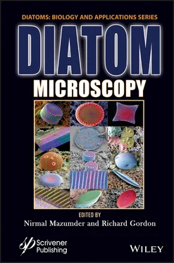
Diatom Microscopy
Здесь есть возможность читать онлайн «Diatom Microscopy» — ознакомительный отрывок электронной книги совершенно бесплатно, а после прочтения отрывка купить полную версию. В некоторых случаях можно слушать аудио, скачать через торрент в формате fb2 и присутствует краткое содержание. Жанр: unrecognised, на английском языке. Описание произведения, (предисловие) а так же отзывы посетителей доступны на портале библиотеки ЛибКат.
- Название:Diatom Microscopy
- Автор:
- Жанр:
- Год:неизвестен
- ISBN:нет данных
- Рейтинг книги:5 / 5. Голосов: 1
-
Избранное:Добавить в избранное
- Отзывы:
-
Ваша оценка:
- 100
- 1
- 2
- 3
- 4
- 5
Diatom Microscopy: краткое содержание, описание и аннотация
Предлагаем к чтению аннотацию, описание, краткое содержание или предисловие (зависит от того, что написал сам автор книги «Diatom Microscopy»). Если вы не нашли необходимую информацию о книге — напишите в комментариях, мы постараемся отыскать её.
The main goal of the book is to demonstrate the wide variety of microscopy methods being used to investigate natural and altered diatom structures. Diatom Microscopy
Diatom Microscopy — читать онлайн ознакомительный отрывок
Ниже представлен текст книги, разбитый по страницам. Система сохранения места последней прочитанной страницы, позволяет с удобством читать онлайн бесплатно книгу «Diatom Microscopy», без необходимости каждый раз заново искать на чём Вы остановились. Поставьте закладку, и сможете в любой момент перейти на страницу, на которой закончили чтение.
Интервал:
Закладка:
Due to the nature of light diffraction, the image resolution of confocal microscopy is limited to roughly 250 nm, which makes it difficult to visualize silica-embedded proteins (ranging from ten to several hundred nanometers) via live-cell imaging. Super-resolution optical microscopy based on photoswitchable fluorescent proteins makes it possible to determine with a high degree of precision the location of proteins at the single-molecule level. Fusion proteins with silaffin-3 (tpSil3) are embedded in biosilica and permanently entrapped inside the valve region during biosilica formation [1.50]. PALM super-resolution optical imaging involves the expression of fusion proteins with photoswitchable fluorescent proteins, which can prevent the problem of silica limiting antibody access to the protein(s) of interest in STORM. Genetic transformation facilitates live-cell imaging of proteins associated with the cell wall, thereby making it possible to use the expression of GFP fusion proteins in Thalassiosira pseudonana to localize distinct regions of biosilica. Gröger et al . [1.22] used the inherently high labeling density of PALM to study proteins in diatom biosilica.
Prior to imaging, it is necessary to select suitable super-resolution probes for biosilica embedded fusion-proteins. Six fluorescent proteins (PATagRFP, PAmCherry1, PA-GFP, mEOS3.2, Dendra2, and Dronpa) have been activated efficiently in cytosol; however, only Dendra2, mEOS3.2, and Dronpa have been activated when embedded in biosilica. Fusing three fluorescent proteins to tpSil3 for PALM imaging has made it possible to achieve average localization precision of roughly 25 nm. Images are reconstructed (stacked) from more than 1000 frames to facilitate localization. Note that the production of pure biosilica requires the extraction of chloroplasts using a detergent-based buffer to eliminate their autofluorescence, which can hinder in vivo single-molecule localization microscopy. Figure 1.18apresents a PALM image of Dendra2 in the biosilica of the valve region of Thalassiosira pseudonana . Figure 1.18dshows the focal plane in the girdle band region. As shown in Figure 1.18b, the average full width at half maximum (FWHM) of 76.0 nm ± 4.6 nm (± S.D.) was derived from multiple line scans perpendicular to the biosilica cylinder ( Figure 1.18a). The 76 nm measurement can be further deconvoluted to 53 nm ± 3 nm by taking into account the localization precision and the linker length of 3 nm between tpSil3 and the chromophore of the fluorescent protein. Fourier ring correlation analysis ( Figure 1.18c) was used to estimate image resolution [1.43], the results of which (74.7 nm) were in good agreement with those in Figure 1.18b. Total internal reflection fluorescence (TIRF) imaging has also been used to determine the locations of tpSil3-Dendra2, tpSil3–mEOS3.2 and tpSil3–Dronpa embedded in biosilica ( Figures 1.18d,e,f; left side). This provides a representative comparison of the enhanced resolution/contrast shown in Figure 1.18d,e,f(right side). Note that tpSil3-Dendra2, tpSil3-mEOS3.2 and tpSil3–Dronpa are shown in the outer region of the fultoportulae basal chamber instead of within the external tubes of the fultoportulae. The appearance of large circular non-fluorescent gaps correlates with the structural elements of fultoportulae observed in SEM images (insets of Figures 1.18d,e,f).

Figure 1.18 PALM analysis of tpSil3. (a) Comparison of epifluorescence image and reconstructed super-resolution image of tpSil3-Dendra2 with z-focus on the girdle band area of the diatom. The position of the line scan is highlighted. (b) Line scan through the silica cell wall showing the fluorescence intensity profiles obtained using the two imaging modalities. The FWHM of the PALM image is indicated. (c) Fourier ring correlation applied to super-resolution image in A revealing an effective resolution of 74.7nm. Comparison of epifluorescence images and reconstructed super-resolution image of Dendra2 (d), mEOS3.2 (e), and Dronpa (f) fused to tpSil3 with thew z-focus on the valve region of the diatom. Enlarged details of the fultoportulae to the right. For comparison, we also present an SEM image at the same scale corresponding to the enlarged details shown in the fluorescence image. Scale bars = 1μm and 100nm in the zoomed images. From [1.22] with permission of Springer Nature.
1.7 Conclusion
The structural, mechanical, and optoelectronic properties of diatoms make them ideal research subjects for biomedical research and other fields. Diatoms have been used in photonics to fabricate optical devices based on the micro-meter and nano-meter pores in silica skeletons. It is also possible to load the diatom structure with specific proteins, enzymes, or antibodies for use as biosensors in disease diagnosis and drug carriers, and their biocompatibility and non-toxicity make them an ideal bone implant material. Nano-patterned diatom frustules can even be used to differentiate cancer cells from normal cells. Nonetheless, further advancements will depend on a comprehensive understanding of diatom structure and morphology, which can only be achieved using non-intrusive methods, such as optical microscopy. Fluorescence-based microscopic methods can also be used to gain deeper insights into the application of diatoms in other fields, such as the monitoring of marine environments in vivo or ex vivo . The use of multidimensional imaging in conjunction with spectroscopic and dynamic information provides the sensitivity and selectivity sufficient to detect events at the molecular level.
Acknowledgement
This work was supported by the India-Taiwan International Cooperative Research Project of Ministry of Science and Technology, Taiwan (grant no. 110-2923-M-039-001-MY3).
References
[1.1] Allen, R.D., Allen, N.S. and Travis, J.L. (1981) Video-enhanced contrast, differential interference contrast (AVEC-DIC) microscopy: a new method capable of analyzing microtubule-related motility in the reticulopodial network of Allogromia laticollaris. Cell Motil 1(3), 291–302.
[1.2] Betzig, E., Patterson, G.H., Sougrat, R., Lindwasser, O.W., Olenych, S., Bonifacino, J.S., Davidson, M.W., Lippincott-Schwartz, J. and Hess, H.F. (2006) Imaging Intracellular Fluorescent Proteins at Nanometer Resolution. Science 313(5793), 1642–1645.
[1.3] Boyd, R.W. and Prato, D. (2008) Nonlinear Optics . Elsevier Science.
[1.4] Buhmann, M., Kroth, P.G. and Schleheck, D. (2012) Photoautotrophicheterotrophic biofilm communities: a laboratory incubator designed for growing axenic diatoms and bacteria in defined mixed-species biofilms. Environ Microbiol Rep 4(1), 133–140.
[1.5] Caputo, A., Nylander, J.A.A. and Foster, R.A. (2019) The genetic diversity and evolution of diatom-diazotroph associations highlights traits favoring symbiont integration. FEMS Microbiol Lett 366(2), fny297.
[1.6] Chauhan, D., Agrawal, G., Deshmukh, S., Roy, S.S. and Priyadarshini, R. (2018) Biofilm formation by Exiguobacterium sp. DR11 and DR14 alter polystyrene surface properties and initiate biodegradation. RSC Advances 8(66), 37590–37599.
[1.7] Débarre, D., Supatto, W., Pena, A.-M., Fabre, A., Tordjmann, T., Combettes, L., Schanne-Klein, M.-C. and Beaurepaire, E. (2006) Imaging lipid bodies in cells and tissues using third-harmonic generation microscopy. Nat Methods 3(1), 47–53.
[1.8] De Stefano, L., Maddalena, P., Moretti, L., Rea, I., Rendina, I., De Tommasi, E., Mocella, V. and De Stefano, M. (2009) Nano-biosilica from marine diatoms: A brand new material for photonic applications. Superlattices Microstruct . 46(1), 84–89.
Читать дальшеИнтервал:
Закладка:
Похожие книги на «Diatom Microscopy»
Представляем Вашему вниманию похожие книги на «Diatom Microscopy» списком для выбора. Мы отобрали схожую по названию и смыслу литературу в надежде предоставить читателям больше вариантов отыскать новые, интересные, ещё непрочитанные произведения.
Обсуждение, отзывы о книге «Diatom Microscopy» и просто собственные мнения читателей. Оставьте ваши комментарии, напишите, что Вы думаете о произведении, его смысле или главных героях. Укажите что конкретно понравилось, а что нет, и почему Вы так считаете.


