Armand Leroi - Mutants
Здесь есть возможность читать онлайн «Armand Leroi - Mutants» весь текст электронной книги совершенно бесплатно (целиком полную версию без сокращений). В некоторых случаях можно слушать аудио, скачать через торрент в формате fb2 и присутствует краткое содержание. Город: New York, Год выпуска: 2005, ISBN: 2005, Издательство: Penguin Books, Жанр: История, Биология, sci_popular, на английском языке. Описание произведения, (предисловие) а так же отзывы посетителей доступны на портале библиотеки ЛибКат.
- Название:Mutants
- Автор:
- Издательство:Penguin Books
- Жанр:
- Год:2005
- Город:New York
- ISBN:0-670-03110-0
- Рейтинг книги:4 / 5. Голосов: 1
-
Избранное:Добавить в избранное
- Отзывы:
-
Ваша оценка:
Mutants: краткое содержание, описание и аннотация
Предлагаем к чтению аннотацию, описание, краткое содержание или предисловие (зависит от того, что написал сам автор книги «Mutants»). Если вы не нашли необходимую информацию о книге — напишите в комментариях, мы постараемся отыскать её.
gives a brilliant narrative account of our genetic code and the captivating people whose bodies have revealed it—a French convent girl who found herself changing sex at puberty; children who, echoing Homer’s Cyclops, are born with a single eye in the middle of their foreheads; a village of long-lived Croatian dwarves; one family, whose bodies were entirely covered with hair, was kept at the Burmese royal court for four generations and gave Darwin one of his keenest insights into heredity. This elegant, humane, and engaging book “captures what we know of the development of what makes us human” (
).
Mutants — читать онлайн бесплатно полную книгу (весь текст) целиком
Ниже представлен текст книги, разбитый по страницам. Система сохранения места последней прочитанной страницы, позволяет с удобством читать онлайн бесплатно книгу «Mutants», без необходимости каждый раз заново искать на чём Вы остановились. Поставьте закладку, и сможете в любой момент перейти на страницу, на которой закончили чтение.
Интервал:
Закладка:
THE CALCULATOR OF FATE
The morphogens that traverse the developing embryo – be they protein or hydrocarbon ring – provide cells with a kind of coordinate grid that they use to find out where they are and so what they should do and be. A cell is thus rather like a navigator who, traversing the wastes of the ocean, labours with sextant and chronometer to find his longitude and latitude. But there is one difference between navigator and cell: while the navigator’s referents, the stars and planets, are always where they should be, the cell’s sometimes are not. Sirenomelia and cyclopia are two instances where mutation has warped the universe that cells refer to or even caused its total collapse.
Yet even bearing this difference (inevitable when comparing the clockwork motions of the physical world with the jerry-built devices of biology) in mind, the analogy still has force. For all the constancy of the heavens, navigators have always lost their way – perhaps because the instruments by which they read the heavens become maladjusted. In the same way, the receptors which allow cells to perceive morphogens and measure their concentrations can also go awry – and any number of congenital disorders are caused by mutations that affect them.
But perhaps the deepest level of the analogy comes when we consider the calculations that navigators must make in order to establish where they are. Cells, too, calculate – and they do so with great precision, absorbing information from their environment, adding it up and arriving at a solution. This calculator – one might call it a calculator of fate – is composed of a vast number of proteins that combine their efforts within each cell to arrive at a solution. Of course, the calculator is not infallible: just as navigators occasionally get their sums wrong, so too, occasionally, do cells.
The consequences of cells making mistakes of this sort are beautifully illustrated by one of the more curious pieces of erotica dug from the ruins of Herculaneum. It is a small marble statue – no larger than a shoebox – that depicts Pan the goat-god, whom the Romans knew as Faunus, raping a nanny goat. Masterfully combining the animal and the human in equal parts, the unknown artist has given his Pan shaggy legs, cloven hooves, thick lips, a flattened snout and an expression of concentrated violence. He has also given the god an unusual anatomical feature. Suspended from his neck, just above the clavicles, are two small pendulous lobes that in life would be no more than a few centimetres long.
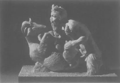
These lobes, which are very distinctive, only appear in Pans of the second or third century bc, or, as in this statue (now in the Secret Cabinet of the Naples Archaeological Museum), in later Roman copies of Greek originals. The innumerable goat-gods who chase across the black- or red-figure vases of the Classical period wooing shepherds or grasping at nymphs do not have them, nor do the allegorical Pans of the Renaissance and Baroque such as those in Sandro Botticelli’s Mars and Venus or Annibale Carracci’s Omnia vincit Amor . Neck lobes would also be quite out of place in the beautiful but vapid Pans of the Pre-Raphaelites.
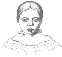
The origin of the god’s lobes is plain enough: they are echoed by an identical pair of appendages on his victim, the neck lobes frequently found on domesticated goats (German goatherds call them Glocken – bells). The sculptor of the original Pan Raping a Goat was clearly an acute observer of nature, and incorporated the lobes as one more detail to signify the goatishness of the god. Neck lobes, however, occur not only in goats but also, albeit rarely, in humans. In 1858 a British physician by the name of Birkett published a short paper describing a seven-year-old girl who had been brought to him with a pair protruding stiffly from either side of her neck. The girl had had them since birth. Birkett was not sure what they were, but he cut them off anyway and put them under the microscope, where he discovered that they were auricles – an extra pair of external ears.
Extra auricles are an instance of a phenomenon called homeosis in which one part of a developing embryo becomes anomalously transformed into another. The particular transformation that causes neck-ears has its origins around five months after conception, when five cartilaginous arches form on either side of the embryo’s head, positioned much where gills would be were the embryo a fish. Indeed, were the embryo a fish, gill arches are what they would become. In humans they form a miscellany of head parts including jaws, the tiny bones of the inner ears, and sundry throat cartilages. The visible, protuberant parts of our ears develop out of the cleft between the first and the second pair of arches. The remaining clefts usually just seal over, leaving our necks smooth, but occasionally in humans and often in goats, one of the lower clefts remains open and develops into something that looks much like an ear. The resemblance, however, is only superficial: the ‘ears’ have none of the internal apparatus that would enable them to hear.
Homeosis was first identified as a distinct phenomenon by the British biologist William Bateson, who in an 1894 book, Materials for the study of variation , coined the term and collected dozens of examples of such transformations. The Materials has something of the flavour of a medieval bestiary – Bateson called it his ‘imaginary museum’ – in which infants with supernumerary ears and heifers with odd numbers of teats jostle for space with five-winged moths, eight-legged beetles and lobsters that have antennae where their eyes should be. A strange book, then. Yet the Materials remains important to, and is cited by, molecular biologists in a way that few nineteenth-century zoological compendia are. This is because the transformations that Bateson identified pointed the way to one of the embryo’s most beautiful devices: the genetic programme that permits cells, and so tissues and organs, to become different from each other. Homeosis pointed the way to the calculator of fate.
The calculator of fate was first discovered in fruit flies. Flies, like earthworms, are divided into repeating units or segments. These segments are especially obvious in maggots, though metamorphosis obscures some of their boundaries. Many segments in the adult fly are specialised in some way. Head segments carry labial palps (with which the fly feeds) and antennae (with which it smells); thoracic segments carry wings, legs, or small balancing organs called halteres; abdominal segments have no appendages at all. The organs of a given segment are established when the fly is only an embryo, long before they can actually be seen. To put it a bit more abstractly, in the embryo each segment is given an identity .
Over the last eighty-odd years, Drosophila geneticists have sought and found dozens of mutations that destroy the identities of segments. Some of these mutations cause flies to grow legs instead of antennae on their heads – and make a fly that cannot smell; others cause halteres to become wings – and make a four-winged dipteran that defies its own definition. Yet other mutations cause wings to become halteres – and leave the fly irredeemably earthbound.
Читать дальшеИнтервал:
Закладка:
Похожие книги на «Mutants»
Представляем Вашему вниманию похожие книги на «Mutants» списком для выбора. Мы отобрали схожую по названию и смыслу литературу в надежде предоставить читателям больше вариантов отыскать новые, интересные, ещё непрочитанные произведения.


Обсуждение, отзывы о книге «Mutants» и просто собственные мнения читателей. Оставьте ваши комментарии, напишите, что Вы думаете о произведении, его смысле или главных героях. Укажите что конкретно понравилось, а что нет, и почему Вы так считаете.