Armand Leroi - Mutants
Здесь есть возможность читать онлайн «Armand Leroi - Mutants» весь текст электронной книги совершенно бесплатно (целиком полную версию без сокращений). В некоторых случаях можно слушать аудио, скачать через торрент в формате fb2 и присутствует краткое содержание. Город: New York, Год выпуска: 2005, ISBN: 2005, Издательство: Penguin Books, Жанр: История, Биология, sci_popular, на английском языке. Описание произведения, (предисловие) а так же отзывы посетителей доступны на портале библиотеки ЛибКат.
- Название:Mutants
- Автор:
- Издательство:Penguin Books
- Жанр:
- Год:2005
- Город:New York
- ISBN:0-670-03110-0
- Рейтинг книги:4 / 5. Голосов: 1
-
Избранное:Добавить в избранное
- Отзывы:
-
Ваша оценка:
Mutants: краткое содержание, описание и аннотация
Предлагаем к чтению аннотацию, описание, краткое содержание или предисловие (зависит от того, что написал сам автор книги «Mutants»). Если вы не нашли необходимую информацию о книге — напишите в комментариях, мы постараемся отыскать её.
gives a brilliant narrative account of our genetic code and the captivating people whose bodies have revealed it—a French convent girl who found herself changing sex at puberty; children who, echoing Homer’s Cyclops, are born with a single eye in the middle of their foreheads; a village of long-lived Croatian dwarves; one family, whose bodies were entirely covered with hair, was kept at the Burmese royal court for four generations and gave Darwin one of his keenest insights into heredity. This elegant, humane, and engaging book “captures what we know of the development of what makes us human” (
).
Mutants — читать онлайн бесплатно полную книгу (весь текст) целиком
Ниже представлен текст книги, разбитый по страницам. Система сохранения места последней прочитанной страницы, позволяет с удобством читать онлайн бесплатно книгу «Mutants», без необходимости каждый раз заново искать на чём Вы остановились. Поставьте закладку, и сможете в любой момент перейти на страницу, на которой закончили чтение.
Интервал:
Закладка:
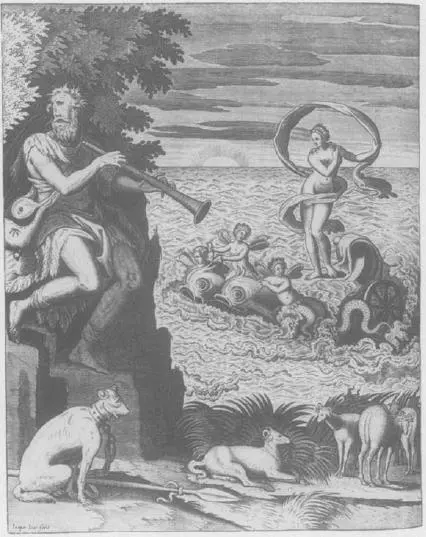
This was the beginning of a tradition of fabulous races that persisted for about fifteen centuries. By the third century ad, Christian writers had adopted the tradition; by the fifth century, St Augustine is wondering whether these races are descended from Adam. In the Middle Ages, the Cyclopes appears essentially unaltered from antiquity in manuscripts of wonder-books such as Thomas a Cantimpré’s De Naturis Rerum which was composed around 1240. In the fourteenth century, their biblical parentage is settled: they are the deformed descendants of Cain and Ham. Around the same time they appear in illuminations of Marco Polo’s travels (the Italian unaccountably fails to mention their existence); in the early 1500s one appears on the wall of a Danish church dressed in the striped pantaloons, floppy hat and leather purse of a late-medieval Baltic dandy. With time, the Cyclops becomes smaller, tamer and moves closer to home.
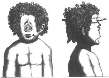
The first illustration of a cyclopic child, as distinct from a Cyclops, was given by Fortunio Liceti. In the 1634 edition of his De monstrorum he describes an infant girl who was born in Firme, Italy, in 1624 and who, he says, had a well-organised body but a head of horrible aspect. In the middle of her face, in place of a nose, there was a mass of skin that resembled a penis or a pear. Below this was a square-shaped piece of reddish skin on which one could see two very close-set eyes like the eyes of a chicken. Although the child died at birth she is depicted with the proportions of a robust ten-year-old, a legacy of the giants that preceded her.
Liceti describes another case of cyclopia as well, this time in a pair of conjoined twins whose crania are fused so that they face away from each other in true Janus style. Conjoined twinning and cyclopia is an unusual combination of anomalies, and one would be inclined to doubt its authenticity but for a 1916 clinical report of a pair of conjoined twins who showed much the same combination of features. And then there is the unusual provenance of Liceti’s drawing. It is, he says, a copy of one preserved in the collection of His Eminence the Reverend Cardinal Barberini at Rome, and the original, which now seems to be lost, was drawn by Leonardo da Vinci.
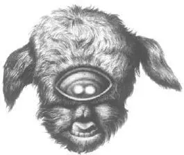
Looking at his bottled babies, Willem Vrolik recognised that some were more severely afflicted than others. Some had only a single eyeball concealed within the eye-orbit, but in others two eyeballs were visible. Some had a recognisable nose, others had none at all. Modern clinicians recognise cyclopia as one extreme in a spectrum of head defects. At the other extreme are people whose only oddity is a single incisor placed symmetrically in their upper jaw instead of the usual two.
The single eye of a cyclopic child is the external sign of a disorder that reaches deep within its skull. All normal vertebrates have split brains. We, most obviously, have left and right cerebral hemispheres that we invoke when speaking of our left or right ‘brains’. Cyclopic infants do not. Instead of two distinct cerebral hemispheres, two optic lobes and two olofactory lobes, their forebrains are fused into an apparently indivisible whole. Indeed, clinicians call this whole spectrum of birth defects the ‘holoprosencephaly series’, from the Greek: holo – whole, prosencephalon – forebrain. It is, in all its manifestations, the most common brain deformity in humans, afflicting 1 in 16,000 live-born children and 1 in 200 miscarried foetuses.
The ease with which foetuses become cyclopic is frightening. Fish embryos will become cyclopic if they are heated, cooled, irradiated, deprived of oxygen, or exposed to ether, chloroform, acetone, phenol, butyric acid, lithium chloride, retinoic acid, alcohol or merely table salt. In the 1950s an epidemic of cyclopic lambs in the western United States was caused by pregnant ewes grazing on corn lilies, a plant of the subalpine meadows which has leaves rich in toxic alkaloids. In humans, diabetic mothers have a two-hundred-fold increased risk of giving birth to cyclopic children, as do alcoholic mothers.
Most cases of cyclopia are not, however, caused by anything the mother did (or did not do) during her pregnancy. Mutations in at least four and perhaps as many as twelve human genes also cause some form of holoprosencephaly. One of these genes encodes a signalling protein called sonic hedgehog. This molecule received its name in the early 1980s when a mutant fruit fly was discovered whose maggot progeny had a surplus of bristles covering their tiny bodies. ‘Hedgehog’ was the obvious name for the gene, and when a related gene was discovered in vertebrates, ‘sonic hedgehog’ seemed the natural choice to a postgraduate student who perhaps loved his gaming-console too much. The sonic hedgehog mutations that cause cyclopia in humans are dominant. This implies that anyone who has just a single copy of the defective gene should have cyclopia or at least some kind of holoprosencephaly. But for reasons that are poorly understood, some carriers of mutant genes are hardly affected at all. They live, and pass the defective gene on to their children.

The fact that sonic hedgehog-defective infants have a single cerebral hemisphere tells us something important. When the forebrain first forms in the normal embryo it is a unitary thing, a simple bulge at the end of the neural tube – only later does it split into a left and right brain. This split is induced by sonic which, like so many signalling molecules, is a morphogen. During the formation of the neural tube, sonic appears in a small piece of mesoderm directly beneath the developing forebrain. Filtering up from one tissue to the next it cleaves the brain in two. This process is especially obvious in the making of eyes. Long before the embryo has eyes, a region of the forebrain is dedicated to their neural wiring. This region – the optic field – first appears as a single band traversing the embryo forebrain. Sonic moulds the optic field’s topography, reducing it to two smaller fields on either side of the head. Mutations or chemicals that inhibit sonic prevent this – thus the single, monstrous, staring eye of the cyclopic infant.
But sonic does more than give us distinct cerebral hemispheres. Mice in which the sonic hedgehog gene has been completely disabled have malformed hearts, lungs, kidneys and guts. They are always stillborn and have no paws. Their faces are malformed beyond cyclopic, reduced to a strange kind of trunk: they have no eyes, ears or mouths. These malformations suggest that sonic is used throughout the developing embryo, almost anywhere it is growing a part. It even seems to be used repeatedly in the making of our heads.
Читать дальшеИнтервал:
Закладка:
Похожие книги на «Mutants»
Представляем Вашему вниманию похожие книги на «Mutants» списком для выбора. Мы отобрали схожую по названию и смыслу литературу в надежде предоставить читателям больше вариантов отыскать новые, интересные, ещё непрочитанные произведения.


Обсуждение, отзывы о книге «Mutants» и просто собственные мнения читателей. Оставьте ваши комментарии, напишите, что Вы думаете о произведении, его смысле или главных героях. Укажите что конкретно понравилось, а что нет, и почему Вы так считаете.