Lynelle R. Johnson - Canine and Feline Respiratory Medicine
Здесь есть возможность читать онлайн «Lynelle R. Johnson - Canine and Feline Respiratory Medicine» — ознакомительный отрывок электронной книги совершенно бесплатно, а после прочтения отрывка купить полную версию. В некоторых случаях можно слушать аудио, скачать через торрент в формате fb2 и присутствует краткое содержание. Жанр: unrecognised, на английском языке. Описание произведения, (предисловие) а так же отзывы посетителей доступны на портале библиотеки ЛибКат.
- Название:Canine and Feline Respiratory Medicine
- Автор:
- Жанр:
- Год:неизвестен
- ISBN:нет данных
- Рейтинг книги:3 / 5. Голосов: 1
-
Избранное:Добавить в избранное
- Отзывы:
-
Ваша оценка:
Canine and Feline Respiratory Medicine: краткое содержание, описание и аннотация
Предлагаем к чтению аннотацию, описание, краткое содержание или предисловие (зависит от того, что написал сам автор книги «Canine and Feline Respiratory Medicine»). Если вы не нашли необходимую информацию о книге — напишите в комментариях, мы постараемся отыскать её.
includes more detail and new information throughout, particularly in the areas of antimicrobial guidelines, anti-fungal therapies, and ant-viral medications in cats.
Logically organized for ease of use in the practice setting,
features problem-based learning to enhance working knowledge of the topics discussed. Chapters cover localization of disease, respiratory diagnostics, respiratory therapeutics, nasal disorders, and diseases of airways. Sections on parenchymal disease; pleural and mediastinal disease; and vascular disorders are also presented.
Offers a complete guide to the diagnosis and treatment of respiratory conditions in canine and feline patients—now expanded to include more detailed and advanced information Focuses on localization of disease, diagnostic testing, and appropriate therapy Thoroughly updated with new antimicrobial guidelines, the latest information on anti-fungal therapy, and new advances in ant-viral medications in cats Includes many new images and topics for discussion Features problem-based learning to support a working understanding of the topics discussed
is an essential resource for veterinary internal medicine specialists, general practitioners, and veterinary students.
Canine and Feline Respiratory Medicine — читать онлайн ознакомительный отрывок
Ниже представлен текст книги, разбитый по страницам. Система сохранения места последней прочитанной страницы, позволяет с удобством читать онлайн бесплатно книгу «Canine and Feline Respiratory Medicine», без необходимости каждый раз заново искать на чём Вы остановились. Поставьте закладку, и сможете в любой момент перейти на страницу, на которой закончили чтение.
Интервал:
Закладка:
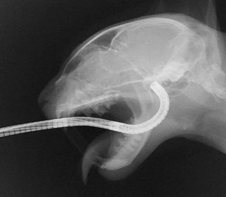
Figure 2.9 Visualization of the choanae is obtained by inserting a flexible endoscope into the oral cavity and flexing the scope maximally (180°) around the soft palate.
If an endoscope is unavailable to complete the retroflexed exam, a bright light source and dental mirror can be used, along with a spay hook to retract the soft palate cranially, although this can be quite challenging in smaller animals. The dental mirror is placed in the back of the pharynx and angled above the soft palate while a light source is used to illuminate the view of the choanae in the mirror.
Foreign material can be removed from the nasopharynx or choanae using the endoscope and samples can be obtained for histopathology. With the endoscope removed from the patient and the tip of the scope in a non‐flexed position, forceps are passed through the biopsy channel until they are at the end of the scope. The scope is then inserted into the mouth and flexed about the soft palate, as previously described. The degree of flexion that the scope can achieve is reduced by the presence of forceps in the channel, making visualization of any lesion more challenging. Forceps are passed forward to grab a foreign body or mass lesion and firm traction is applied. It is most efficient and safest to unflex the scope, withdraw it from the patient, and extrude the forceps to remove the sample, rather than pulling the sample and instrument back through the scope.
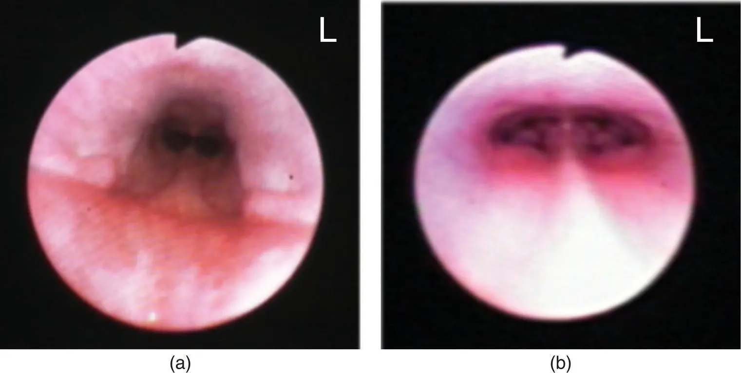
Figure 2.10 Image of the normal nasopharynx in (a) a dog and (b) a cat. The soft palate is dorsal in these images.
After the caudal nasopharynx is examined and samples are obtained for histopathology as indicated, a moistened surgical lap pad is used to pack the throat in order to prevent aspiration during rostral rhinoscopy. Equipment available for rostral rhinoscopy includes rigid endoscopes (with or without an external sheath), otoscopes, and small flexible endoscopes. Rigid scopes have better optics, are easier to maneuver, and come in smaller sizes, while flexible scopes allow greater access to the nasal cavity and can permit examination of the frontal sinuses when there is marked turbinate destruction (such as in nasal aspergillosis). Rigid scopes are available with viewing angles from 0 to 30°. Rhinoscopy can be performed using the telescope portion of the rigid scope alone (∼2.8 mm outer diameter) or by using the sheathed scope (∼5 mm outer diameter), which has flush and suction ports as well as a biopsy port available ( Figure 2.11). The biopsy port of the sheath will accept standard endoscopic biopsy or foreign body retrieval instruments.
Before entering the nasal cavity, the endoscope is placed against the skull and measured to the level of the medial canthus of the eye to approximate the position of the cribriform plate. A piece of tape is applied to the instrument at this length, and the equipment should not be passed further than this point to avoid penetrating through the cribriform plate into the brain case. The normal nasal cavity is made up of scrolls of turbinates comprising the dorsal, middle, and ventral concha. The mucosa is generally pink, with a pale sheen of serous secretions coating the smooth epithelial surface ( Figure 2.12). The primary changes to look for during rhinoscopy are mucosal hyperemia, mucus accumulation, epithelial irregularities, turbinate destruction that is visualized as increased space between turbinates, or a mass effect that reduces space in the nasal cavity. After the initial visual inspection and when the cribriform plate is intact, nasal drops containing oxymetazoline or phenylephrine can be dropped or sprayed into the nasal cavity to create vasoconstriction. This will cause swollen and hyperemic mucosa to blanch and shrink, thus changing the overall appearance of the mucosa, but allowing improved access in some situations. Topical anesthetic drops or a misted spray of tetracaine can also be used to help reduce the stimulation caused by rhinoscopy, although this is variably effective.
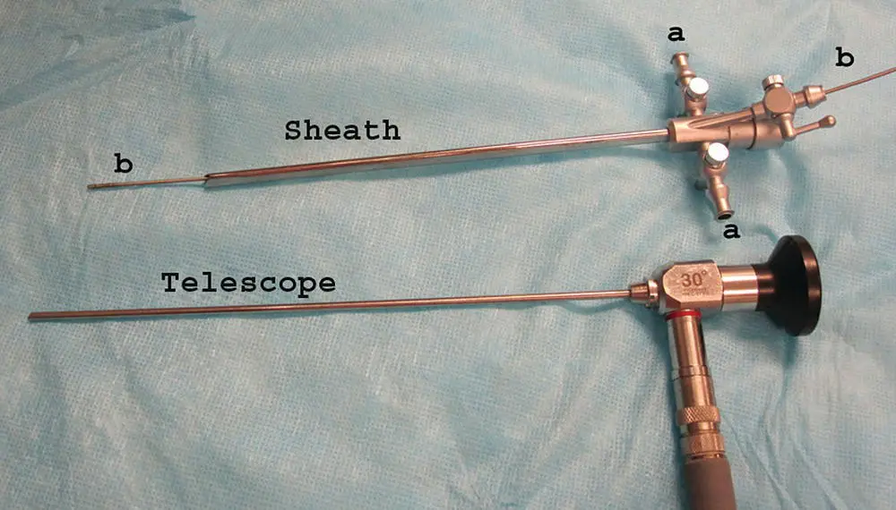
Figure 2.11 The sheath (∼5 mm outer diameter) for the telescope portion of the rigid scope (∼2.8 mm outer diameter) has flush and suction ports (a) as well as a biopsy port (b) available.
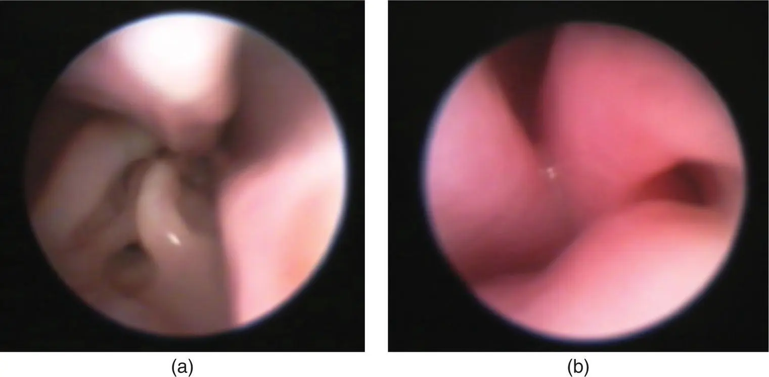
Figure 2.12 Normal nasal turbinates in (a) the dog and (b) the cat.
Mass lesions, fungal plaques, and specific mucosal lesions should be biopsied with visualization to improve the probability of achieving a diagnostic sample. With diffuse or generalized disease, an attempt should also be made to obtain at least the initial nasal biopsy with visualization of the area ( Figure 2.13). This can be accomplished either by using a biopsy instrument that fits over the scope, by using a flexible biopsy instrument that passes through the sheathed scope, or by advancing a rigid biopsy instrument along the outside of the rigid telescope. Once bleeding starts, the visual field is usually lost, although intermittent flush and suction can be used to clear the field. Nasal flush can be achieved by attaching a bag of chilled fluids to the biopsy port on the sheath of the scope, or by inserting a red rubber catheter into the nasal cavity and using syringes to obtain a pulsatile pressurized flush. In cats, a 20–60 ml syringe inserted into each naris can be used for nasal flush ( Figure 2.14). Nasal flush should also be used if excessive mucus obscures visualization of the nasal cavity. If a foreign body is highly suspected but is not visualized or removed during the procedure, nasal flush will sometimes dislodge the material. The surgical lap pad protecting the endotracheal tube should be removed, examined for foreign material periodically, and replaced with a new one. Also, if a foreign body is suspected, consider placing a Foley catheter in the caudal nasopharynx and using retrograde flush of the nasal cavities to dislodge items ( Figure 2.15).
After removing the moistened lap pad from the oral cavity, the airway above the endotracheal tube is suctioned to remove any additional fluid. Before recovery from anesthesia, a complete oral exam and dental probing are recommended to rule out dental disease as a cause for nasal discharge ( Figure 2.16).
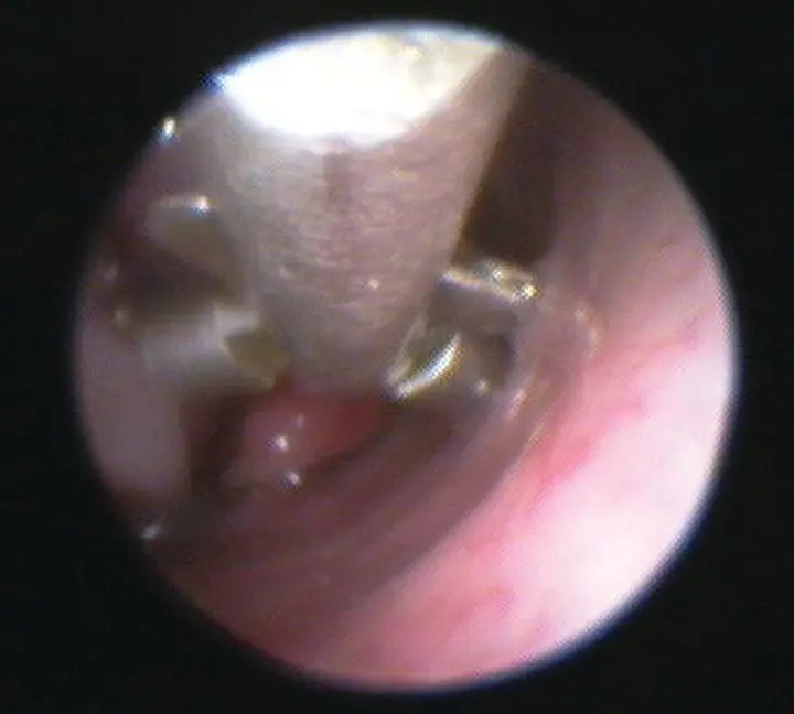
Figure 2.13 Diagnostic yield of a nasal biopsy sample is improved when the site to be sampled is visualized during the biopsy procedure.
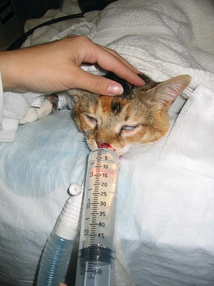
Figure 2.14 Nasal flush can be performed by inserting a syringe tip into the nostril and injecting pulses of chilled saline through each nostril. The animal must be intubated and have a moistened lap pad placed in the oral cavity to protect against aspiration.
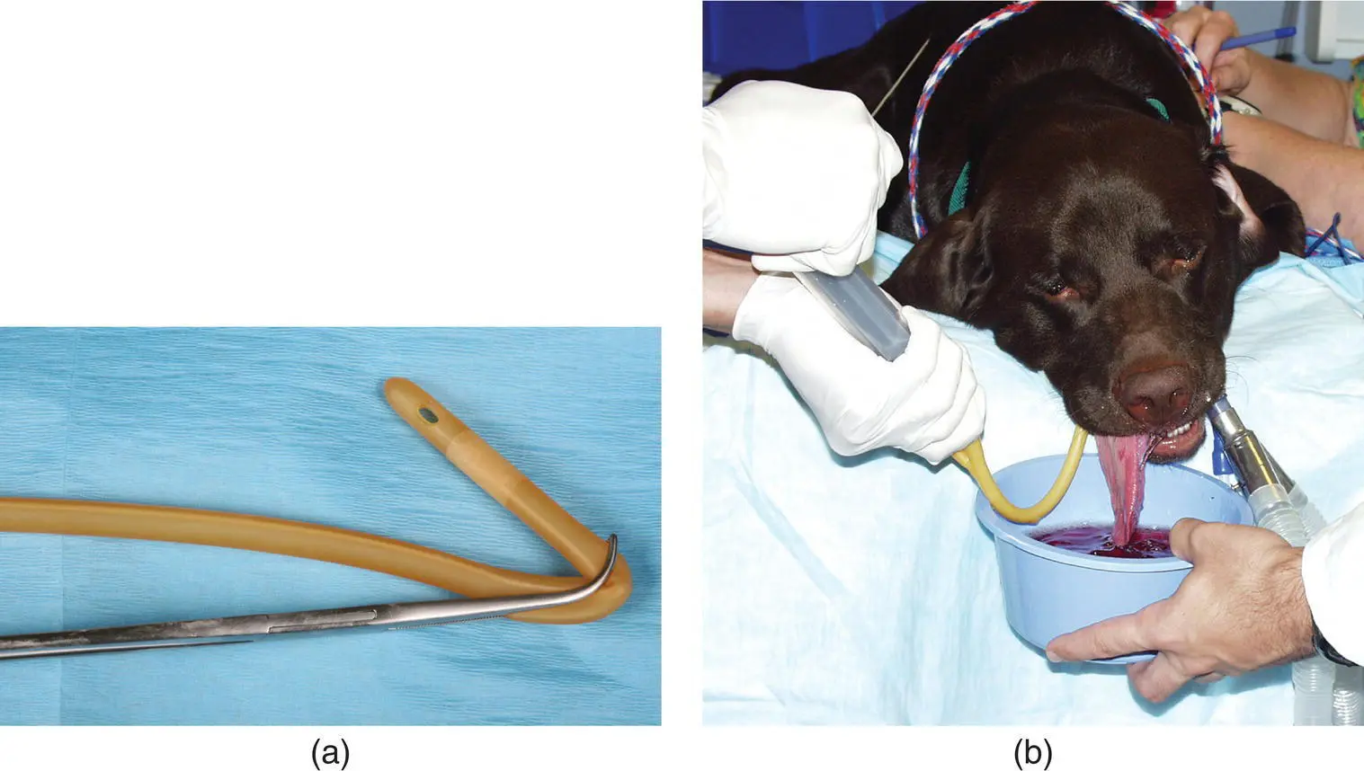
Figure 2.15 (a) Right‐angle forceps can be used to place a Foley catheter above the soft palate. (b) The forceps with Foley are inserted into the oral cavity and a spay hook can be used to retract the palate over the balloon portion of the Foley. The balloon is fully inflated and nasal flush can be used to dislodge foreign material or exudate into a collection bowl.
Читать дальшеИнтервал:
Закладка:
Похожие книги на «Canine and Feline Respiratory Medicine»
Представляем Вашему вниманию похожие книги на «Canine and Feline Respiratory Medicine» списком для выбора. Мы отобрали схожую по названию и смыслу литературу в надежде предоставить читателям больше вариантов отыскать новые, интересные, ещё непрочитанные произведения.


Обсуждение, отзывы о книге «Canine and Feline Respiratory Medicine» и просто собственные мнения читателей. Оставьте ваши комментарии, напишите, что Вы думаете о произведении, его смысле или главных героях. Укажите что конкретно понравилось, а что нет, и почему Вы так считаете.