Lynelle R. Johnson - Canine and Feline Respiratory Medicine
Здесь есть возможность читать онлайн «Lynelle R. Johnson - Canine and Feline Respiratory Medicine» — ознакомительный отрывок электронной книги совершенно бесплатно, а после прочтения отрывка купить полную версию. В некоторых случаях можно слушать аудио, скачать через торрент в формате fb2 и присутствует краткое содержание. Жанр: unrecognised, на английском языке. Описание произведения, (предисловие) а так же отзывы посетителей доступны на портале библиотеки ЛибКат.
- Название:Canine and Feline Respiratory Medicine
- Автор:
- Жанр:
- Год:неизвестен
- ISBN:нет данных
- Рейтинг книги:3 / 5. Голосов: 1
-
Избранное:Добавить в избранное
- Отзывы:
-
Ваша оценка:
Canine and Feline Respiratory Medicine: краткое содержание, описание и аннотация
Предлагаем к чтению аннотацию, описание, краткое содержание или предисловие (зависит от того, что написал сам автор книги «Canine and Feline Respiratory Medicine»). Если вы не нашли необходимую информацию о книге — напишите в комментариях, мы постараемся отыскать её.
includes more detail and new information throughout, particularly in the areas of antimicrobial guidelines, anti-fungal therapies, and ant-viral medications in cats.
Logically organized for ease of use in the practice setting,
features problem-based learning to enhance working knowledge of the topics discussed. Chapters cover localization of disease, respiratory diagnostics, respiratory therapeutics, nasal disorders, and diseases of airways. Sections on parenchymal disease; pleural and mediastinal disease; and vascular disorders are also presented.
Offers a complete guide to the diagnosis and treatment of respiratory conditions in canine and feline patients—now expanded to include more detailed and advanced information Focuses on localization of disease, diagnostic testing, and appropriate therapy Thoroughly updated with new antimicrobial guidelines, the latest information on anti-fungal therapy, and new advances in ant-viral medications in cats Includes many new images and topics for discussion Features problem-based learning to support a working understanding of the topics discussed
is an essential resource for veterinary internal medicine specialists, general practitioners, and veterinary students.
Canine and Feline Respiratory Medicine — читать онлайн ознакомительный отрывок
Ниже представлен текст книги, разбитый по страницам. Система сохранения места последней прочитанной страницы, позволяет с удобством читать онлайн бесплатно книгу «Canine and Feline Respiratory Medicine», без необходимости каждый раз заново искать на чём Вы остановились. Поставьте закладку, и сможете в любой момент перейти на страницу, на которой закончили чтение.
Интервал:
Закладка:
Thoracic ultrasound is highly useful in detecting pleural fluid and evaluating parenchymal mass lesions, and has also proven effective in evaluating pulmonary infiltrates, particularly in the emergency situation when the clinician needs to distinguish congestive heart failure from a respiratory condition. The presence of fluid in the interstitial–alveolar space in association with pulmonary edema results in the artifact of comet tails, referred to as B‐lines. In dogs and cats with pulmonary edema due to congestive heart failure, there is an increase in the number of detectable B‐lines visible in multiple lung fields (Lisciandro et al. 2017; Ward et al. 2017). Because this technique relies on attenuation of the ultrasound beam by thickening of the interstitial–alveolar junctions, other diseases that result in this process and extend to the lung periphery could lead to the generation of B‐lines on ultrasound. Therefore, the use of lung ultrasound in large numbers of patients with respiratory conditions such as interstitial lung disease, pneumonia, and non‐cardiogenic pulmonary edema requires additional study. Nonetheless, it appears that ultrasound of the lung could be used to evaluate patients for fluid overload or congestive failure and assess response to therapy. This technique would likely be less stressful to the patient than recheck radiographs.
Fluoroscopy
Airway fluoroscopy uses air as the contrast medium to evaluate dynamic changes in the luminal diameter of the airways during normal breathing and during cough. Fluoroscopy is typically performed in lateral recumbency. It most readily recognizes cervical tracheal collapse because of the large airway diameter, but can also provide information on lobar bronchial collapse. A fluoroscopic study performed in right lateral recumbency will highlight the left lung lobes, while a study in left lateral recumbency will examine the right lobar bronchi, although right lateral recumbency appears to be used most commonly. Dorsoventral positioning would likely be required to identify collapse of the accessory lobar bronchus. Fluoroscopy has poorer resolution than radiographs and the lower image quality can hamper interpretation.
Given the intricate relationship between neural pathways of the respiratory and digestive tracts as well as their close anatomic association, videofluoroscopic swallowing studies have a role in investigating the underlying etiology of some respiratory diseases, as well as detecting risk factors for disease in other cases. Esophageal dysfunction and gastroesophageal reflux are common and can lead to chronic recurrent pneumonia or pneumonitis. Increasing evidence indicates that laryngeal paralysis in the geriatric Labrador is accompanied by esophageal dysfunction as part of a generalized neuropathy, and swallowing studies can help assess the risk for lower respiratory complications in affected dogs. Weak pharyngeal contractions in other breeds of dogs also can predispose to aspiration events. Interestingly, videofluoroscopy has revealed that brachycephalic breeds commonly display pharyngeal collapse (Pollard et al. 2018), likely due to anatomic issues and pressure fluctuations associated with respiratory effort. In addition to perpetuating aspiration injury to the lower respiratory tract, nasopharyngeal aspiration events can lead to nasal discharge and might be a cause for upper respiratory tract inflammation.
Computed Tomography
Imaging the respiratory tract with CT provides superior detail in comparison to standard radiography because of the removal of superimposed structures and the ability to reformat images in three dimensions. When a helical CT is available, image acquisition is very fast; however, anesthesia is employed in many instances to obtain optimal images. Some facilities perform CT in awake or sedated patients using a plexiglass holding chamber (VetMousetrap™; Oliveira et al. 2011). Motion artifact can obscure detail, particularly when respirations interfere with the evaluation of thoracic structures. It is a clinical decision whether the risks of anesthesia outweigh the value of images obtained, but often an intervention is planned after CT (e.g., endoscopy or surgery) which will also require anesthesia. For the thorax, the animal is typically ventilated several times and a breath hold at 15 cm water (H 2O) is employed to expand the chest during the 10–30 seconds of image acquisition.
Airway Sampling
Transoral Tracheal Wash
A transoral tracheal wash is appropriate for use in large and small dogs or in cats. A sterile endotracheal tube and a sterile polypropylene or open‐ended red rubber catheter (3.5–8 French) are needed. Prior to doing a tracheal wash, the catheter should be measured against the animal and marked at a position that estimates the location of the carina, which is approximately at the fourth intercostal space ( Figure 2.3). Passing the catheter too far distally can result in airway damage. The animal is anesthetized with a short‐acting anesthetic agent. Options include propofol or alfaxalone with midazolam, or ketamine with valium (a 1 : 1 mixture). Lubricant should not be used on the endotracheal tube because it can adversely affect cytology results. Prior to intubation, the function of the larynx is assessed to ensure normal abduction on inspiration. In the cat, local lidocaine can be used to facilitate intubation and avoid contamination of the tube through contact with oropharyngeal or laryngeal mucosa, although being patient while drugs take effect generally allows a clean intubation. An assistant holds the endotracheal tube in place during the lavage.
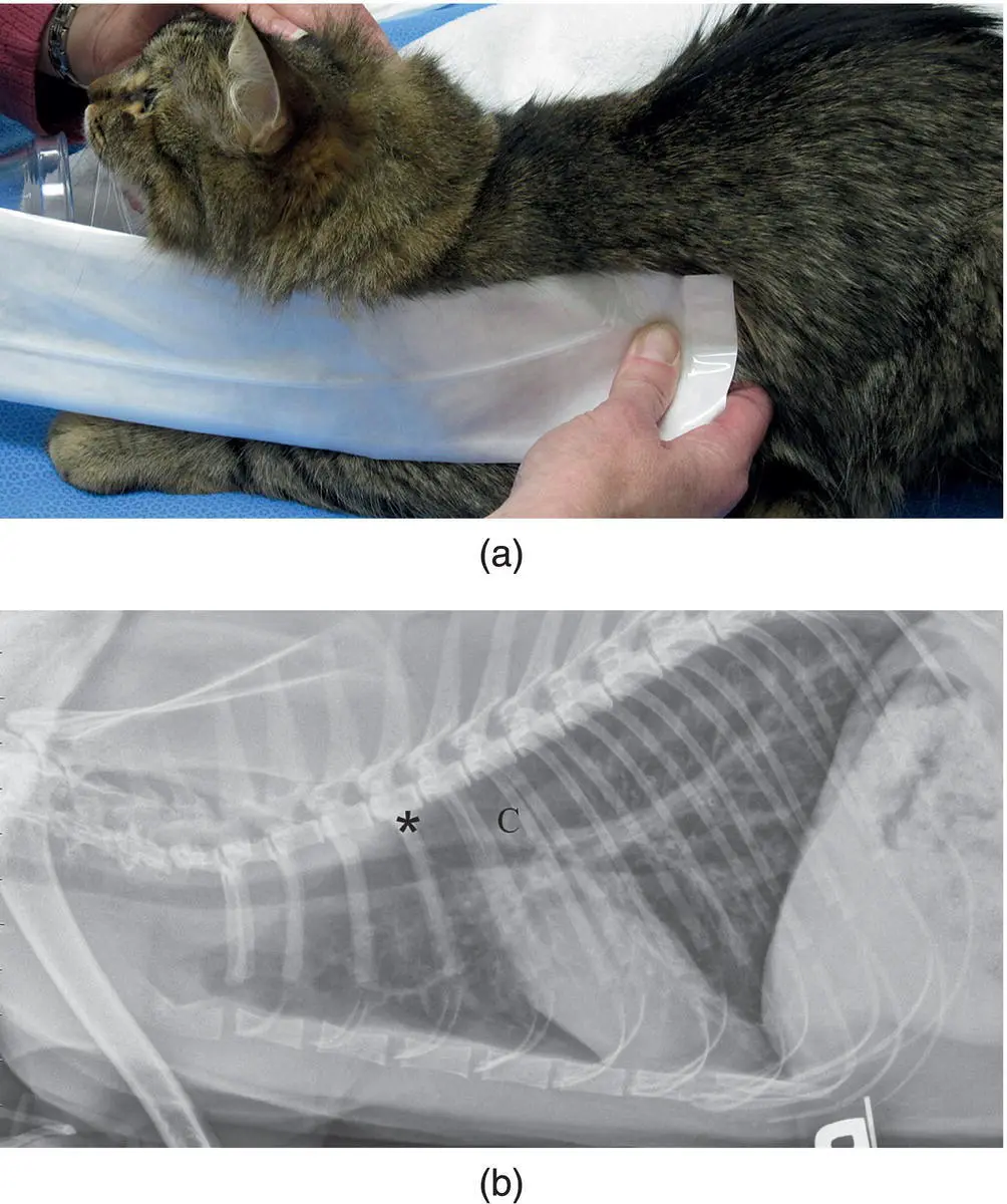
Figure 2.3 (a and b) The urinary catheter used to collect an airway sample during a tracheal wash is measured to approximately the fourth rib (*) to avoid passing the catheter beyond the level of the carina (C).
With the endotracheal tube held in place, the polypropylene or red rubber catheter is passed sterilely through the tube to the fourth intercostal space, and the three‐way stopcock with syringe is attached to the outer port. An aliquot of saline (4–6 ml) is instilled into the airway followed by 2–3 ml of air to ensure that all fluid has exited the catheter ( Figure 2.4). The stopcock is turned off to the syringe and either hand suction or house suction can be applied to aspirate fluid and cells from the lower airway. Use of a sterile suction trap ( Figure 2.5) can be beneficial in collecting the sample when house suction is used. If retrieval of fluid is less than desired, the cuff of the endotracheal tube can be inflated to improve suction. Retrieval of fluid can also be enhanced by having the assistant compress the chest, perform coupage, or stimulate a cough during suction. Instillation and aspiration of fluid can be repeated several times until an adequate sample has been retrieved. Typically ~1.0 ml is sufficient for culture and cytology.
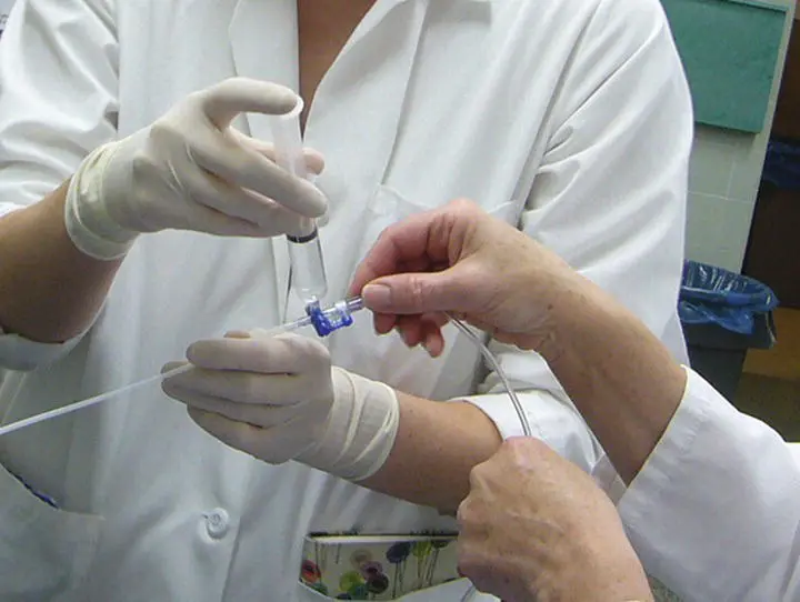
Figure 2.4 A three‐way stopcock is turned on to the urinary catheter used to collect the airway wash and off to the suction catheter.
A modification of the transoral tracheal wash can be performed, which yields a sample more closely approximating that of a bronchoalveolar lavage (BAL). In the dog, this is achieved using a 16‐French Argyle™ stomach tube (Sherwood Medical Co./Tyco Healthcare Kendall, Deland, FL; Hawkins and Berry 1999). The dog must be large enough to accommodate an endotracheal tube that will not be fully occluded by the stomach tube. The stomach tube is shortened to remove any side holes on the distal end and to create a length that approximates the distance from the endotracheal tube to the last rib. The distal end of the tube can be tapered with a sterile pencil sharpener to improve the ability to wedge within the airway, and the tube should be sterilized prior to use. Endotracheal intubation is carried out using a short‐acting anesthetic agent (e.g. propofol or alfaxalone with midazolam) and a sterile endotracheal tube. The dog is placed in dorsal recumbency, and the modified stomach tube is passed slowly through the lumen of the endotracheal tube until it meets resistance. Gentle pressure should be used when passing and wedging this tube to avoid perforating the lung. As soon as slight resistance is encountered, the tube is withdrawn 1–2 cm and lavage is initiated with 20 ml of sterile saline followed by 5 ml of air. An adaptor might be needed to fit the syringe to the stomach tube. Gentle hand suction is applied to retrieve the fluid, and a second aliquot can be instilled as needed.
Читать дальшеИнтервал:
Закладка:
Похожие книги на «Canine and Feline Respiratory Medicine»
Представляем Вашему вниманию похожие книги на «Canine and Feline Respiratory Medicine» списком для выбора. Мы отобрали схожую по названию и смыслу литературу в надежде предоставить читателям больше вариантов отыскать новые, интересные, ещё непрочитанные произведения.


Обсуждение, отзывы о книге «Canine and Feline Respiratory Medicine» и просто собственные мнения читателей. Оставьте ваши комментарии, напишите, что Вы думаете о произведении, его смысле или главных героях. Укажите что конкретно понравилось, а что нет, и почему Вы так считаете.