An Illustrated Guide to Oral Histology
Здесь есть возможность читать онлайн «An Illustrated Guide to Oral Histology» — ознакомительный отрывок электронной книги совершенно бесплатно, а после прочтения отрывка купить полную версию. В некоторых случаях можно слушать аудио, скачать через торрент в формате fb2 и присутствует краткое содержание. Жанр: unrecognised, на английском языке. Описание произведения, (предисловие) а так же отзывы посетителей доступны на портале библиотеки ЛибКат.
- Название:An Illustrated Guide to Oral Histology
- Автор:
- Жанр:
- Год:неизвестен
- ISBN:нет данных
- Рейтинг книги:5 / 5. Голосов: 1
-
Избранное:Добавить в избранное
- Отзывы:
-
Ваша оценка:
An Illustrated Guide to Oral Histology: краткое содержание, описание и аннотация
Предлагаем к чтению аннотацию, описание, краткое содержание или предисловие (зависит от того, что написал сам автор книги «An Illustrated Guide to Oral Histology»). Если вы не нашли необходимую информацию о книге — напишите в комментариях, мы постараемся отыскать её.
An introduction to tooth development, including the bud, cap, early bell, and late bell stages A thorough exploration of enamel, dentin, cementum and dental pulp A discussion of the periodontal ligament, including alveolar crest fibers, horizontal, oblique, apical, and inter-radicular fibers, transseptal fibers, and gingival fibers A guide to alveolar bone, oral mucosa, and salivary glands Perfect for postgraduate dental students,
will also be useful to undergraduate dental students, and those looking to improve their understanding of the microscopic structure of dental tissues and their pathologies.
An Illustrated Guide to Oral Histology — читать онлайн ознакомительный отрывок
Ниже представлен текст книги, разбитый по страницам. Система сохранения места последней прочитанной страницы, позволяет с удобством читать онлайн бесплатно книгу «An Illustrated Guide to Oral Histology», без необходимости каждый раз заново искать на чём Вы остановились. Поставьте закладку, и сможете в любой момент перейти на страницу, на которой закончили чтение.
Интервал:
Закладка:
1.2.2 Key Identifying Features
The enamel organ resembles a cap present on top of the dental papilla. The dental follicle and dental papilla become more recognizable during this stage compared to the bud stage.
1.2.3 Clinical Significance
It is believed that the blood supply of the tooth is established during the cap stage. The blood vessels first enter through the dental follicle, then move into the dental papilla [2]. Any disruptions during this stage could severely affect the vascular supply of the tooth and in turn, its maturation, vitality, and eruption.
1.3 Early Bell Stage
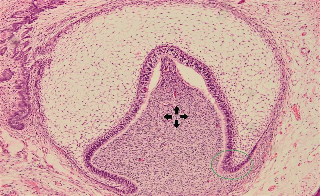
Figure 1.6H and E stained section showing the early bell stage of tooth development (green circle, cervical loop; arrows, dental papilla).
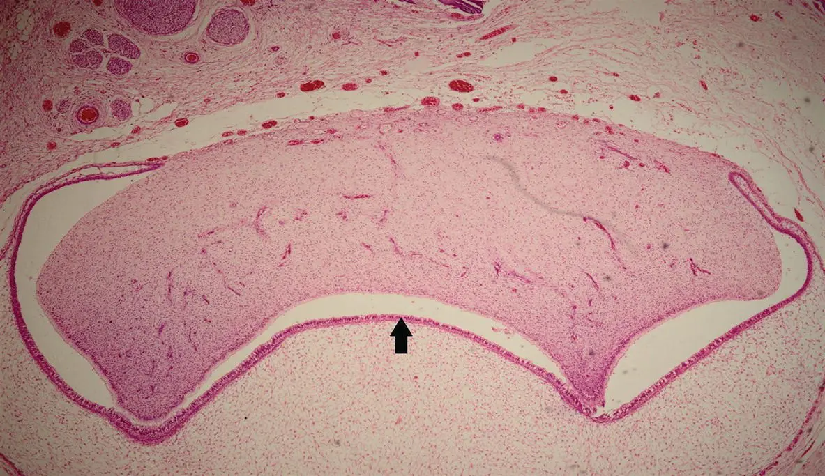
Figure 1.7H and E stained section showing the early bell stage of tooth development (arrow, dental follicle).
1.3.1 Description
During this stage, the enamel organ resembles a bell. It is during this stage that the tooth crown will undertake its final shape (morphodifferentiation) and ameloblasts along with odontoblasts are histodifferentiated. The region where the outer and inner enamel epithelial cells meet at the border of enamel organ is called the cervical loop . In between the stellate reticulum and the inner enamel epithelial cells, some of the cell population differentiates and forms a new layer of cells called stratum intermedium . The cells of the inner enamel epithelium and stratum intermedium work collaboratively to form the enamel tissue. The enamel organ during the early bell stage clearly shows its four diverse layers: outer enamel epithelium, inner enamel epithelium, stellate reticulum, and stratum intermedium. During this stage, the enamel organ loses its contact with the oral epithelium as the dental lamina is broken down. This connection is restored during the process of tooth eruption. The remnants of dental lamina are called epithelial rest of Serres. In the early bell stage, the enamel knot disappears and the enamel cord appears between the stratum intermedium and stellate reticulum.
1.3.2 Key Identifying Features
On histological sections, bell‐shaped enamel organ dissociated from the oral epithelium can be seen clearly. The tooth germ during this stage is enclosed by dental follicle. The cervical loop is also very prominent and easily recognizable.
1.3.3 Clinical Significance
Many important structures appear during this stage. It is believed that enamel cord facilitates the change from the cap to bell stage [1]. The cervical loop is responsible for the formation of Hertwig's epithelial root sheath (HERS) [3].
1.4 Late Bell Stage
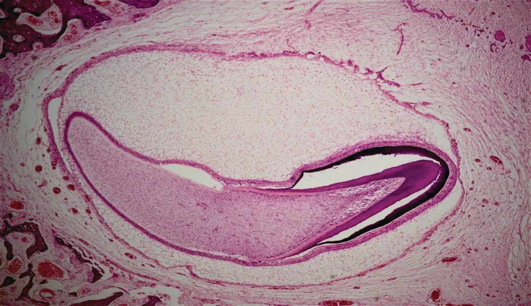
Figure 1.8H and E stained decalcified section showing the late bell stage of tooth development.
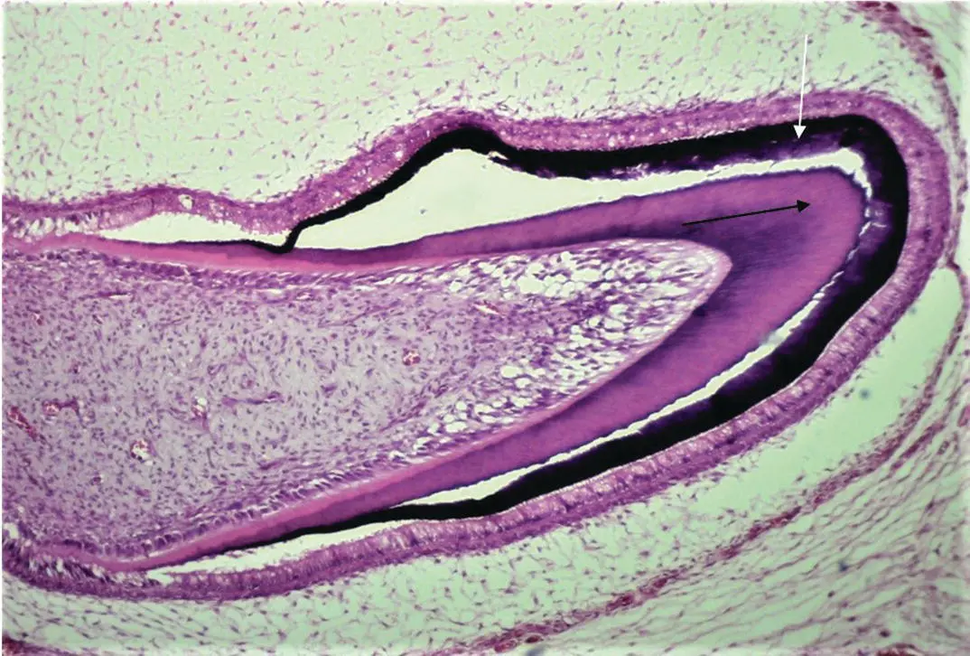
Figure 1.9H and E stained decalcified section showing the late bell stage of tooth development (white arrow, enamel; black arrow, dentin).
1.4.1 Description
In the late bell stage, the tooth germ increases in size, and the hard tissues of the teeth start forming. The process of dentin formation is called dentinogenesis and it always precedes the process of enamel formation, i.e. amelogenesis . It is beyond the scope of this book to go into details of these processes but briefly, under the influence of inner enamel epithelium (which changes into pre‐ameloblasts), the adjacent peripheral cells of dental papilla become odontoblasts. These odontoblasts start secreting of pre‐dentin followed by dentin; this secretion stimulates pre‐ameloblasts to change into ameloblasts which start secreting the enamel matrix (which mineralizes and becomes dental enamel later). While secreting, odontoblasts move away from the secretion area, leaving behind their odontoblastic processes. Similarly, ameloblasts migrate away from dentin while secreting enamel matrix. It should be noted that the formation of these two tissues begins in the area of future cusps/incisal edges and then slopes downward. This is the stage where the commencement of root formation begins as well.
1.4.2 Key Identifying Features
On the histological sections, prominently visible dental hard tissues (enamel and dentin) can be seen. Ameloblasts (on top of the newly formed enamel) and odontoblasts (just below the newly formed dentin) are also evident.
1.4.3 Clinical Significance
The visible development of HERS begins during this stage. The HERS is responsible for determining the shape, size, and number of roots [4]. Disruption to this stage could affect amelogenesis and dentinogenesis, leading to the formation of abnormal enamel and dentin, respectively (or non‐formation) [5].
1.5 Root Formation
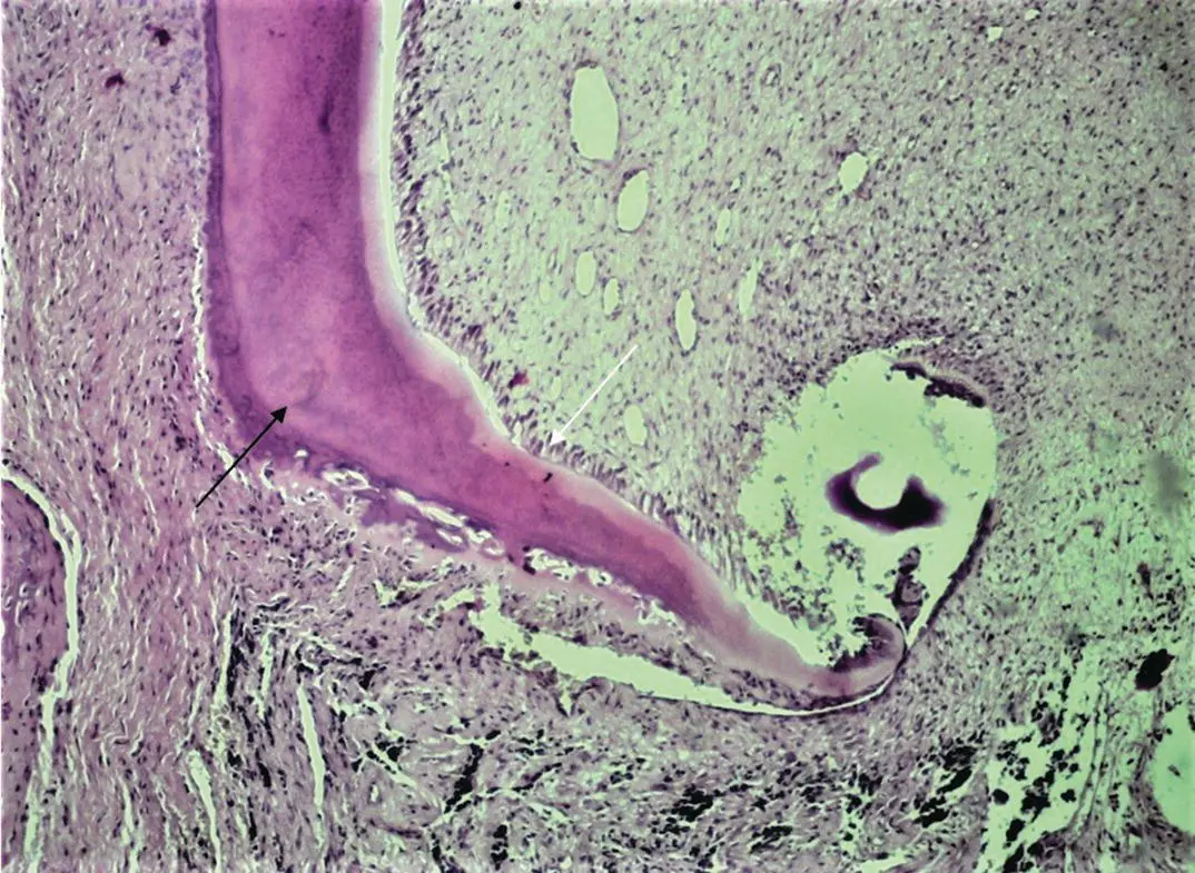
Figure 1.10H and E stained section showing a tooth's root formation (white arrow, odontoblasts; black arrow, dentin).
1.5.1 Description
The tooth root has many important functions including anchorage of the tooth in maxilla/mandible and facilitating provision of blood supply (through apical foramina). The inside of the root is composed of radicular dentin and pulp canals whereas, on the outside, it is covered by a thin calcified layer of cementum. Root formation occurs because of the interaction between HERS, dental papilla, and dental follicle. After crown formation, the cervical loop grows apically as HERS circling dental papilla. The ectomesenchymal cells of dental papilla near the HERS change into odontoblasts and start secreting radicular dentin. The root dentin comes in contact with the dental follicle due to the perforation of HERS which leads to its mesh‐network appearance. This contact changes dental follicular cells into cementoblasts (forming cementum), fibroblasts (forming PDL), and osteoblasts (forming alveolar bone). It should be noted that the HERS only maps the shape of the root and then disintegrates. Its remnants are known as epithelial cell rests of Malassez.
1.5.2 Key Identifying Features
On histological sections, developing root with prominent radicular dentin can be clearly seen just below a complete crown.
1.5.3 Clinical Significance
The HERS is responsible for determining the number of roots by forming a pair of tongue‐shaped extensions that fuse [6]. Root formation plays an important role in tooth eruption. It is believed that with the pressure of the developing root, the crown of the tooth starts moving vertically to erupt in the oral cavity [7]. It should be noted, however, that there is evidence for rootless teeth to erupt [8] suggesting that it is a multifactorial process where root formation has a role, but it is not the only mechanism involved.
Читать дальшеИнтервал:
Закладка:
Похожие книги на «An Illustrated Guide to Oral Histology»
Представляем Вашему вниманию похожие книги на «An Illustrated Guide to Oral Histology» списком для выбора. Мы отобрали схожую по названию и смыслу литературу в надежде предоставить читателям больше вариантов отыскать новые, интересные, ещё непрочитанные произведения.


Обсуждение, отзывы о книге «An Illustrated Guide to Oral Histology» и просто собственные мнения читателей. Оставьте ваши комментарии, напишите, что Вы думаете о произведении, его смысле или главных героях. Укажите что конкретно понравилось, а что нет, и почему Вы так считаете.