An Illustrated Guide to Oral Histology
Здесь есть возможность читать онлайн «An Illustrated Guide to Oral Histology» — ознакомительный отрывок электронной книги совершенно бесплатно, а после прочтения отрывка купить полную версию. В некоторых случаях можно слушать аудио, скачать через торрент в формате fb2 и присутствует краткое содержание. Жанр: unrecognised, на английском языке. Описание произведения, (предисловие) а так же отзывы посетителей доступны на портале библиотеки ЛибКат.
- Название:An Illustrated Guide to Oral Histology
- Автор:
- Жанр:
- Год:неизвестен
- ISBN:нет данных
- Рейтинг книги:5 / 5. Голосов: 1
-
Избранное:Добавить в избранное
- Отзывы:
-
Ваша оценка:
An Illustrated Guide to Oral Histology: краткое содержание, описание и аннотация
Предлагаем к чтению аннотацию, описание, краткое содержание или предисловие (зависит от того, что написал сам автор книги «An Illustrated Guide to Oral Histology»). Если вы не нашли необходимую информацию о книге — напишите в комментариях, мы постараемся отыскать её.
An introduction to tooth development, including the bud, cap, early bell, and late bell stages A thorough exploration of enamel, dentin, cementum and dental pulp A discussion of the periodontal ligament, including alveolar crest fibers, horizontal, oblique, apical, and inter-radicular fibers, transseptal fibers, and gingival fibers A guide to alveolar bone, oral mucosa, and salivary glands Perfect for postgraduate dental students,
will also be useful to undergraduate dental students, and those looking to improve their understanding of the microscopic structure of dental tissues and their pathologies.
An Illustrated Guide to Oral Histology — читать онлайн ознакомительный отрывок
Ниже представлен текст книги, разбитый по страницам. Система сохранения места последней прочитанной страницы, позволяет с удобством читать онлайн бесплатно книгу «An Illustrated Guide to Oral Histology», без необходимости каждый раз заново искать на чём Вы остановились. Поставьте закладку, и сможете в любой момент перейти на страницу, на которой закончили чтение.
Интервал:
Закладка:
List of Contributors
Amr Bugshan Department of Biomedical Dental Sciences College of Dentistry Imam Abdulrahman Bin Faisal University Dammam, Saudi Arabia
Saqlain Gilani Department of Oral Biology Islamic International Dental College Riphah International University Islamabad, Pakistan
Shehriar Husain Department of Dental Materials Science Sindh Institute of Oral Health Sciences Jinnah Sindh Medical University Karachi, Pakistan
Erum Khan CODE‐M Center of Dental Education & Medicine Karachi, Pakistan and Bhitai Dental and Medical College Liaquat University of Medical and Health Sciences, Jamshoro, Pakistan
Syed Ali Khurram Unit of Oral and Maxillofacial Pathology School of Clinical Dentistry University of Sheffield Sheffield, United Kingdom
Zohaib Khurshid Department of Prosthodontics and Dental Implantology College of Dentistry King Faisal University Al Ahsa, Saudi Arabia
Juzer Shabbir Department of Operative Dentistry and Endodontics Liaquat College of Medicine and Dentistry Karachi, Pakistan
Faraz Mohammed Department of Biomedical Dental Sciences College of Dentistry Imam Abdulrahman Bin Faisal University Dammam, Saudi Arabia
Fizza Saher Department of Oral Biology College of Dentistry Ziauddin University Karachi, Pakistan
Rizwan Ullah Department of Oral Biology Sindh Institute of Oral Health Sciences Jinnah Sindh Medical University Karachi, Pakistan
Muhammad Sohail Zafar Department of Restorative Dentistry College of Dentistry Taibah University, Al Madinah Al Munawwarah, Saudi Arabia and Department of Dental Materials Islamic International Dental College Riphah International University Islamabad, Pakistan
About the Companion Website
This book is accompanied by a website at:
www.wiley.com/go/farooq/oral_histology 
Scan the QR code:

The website includes:
PowerPoint of figures
Sample preparation
1 Tooth Development
Saqib Ali1, Imran Farooq1, and Syed Ali Khurram2
1 Department of Biomedical Dental Sciences, College of Dentistry, Imam Abdulrahman Bin Faisal University, Dammam, Saudi Arabia
2 Unit of Oral and Maxillofacial Pathology, School of Clinical Dentistry, University of Sheffield, Sheffield, United Kingdom
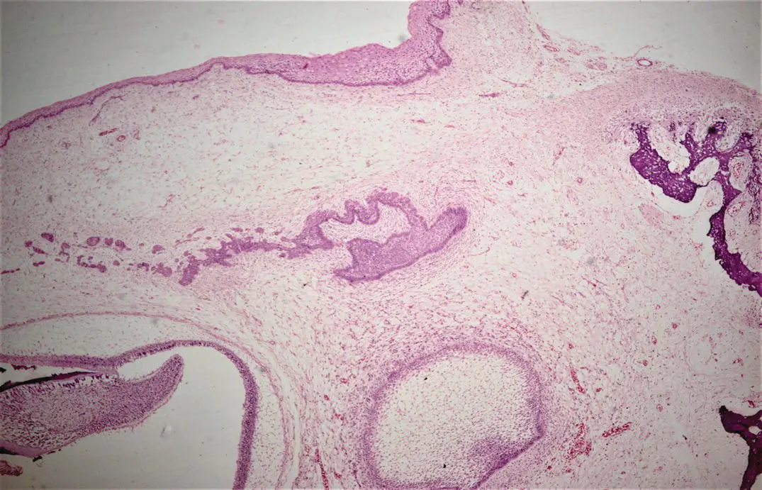
Figure 1.1H and E stained section showing tooth development.
Tooth development starts on the 37th day of gestation with the formation of primary epithelial bands in the place of future upper and lower jaws. These horse‐shoe‐shaped bands correspond to the future dental arches. These epithelial bands then form two ingrowths called dental lamina (lingually positioned) and vestibular lamina (buccally positioned). These ingrowths extend into the mesenchyme which is surrounded by the neural crest cells. The vestibular lamina proliferates within the mesenchyme and leads to the formation of the vestibule (between the cheek and tooth‐bearing portion of the jaw). The dental lamina gives rise to epithelial outgrowths toward the mesenchyme due to continuous proliferative activity which correspond to the location of forthcoming deciduous teeth. The tooth development is divided into the following stages: bud, cap, and bell (early and late). These stages along with the changes happening in the tooth germ are discussed in detail in the following sections.
1.1 Bud Stage
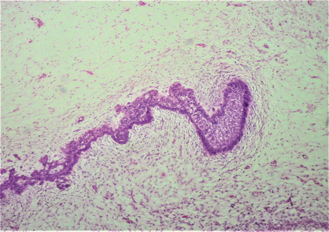
Figure 1.2H and E stained section showing the bud stage of tooth development.
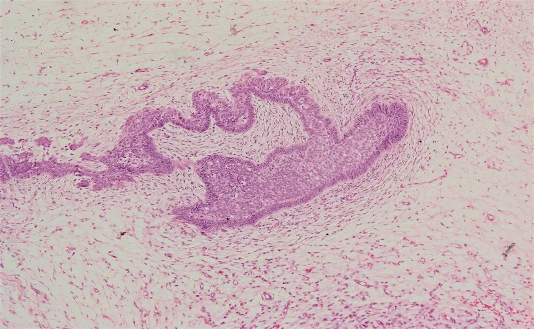
Figure 1.3H and E stained section showing the bud stage of tooth development.
1.1.1 Description
The bud stage is the first stage of tooth development. It represents the first epithelial intrusion into the ectomesenchyme. The cells of the epithelium show minimal changes and the ectomesenchymal cells surround the epithelial bud. Due to the ectomesenchymal condensation (a process in which the epithelial bud propagates into the ectomesenchyme), the density of the cells increases near the epithelial outgrowth. This condensation is owed to the increased mitotic activity carried in the cells of the tooth bud and mesenchymal cells surrounding it. These buds develop at the distal side of the dental lamina and each bud represents a group of cells at dental lamina's end. The epithelial part is separated from the mesenchyme by a basement membrane. The ectomesenchyme surrounding the tooth bud is called the dental follicle or sac whereas the area directly subjacent to the condensation is called the dental papilla . The dental follicle is ultimately responsible for the formation of cementum, periodontal ligament (PDL), and alveolar bone. The dental papilla is responsible for the formation of dental pulp and dentin.
1.1.2 Key Identifying Features
The enamel organ at this stage appears roughly ovoid to spherical with poor histodifferentiation and morphodifferentiation. A typical tooth bud consists of centrally located polygonal (multiple‐shaped) cells and peripherally arranged columnar cells.
1.1.3 Clinical Significance
The successful development of the tooth depends on the interaction of epithelial and mesenchymal components. If these parts grow individually, neither will differentiate further [1]. This epithelial–mesenchymal interaction starts in the bud stage; therefore, any problem affecting the bud stage could seriously affect the development of teeth.
1.2 Cap Stage
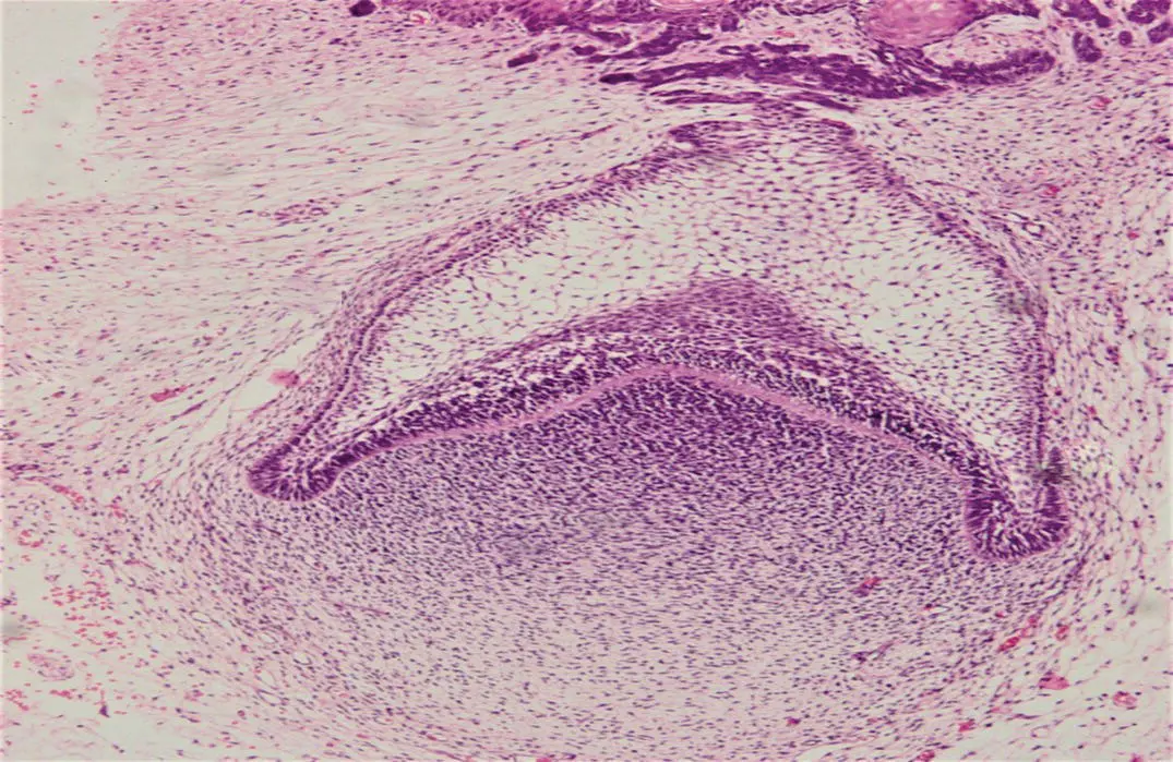
Figure 1.4H and E stained section showing the cap stage of tooth development.
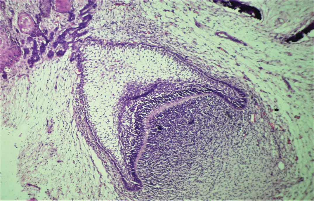
Figure 1.5H and E stained section showing the cap stage of tooth development.
1.2.1 Description
The cap stage is the second stage of tooth development. As the tooth bud matures, it takes part of dental lamina along with it, which is called the lateral lamina. The tooth bud grows non‐uniformly, and the growth is more in certain areas and less in others. This stage is called the cap stage as the epithelial outgrowth looks like a cap which is sitting on top of the condensed ectomesenchyme (dental papilla). During this stage, greater differentiation is seen in the central and peripheral cells. The central polygonal cells change into the stellate reticulum cells which have a somewhat star‐shaped appearance due to greater intake of water, pushing the cells apart but retaining their desmosomal attachments. The peripheral cells change into external and inner enamel epithelium. The outer enamel epithelium cells are cuboidal whereas the inner epithelial cells are tall and columnar. These layers of epithelial cells are separated from the dental follicle and dental papilla by a basement membrane. Another structure called the enamel knot is formed during this stage which represents a collection of cells in the center of the inner enamel epithelium. It is a transitory structure which is believed to contribute cells to the enamel cord (strand of cells) .
Читать дальшеИнтервал:
Закладка:
Похожие книги на «An Illustrated Guide to Oral Histology»
Представляем Вашему вниманию похожие книги на «An Illustrated Guide to Oral Histology» списком для выбора. Мы отобрали схожую по названию и смыслу литературу в надежде предоставить читателям больше вариантов отыскать новые, интересные, ещё непрочитанные произведения.


Обсуждение, отзывы о книге «An Illustrated Guide to Oral Histology» и просто собственные мнения читателей. Оставьте ваши комментарии, напишите, что Вы думаете о произведении, его смысле или главных героях. Укажите что конкретно понравилось, а что нет, и почему Вы так считаете.