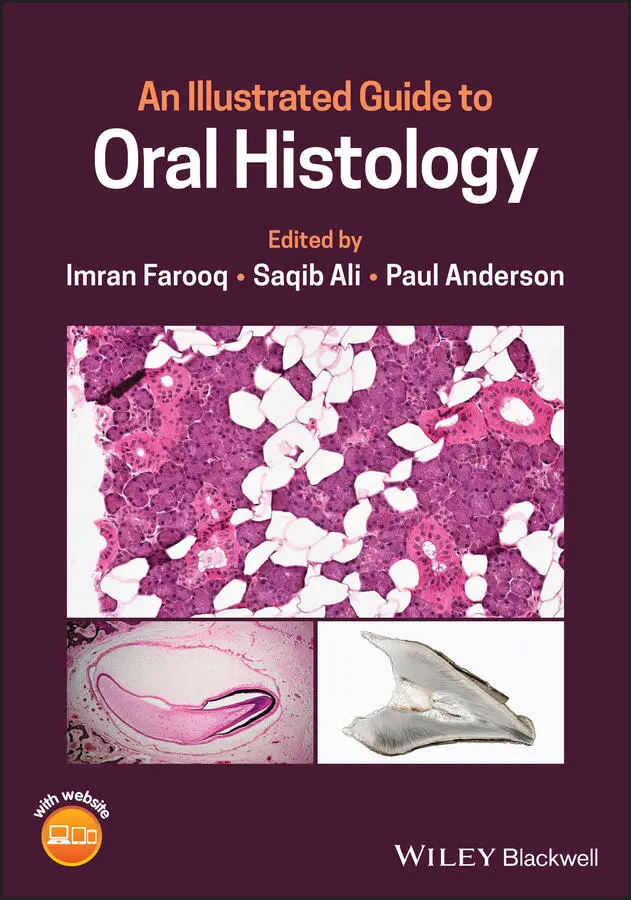
An Illustrated Guide to Oral Histology
Здесь есть возможность читать онлайн «An Illustrated Guide to Oral Histology» — ознакомительный отрывок электронной книги совершенно бесплатно, а после прочтения отрывка купить полную версию. В некоторых случаях можно слушать аудио, скачать через торрент в формате fb2 и присутствует краткое содержание. Жанр: unrecognised, на английском языке. Описание произведения, (предисловие) а так же отзывы посетителей доступны на портале библиотеки ЛибКат.
- Название:An Illustrated Guide to Oral Histology
- Автор:
- Жанр:
- Год:неизвестен
- ISBN:нет данных
- Рейтинг книги:5 / 5. Голосов: 1
-
Избранное:Добавить в избранное
- Отзывы:
-
Ваша оценка:
An Illustrated Guide to Oral Histology: краткое содержание, описание и аннотация
Предлагаем к чтению аннотацию, описание, краткое содержание или предисловие (зависит от того, что написал сам автор книги «An Illustrated Guide to Oral Histology»). Если вы не нашли необходимую информацию о книге — напишите в комментариях, мы постараемся отыскать её.
An introduction to tooth development, including the bud, cap, early bell, and late bell stages A thorough exploration of enamel, dentin, cementum and dental pulp A discussion of the periodontal ligament, including alveolar crest fibers, horizontal, oblique, apical, and inter-radicular fibers, transseptal fibers, and gingival fibers A guide to alveolar bone, oral mucosa, and salivary glands Perfect for postgraduate dental students,
will also be useful to undergraduate dental students, and those looking to improve their understanding of the microscopic structure of dental tissues and their pathologies.
An Illustrated Guide to Oral Histology — читать онлайн ознакомительный отрывок
Ниже представлен текст книги, разбитый по страницам. Система сохранения места последней прочитанной страницы, позволяет с удобством читать онлайн бесплатно книгу «An Illustrated Guide to Oral Histology», без необходимости каждый раз заново искать на чём Вы остановились. Поставьте закладку, и сможете в любой момент перейти на страницу, на которой закончили чтение.
Интервал:
Закладка:
Library of Congress Cataloging‐in‐Publication Data
Names: Farooq, Imran, 1984– editor. | Ali, Saqib, 1985– editor. | Anderson, Paul (Professor of oral biology), 1959– editor.
Title: An illustrated guide to oral histology / edited by Imran Farooq, Saqib Ali, Paul Anderson.
Description: First edition. | Hoboken, NJ : Wiley, 2021. | Includes bibliographical references and index.
Identifiers: LCCN 2020038283 (print) | LCCN 2020038284 (ebook) | ISBN 9781119669449 (cloth) | ISBN 9781119669548 (adobe pdf) | ISBN 9781119669609 (epub)
Subjects: MESH: Mouth–anatomy & histology | Atlas
Classification: LCC QP146 (print) | LCC QP146 (ebook) | NLM WU 17 | DDC 612.3/1–dc23
LC record available at https://lccn.loc.gov/2020038283LC ebook record available at https://lccn.loc.gov/2020038284
Cover Design: Wiley
Cover Images: Imran Farooq
Preface
It gives us great pleasure to present our book that addresses problems with respect to the teaching and learning of oral histology. The idea to write a book focused on important details of oral histological features popped up around two years ago, when we felt there are many deficient areas in the present literature concerning the said topic. This book gives information about these features in a user‐friendly format. It contains high‐definition (HD) histological images of oral tissues with integrated text containing their introduction, key identifying histological features, and clinical significance. The textbook is intended for dental undergraduate and postgraduate students, license examination aspirants, and oral histology instructors. We strongly believe that the book will suit the needs of professionals in each of these disciplines.
We would like to mention here that we do not wish the present book to be a substitution of more general textbooks in oral histology. It is our belief that a good dental practitioner not only needs strong clinical skills, but also a solid understanding of basic sciences. Consequently, our book should be considered as providing the first step of the ladder in learning oral histology. This book is aimed at encouraging students to pursue a more exhaustive appreciation of the subject. To counter technology needs and in‐line with the digital age, a companion website for the book has also been developed.
Finally, we do not imagine ourselves to be error‐free, and would always be open to criticism. Your suggestions to improve the book are greatly appreciated.
Imran FarooqSaqib AliPaul Anderson
Sample Preparation
This atlas contains images obtained through hematoxylin and eosin (H and E) staining, micro‐computed tomography (micro‐CT), ground sectioning, and scanning electron microscopy (SEM). The steps for the preparation of samples and collection of images are as follows.
Hematoxylin and Eosin (H and E) Stained Sections
The tissues were stored in 10% buffered formalin prior to their use. The hard tissue samples were decalcified in 8% formic acid. The tissues (hard and soft) were washed with distilled water and then transferred into alcohol solution for the dehydration procedure. Post‐dehydration, the samples were cleared in xylene solution. The tissues after clearing were shifted into soft paraffin and hard paraffin baths. The sections were blocked by embedding in hard paraffin and thin sections of 7 μm were taken from blocked tissues using microtome. H and E staining was performed, samples were dehydrated, and cover slips were placed using DPX mounting medium.
Micro‐computed Tomography (Micro‐CT)
The tissue blocks to observe enamel and dentin were prepared by cutting the roots with a high‐speed handpiece. The anatomical crown portion was retained and a micro‐CT machine (SkyScan 1172, version 1.5; Bruker Micro‐CT, Kontich, Belgium) was used to obtain images of enamel and dentin. The images were obtained by scanning the samples using a voltage source of 100‐KV, source current of 100‐μA, pixel size of 27.45‐μm, and exposure time of 1600 msec. In addition, 360° rotation, filter of Al + Cu and, Tagged Image File Format (TIFF) were used. The raw images were recreated using the NRecon software (Bruker SkyScan, Aartselaar, Belgium). The TIFF images were later converted to Joint Photographic Experts Group (JPEG) format using Microsoft Paint ®software.
Ground Sections
For each ground section preparation, freshly extracted teeth were collected and fixed in acrylic blocks. The teeth were sectioned using a water‐cooled diamond saw (Isomet® 5000 Linear Precision Saw, Buehler Ltd, IL, USA) longitudinally (for longitudinal sections) and horizontally (for transverse sections) to split the tooth into two parts. Each thick section of tooth was then grinded on carborundum stone with equal digital pressure to make the sections paper thin. The sections were then dehydrated in absolute alcohol for 10 minutes and clearing was then performed in xylene solution for 15 minutes. The section was mounted on to a glass slide using DPX mounting medium. Coverslip was then placed on top of the section carefully to avoid air entrapment.
Scanning Electron Microscopy
To observe dentinal tubules, SEM was performed. Dentin discs of 1.5 mm were first made by cutting the teeth horizontally over cemento‐enamel junction using a precision saw (Isomet® 5000 Linear Precision Saw, Buehler Ltd, IL, USA). The discs were exposed to ethylenediaminetetraacetic acid (EDTA) for one minute to unblock the dentinal tubules. After washing them with distilled water for one minute and post air drying, these discs were mounted on stubs and sputter coated with gold. The discs were observed in an SEM (FEI, Inspect F50, The Netherlands) to obtain micrographs of dentinal tubules.
About the Editors
Dr. Imran FarooqDr. Imran Farooq graduated from Baqai Dental College, Karachi, Pakistan in 2007, and then worked in the same institute as a Clinical Demonstrator (Restorative Dentistry) for two years before joining Queen Mary, University of London (QMUL), UK, for his postgraduation in Oral Biology. After graduation from QMUL in 2011, he joined the College of Dentistry, Imam Abdulrahman Bin Faisal University, Dammam, Saudi Arabia, in 2012. He is currently working as an Assistant Professor in the Oral Biology Division. He has 10+ years’ experience in teaching oral biology and histology to dental students. He has published 70+ research articles and book chapters that have received 700+ citations to date. His research interests include biomaterials, dental materials, hard tissue mineralization, and dental education.
Dr. Saqib AliDr. Saqib Ali graduated from Baqai Dental College, Karachi, Pakistan in 2009, and then joined QMUL, UK, to pursue his postgraduation in Oral Biology. After graduating from QMUL in 2011, he worked as an Assistant Professor in the Oral Biology Department of Sardar Begum Dental College and Khyber College of Dentistry, Peshawar, Pakistan, and then, since 2016, he has been working as a faculty member (Course Director Oral Biology) in College of Dentistry, Imam Abdulrahman Bin Faisal University, Dammam, Saudi Arabia. He has 10+ years’ experience in teaching oral biology and histology to dental students. He has published 40+ research articles and book chapters that have received 200+ citations to date. His research interests include biomaterials, dental materials, hard tissue mineralization, and dental education.
Prof. Paul AndersonProf. Paul Anderson graduated from Leeds in Biophysics and then completed a PhD in Biophysics in relation to Dentistry at the London Hospital Medical College (now part of QMUL). He has supervised 15 PhD students to completion, and has given invited research seminars in the Europe, the United States, Australia, New Zealand, Beijing, Hong Kong, and Malaysia. He is currently an associate editor of Caries Research, and Hon. Asst. Secretary of British Society for Oral and Dental Research (BSODR). He is also chair of the Institute of Dentistry Dental Postgraduate Committee. In 2011, he started the UK’s first ever MSc in Oral Biology program in the United Kingdom, with now over 100 graduates. He has published 150+ research articles and book chapters that have received 2000+ citations to date. His research interests include X‐ray microscopy, enamel and hydroxyapatite chemistry, and the biophysical chemical effects of salivary proteins in dental hard tissue protection.
Читать дальшеИнтервал:
Закладка:
Похожие книги на «An Illustrated Guide to Oral Histology»
Представляем Вашему вниманию похожие книги на «An Illustrated Guide to Oral Histology» списком для выбора. Мы отобрали схожую по названию и смыслу литературу в надежде предоставить читателям больше вариантов отыскать новые, интересные, ещё непрочитанные произведения.


Обсуждение, отзывы о книге «An Illustrated Guide to Oral Histology» и просто собственные мнения читателей. Оставьте ваши комментарии, напишите, что Вы думаете о произведении, его смысле или главных героях. Укажите что конкретно понравилось, а что нет, и почему Вы так считаете.