Digital Workflow in Reconstructive Dentistry
Здесь есть возможность читать онлайн «Digital Workflow in Reconstructive Dentistry» — ознакомительный отрывок электронной книги совершенно бесплатно, а после прочтения отрывка купить полную версию. В некоторых случаях можно слушать аудио, скачать через торрент в формате fb2 и присутствует краткое содержание. Жанр: unrecognised, на английском языке. Описание произведения, (предисловие) а так же отзывы посетителей доступны на портале библиотеки ЛибКат.
- Название:Digital Workflow in Reconstructive Dentistry
- Автор:
- Жанр:
- Год:неизвестен
- ISBN:нет данных
- Рейтинг книги:4 / 5. Голосов: 1
-
Избранное:Добавить в избранное
- Отзывы:
-
Ваша оценка:
Digital Workflow in Reconstructive Dentistry: краткое содержание, описание и аннотация
Предлагаем к чтению аннотацию, описание, краткое содержание или предисловие (зависит от того, что написал сам автор книги «Digital Workflow in Reconstructive Dentistry»). Если вы не нашли необходимую информацию о книге — напишите в комментариях, мы постараемся отыскать её.
Contributors
Amirah M. R. Alammar • Abdulaziz Alsahaf • Wael Att • Maria Bateli • Jasmin Bernhart • Shaza Bishti • Sarah Blattner • Miha Brezavšček • Sandy Cepa • Nadine Emmanoulidi • Ahmed Fawzy • Manrique Fonseca • Michele Frapporti • Rumpa Ganguly • Yousef Al-Ghamdi • Petra Ch. Gierthmuehlen • Aiste Gintaute • Ulrich Lamott • Christos Lamprinos • Matthias Petsch • Udo Plaster • Aikaterini Ploumaki • Hanna Rauberger • Elisabeth Schwartzkopff • Christian F. Selz • Thamer Al-Sharif • Benedikt Spies • Frank A. Spitznagel • Jörg R. Strub • Michael Swain • Taskin Tuna • Alexander Vuck • Siegbert Witkowski
Digital Workflow in Reconstructive Dentistry — читать онлайн ознакомительный отрывок
Ниже представлен текст книги, разбитый по страницам. Система сохранения места последней прочитанной страницы, позволяет с удобством читать онлайн бесплатно книгу «Digital Workflow in Reconstructive Dentistry», без необходимости каждый раз заново искать на чём Вы остановились. Поставьте закладку, и сможете в любой момент перейти на страницу, на которой закончили чтение.
Интервал:
Закладка:
A combination of the aforementioned imaging techniques facilitates continuous image capture to create an accurate 3D representation of the prepared teeth. This technology is currently implemented in the CS 3500 (Carestream Dental, Atlanta, GA, USA), which utilizes a green laser and four light emitting diodes (LEDs; UV, blue, green, and red) for the acquisition and the illumination of the object, respectively, while a complementary metal oxide semiconductor (CMOS) sensor receives the acquired data. According to the manufacturer, the scanner has a field of view of approximately 16 mm × 12 mm and a working depth of –1 mm to 15 mm. Although information about the capturing speed is not disclosed, the scanner is completely powder-free and enables full-arch scanning. The digital data acquired by the CS 3500 can be used to render a multicolored model.
Light Beam-based IOS Systems
This group of IOS systems employs visible light beam for image capture. Here, the image capture techniques can vary between still image, video capture, and real-time image capture. Although system dependent, the majority of IOS systems belonging to this group require the application of titanium dioxide as a reflecting agent.
Still Image Capture Technique
The still image capture technique utilizes a technology known as active triangulation, in which the intersection of three linear light beams is used to locate a given point in a 3D space. 23This concept has been used in a variety of industrial measuring devices. However, surfaces that disperse light irregularly do not reflect it evenly, and surfaces that are irregular, such as tooth surfaces, adversely affect the accuracy of the scan based on triangulation. 24Consequently, an opaque powder coating (titanium dioxide) is required to provide uniform light dispersion and to enhance the accuracy of the scan. 25The active triangulation techniques are implemented in the Cerec AC Bluecam system (Sirona Dental Systems, Bensheim, Germany). The contemporary versions of Cerec Bluecam utilize a blue light technology to scan the dentition.
Prior versions of Cerec utilized infrared light technology with a longer wavelength (820 nm) than the blue (470 nm) associated with the Cerec Bluecam. This development enables for an increased depth-of-field and is claimed to improve scanning accuracy by around 60%. 20,22The shutter speed of 17 ms per image further reduces the blurring risk, as does the option to rest the camera on the teeth (use of the Cerec camera support is recommended). The depth-of-field range itself, at 14 mm, is sufficient to allow operators to disregard this parameter entirely, because the image will always be accurately focused from cusp to preparation margin. The wavelength of the blue light also facilitates a nearly distortion-free representation, even close to the edges, of the image. 22,26MIA 3D (DynSys3D, Migdal Ha’Emeq, Israel) is another system that uses the active triangulation technique. Both systems capture and render a monochromatic 3D model.
As a further evolution of the Cerec Bluecam, Sirona launched the Cerec Omnicam. The new camera is able to capture images combining continuous and static stripe projection techniques. The main feature of this IOS system is that the teeth in most cases do not necessarily require prescan powdering. Furthermore, during the scan procedure the image data are reproduced, with natural color and in real time, on the screen, while a full-color 3D model is generated. 27
Video Capture Technique
While all other technologies presented in light beam-based IOS systems use some form of still image acquisition, the active wavefront sampling (AWS) technology is the only technique which captures 3D data in a video sequence 28and models in real time. 9AWS refers to getting 3D information from a single lens imaging system by measuring depth based on the defocusing of the primary optical system. 29The technique is implemented in the Lava Chairside Oral Scanner (COS) and its successor, the True Definition Scanner (3M Espe, Seefeld, Germany). 30,31Lava COS incorporates 192 blue LEDs for illumination, three sensors, and 22 lenses that capture the object simultaneously from different perspectives. 9,20Then, the 3D surface patches are generated in real time by means of a proprietary image processing algorithm using the in-focus and out-of-focus information. 29The system captures 20 3D data sets per second and each data set contains 10,000 data points of information, resulting in roughly 24 million data points (or 2,400 data sets) captured per arch. 20The operator has a field of view of approximately 10 mm × 13.5 mm in which data is being captured. 9Similar to the systems that use active triangulation, and due to the application of a reflecting agent, the rendered models are monochromatic. The successor system, True Definition Scanner, implements the same technology as Lava COS, but with fewer LEDs and lenses. The scanning wand of True Definition Scanner consists of six LEDs for illumination, three optical lenses, and a CMOS sensor for image capturing and data acquisition.
Ultrafast Optical Sectioning Technique
The ultrafast optical sectioning technology is similar to the video capture technique and facilitates continuous image capture. Rather than artificially forming interpolated surfaces, this technique utilizes up to 1,000 3D images to create true geometries based on real data. The technology is being implemented in the 3Shape TRIOS IOS system (3Shape, Copenhagen, Denmark). According to the manufacturer, the scanner captures over 3,000 two-dimensional (2D) images per second, which is 100 times faster than a conventional video camera. 32The TRIOS (3Shape/Cara) has a field of view of approximately 17 mm × 20 mm and a working depth of 0 to 18 mm. Additionally, it does not require the application of a reflecting agent, compared to other light beam-based IOS systems. The scanner is completely motion- and position-free and can be placed on the teeth for support during scanning. The software and hardware of TRIOS have the capability to capture and render a fully colored model ( Fig 2.5). The evolution of the existing system led to the third generation of TRIOS, the TRIOS 3. The new IOS system integrates an intraoral camera for taking high-definition pictures, facilitating practitioner–patient communication. TRIOS 3 is also capable of matching and saving the teeth shades during a digital impression. A new version with a wireless scanning wand that facilitates a significant faster scanning capability was introduced at the 2017 IDS.
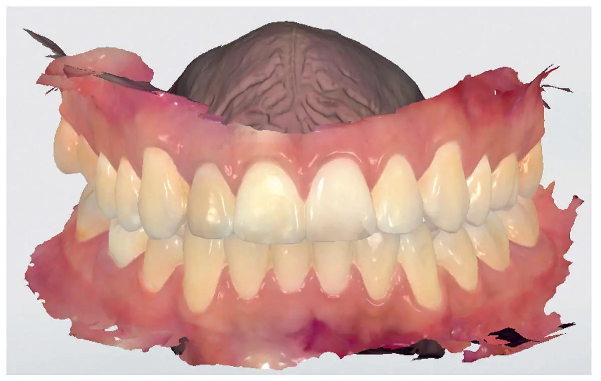
Fig 2.5Rendered model in RealColor after scanning with Trios (3Shape, Copenhagen, Denmark).
Efficacy of IOS Systems
IOS systems were developed to overcome the problems and disadvantages of the traditional impression fabrication process, particularly, mold instability, plaster pouring, laceration on margins, as well as geometrical and dimensional discrepancies between the die and the mold. Furthermore, IOS systems provide several advantages over conventional impressions. These include the elimination of the need for impression trays and materials, the immediate evaluation and quality control after the scan, and the digital archiving of patient records. On the other hand, studies have shown that the accuracy of IOS systems seems to be similar to that of conventional impression materials. The accuracy of a full-arch impression using conventional materials is approximately 55 µm, while that of IOS systems is between 40 and 49 µm. 26,33,34Furthermore, the accuracy of elastomeric impressions seems to be negatively affected by particular angulation of the axial walls of abutment teeth, while the IOSs can accurately reproduce a tooth abutment irrespectively of its geometry. 35Comparing the different steps necessary for the fabrication of a restoration, IOS systems seem to require fewer steps than the conventional impression techniques, being significantly faster than the conventional procedure. 36-39Particularly, an in vitro study found digital impression-making methods were significantly faster compared to the conventional technique, after contrasting them in three realistic clinical scenarios, such as impression making of one, two, and full-arch abutments for the fabrication of a crown, a three-unit fixed partial denture, and full-arch single crowns, respectively ( Fig 2.6). 36
Читать дальшеИнтервал:
Закладка:
Похожие книги на «Digital Workflow in Reconstructive Dentistry»
Представляем Вашему вниманию похожие книги на «Digital Workflow in Reconstructive Dentistry» списком для выбора. Мы отобрали схожую по названию и смыслу литературу в надежде предоставить читателям больше вариантов отыскать новые, интересные, ещё непрочитанные произведения.


Обсуждение, отзывы о книге «Digital Workflow in Reconstructive Dentistry» и просто собственные мнения читателей. Оставьте ваши комментарии, напишите, что Вы думаете о произведении, его смысле или главных героях. Укажите что конкретно понравилось, а что нет, и почему Вы так считаете.