Digital Workflow in Reconstructive Dentistry
Здесь есть возможность читать онлайн «Digital Workflow in Reconstructive Dentistry» — ознакомительный отрывок электронной книги совершенно бесплатно, а после прочтения отрывка купить полную версию. В некоторых случаях можно слушать аудио, скачать через торрент в формате fb2 и присутствует краткое содержание. Жанр: unrecognised, на английском языке. Описание произведения, (предисловие) а так же отзывы посетителей доступны на портале библиотеки ЛибКат.
- Название:Digital Workflow in Reconstructive Dentistry
- Автор:
- Жанр:
- Год:неизвестен
- ISBN:нет данных
- Рейтинг книги:4 / 5. Голосов: 1
-
Избранное:Добавить в избранное
- Отзывы:
-
Ваша оценка:
Digital Workflow in Reconstructive Dentistry: краткое содержание, описание и аннотация
Предлагаем к чтению аннотацию, описание, краткое содержание или предисловие (зависит от того, что написал сам автор книги «Digital Workflow in Reconstructive Dentistry»). Если вы не нашли необходимую информацию о книге — напишите в комментариях, мы постараемся отыскать её.
Contributors
Amirah M. R. Alammar • Abdulaziz Alsahaf • Wael Att • Maria Bateli • Jasmin Bernhart • Shaza Bishti • Sarah Blattner • Miha Brezavšček • Sandy Cepa • Nadine Emmanoulidi • Ahmed Fawzy • Manrique Fonseca • Michele Frapporti • Rumpa Ganguly • Yousef Al-Ghamdi • Petra Ch. Gierthmuehlen • Aiste Gintaute • Ulrich Lamott • Christos Lamprinos • Matthias Petsch • Udo Plaster • Aikaterini Ploumaki • Hanna Rauberger • Elisabeth Schwartzkopff • Christian F. Selz • Thamer Al-Sharif • Benedikt Spies • Frank A. Spitznagel • Jörg R. Strub • Michael Swain • Taskin Tuna • Alexander Vuck • Siegbert Witkowski
Digital Workflow in Reconstructive Dentistry — читать онлайн ознакомительный отрывок
Ниже представлен текст книги, разбитый по страницам. Система сохранения места последней прочитанной страницы, позволяет с удобством читать онлайн бесплатно книгу «Digital Workflow in Reconstructive Dentistry», без необходимости каждый раз заново искать на чём Вы остановились. Поставьте закладку, и сможете в любой момент перейти на страницу, на которой закончили чтение.
Интервал:
Закладка:
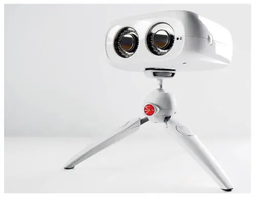
Fig 2.13PIC camera for implant position capture via photogrammetry. The capturing device is equipped with two CCD cameras. Illumination of the scan abutments is performed by infrared flashes surrounding the lenses.
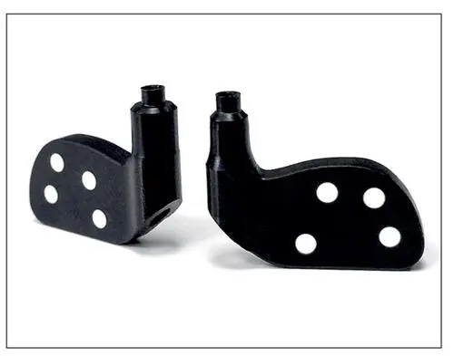
Fig 2.14Flag-shaped PIC abutments, which are screw-retained onto the implants for the capturing procedure. Each abutment is uniquely dot-coded to allow easy identification of each implant.
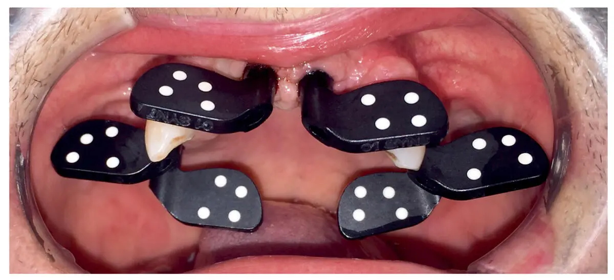
Fig 2.15Clinical example demonstrating the positioning of PIC abutments onto different implants. The PIC camera captures the 3D positions and angulations of implants and via the coded abutments.
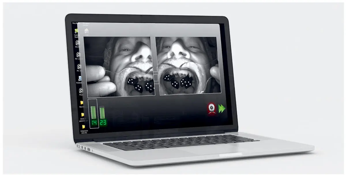
Fig 2.16The PIC software processes information about the angles and distances between implants. The data is then interrelated and treated as a unit to create a PIC file. Further steps include soft issue registration (e.g., IOS scan) and matching with the PIC file.
Based on clinical experience, the photogrammetry technique seems to provide reliable, fast, and comfortable digital acquisition of implant information. While the manufacturer claims an error range of less than 10 microns with the technology, studies about the accuracy of the data acquired from different clinical scenarios are still lacking.
Current Indications and Future Possibilities of IOS Systems
While the first generation of IOS systems was only indicated for the fabrication of inlays, contemporary IOS systems offer the possibility to fabricate a wide spectrum of fixed restorations, including inlays, onlays, veneers, single crowns, and fixed partial dentures (FPDs) ( Table 2.3). Due to the current software and hardware configurations, the number of fixed partial denture units varies from one IOS system to the next. Depending on the system, the options of restorative materials can also vary from resins, nonprecious/precious metal alloys, and ceramics, to high-strength ceramics. The fabrication process can be either subtractive (milling or grinding) or additive (stereolithography, 3D printing, selective laser sintering, and so on). For the fabrication of high-strength ceramic restorations, subtractive procedures are the currently available manufacturing techniques. On the other hand, a limited number of IOS systems, including related CAD/CAM techniques, claim that they have expanded the indications from the fabrication of fixed restorations to removable partial dentures or even complete dentures (e.g., Omnicam and True Definition Scanner; see Table 2.4). Other systems can provide the possibility of implementing CAI for the production of implant surgical splints (e.g., 3Shape TRIOS). Regarding implant restorations, the majority of IOS systems support such an option with the help of scan bodies. The number and type of implant restorations are completely software dependent. The same concept follows the 3Shape TRIOS Scan IOS system to fabricate post-and-core restorations, via optical capturing of scanning flacks (3Shape Scan Post, 3Shape), which are provided in different lengths and diameters ( Fig 2.17). Finally, some IOS systems, such as 3Shape TRIOS and iTero, extend their indication range to orthodontics, allowing not only the digitization of the procedure through the fabrication of an orthodontic appliance, but also digital analysis of the pretreatment situation and the treatment planning.
Table 2.3Overview of different features and indications of commercially available intraoral scanner systems (information provided by the manufacturer may vary depending of device/software version and service strategy of manufacturer)
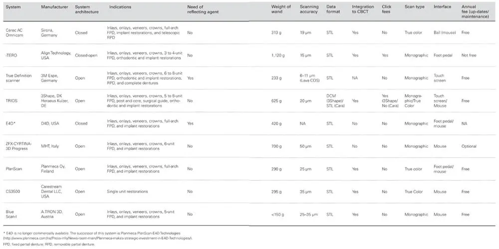
Table 2.4Guide to available intraoral scanners with links to manufacturers (modified from Jokstad 17)
| Product name | Manufacturer | URL |
| 3D Progress Plus | MHT (Medical High Technologies, Italy/Switzerland) | https://www.mht.it |
| Aadva ← IOS Bluescan-I ← a.tron 3D | GC, Belgium 2016 a.tron 3D, Klagenfurt, Austria | https://www.gceurope.com |
| Cerec OmniCam/BlueCam | Dentsply Sirona, Germany | https://www.dentsplysirona.com |
| Condor | Clon3D, Belgium | https://condorscan.com |
| CS3500/CS3600 | Carestream Dental, USA | https://www.carestreamdental.com |
| Dentium Rainbow iOS | Dentium, Korea | http://dentium.com |
| DWIO ← Diglmprint Steinbichler | Dental Wings, Canada ← 2013 Steinbichler | http://www.dentalwings.com |
| IntraScan Zfx | zfx, Germany | http://www.zfx-dental.com |
| i/s/can oral | Goldquadrat, Germany | http://goldquadrat.de |
| Itero Element/Itero | Align Technology, USA ← 2011 Cadent, Israel | http://www.itero.com |
| MIA3D | Densys, Israel | https://densys3d.com |
| Organical Scan Oral | R+K CAD/CAM Technologie, Germany | http://www.organical-cadcam.com |
| PlanScan ← E4D | PlanMeca, Finland ← 2015 E4D Tech, USA | http://www.planmeca.com |
| Primescan | Dentsply Sirona, Germany | https://www.dentsplysirona.com |
| Progress IODIS | Clon 3D / IODIS / Intellidenta (USA?) | https://clon3d.com |
| TRIOS 3 / TRIOS Color / Standard | 3Shape, Denmark | https://www.3shape.com |
| True Definition Scanner ← Lava COS (Chairside Oral Scanner) | 3M Espe, USA ← 2006 Brontes Technology | https://www.3mespe.com |
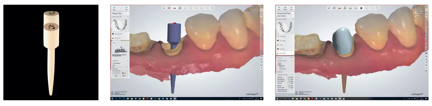
Fig 2.17The 3Shape Scan Post system is approved for both intraoral application in the clinic and for model scanning in the laboratory; courtesy of Nelson Silva, Rodrigo Albuquerque, Luis Morgan, UFMG, Belo Horizonte, Bazil.
In summary, the current possibilities of IOS systems and their relatively wide indication spectrum, as well as software compatibility (open STL format), are gaining widespread acceptance in the dental community.
Hence, the current IOS systems can still be considered as a blueprint for future systems. Current research focuses on the extension of the indication range of IOS systems to include edentulous jaws. Once established, the fabrication of removable partial dentures, including complete dentures, would be possible using IOS systems. More interesting, the introduction of advanced imaging technologies such as optical coherence tomography (OCT) that are being used in other medical specialties (e.g., cardiology, ophthalmology, and dermatology) will facilitate the possibility for IOS systems to perform transgingival scans. In other words, this technology will allow CAI to be performed without placing retraction cords. 61,62A further technology that is still under development implements high-frequency ultrasound for intraoral scans (WhiteSonic, Mannheim, Germany). The manufacturer claims improved signal processing compared to existing IOS systems. Model-based concepts combined with powerful and intelligent algorithms make it possible to scan subgingival structures, such as preparation margins and bone. With such innovations, CAI will rapidly replace conventional techniques.
Читать дальшеИнтервал:
Закладка:
Похожие книги на «Digital Workflow in Reconstructive Dentistry»
Представляем Вашему вниманию похожие книги на «Digital Workflow in Reconstructive Dentistry» списком для выбора. Мы отобрали схожую по названию и смыслу литературу в надежде предоставить читателям больше вариантов отыскать новые, интересные, ещё непрочитанные произведения.


Обсуждение, отзывы о книге «Digital Workflow in Reconstructive Dentistry» и просто собственные мнения читателей. Оставьте ваши комментарии, напишите, что Вы думаете о произведении, его смысле или главных героях. Укажите что конкретно понравилось, а что нет, и почему Вы так считаете.