Digital Workflow in Reconstructive Dentistry
Здесь есть возможность читать онлайн «Digital Workflow in Reconstructive Dentistry» — ознакомительный отрывок электронной книги совершенно бесплатно, а после прочтения отрывка купить полную версию. В некоторых случаях можно слушать аудио, скачать через торрент в формате fb2 и присутствует краткое содержание. Жанр: unrecognised, на английском языке. Описание произведения, (предисловие) а так же отзывы посетителей доступны на портале библиотеки ЛибКат.
- Название:Digital Workflow in Reconstructive Dentistry
- Автор:
- Жанр:
- Год:неизвестен
- ISBN:нет данных
- Рейтинг книги:4 / 5. Голосов: 1
-
Избранное:Добавить в избранное
- Отзывы:
-
Ваша оценка:
Digital Workflow in Reconstructive Dentistry: краткое содержание, описание и аннотация
Предлагаем к чтению аннотацию, описание, краткое содержание или предисловие (зависит от того, что написал сам автор книги «Digital Workflow in Reconstructive Dentistry»). Если вы не нашли необходимую информацию о книге — напишите в комментариях, мы постараемся отыскать её.
Contributors
Amirah M. R. Alammar • Abdulaziz Alsahaf • Wael Att • Maria Bateli • Jasmin Bernhart • Shaza Bishti • Sarah Blattner • Miha Brezavšček • Sandy Cepa • Nadine Emmanoulidi • Ahmed Fawzy • Manrique Fonseca • Michele Frapporti • Rumpa Ganguly • Yousef Al-Ghamdi • Petra Ch. Gierthmuehlen • Aiste Gintaute • Ulrich Lamott • Christos Lamprinos • Matthias Petsch • Udo Plaster • Aikaterini Ploumaki • Hanna Rauberger • Elisabeth Schwartzkopff • Christian F. Selz • Thamer Al-Sharif • Benedikt Spies • Frank A. Spitznagel • Jörg R. Strub • Michael Swain • Taskin Tuna • Alexander Vuck • Siegbert Witkowski
Digital Workflow in Reconstructive Dentistry — читать онлайн ознакомительный отрывок
Ниже представлен текст книги, разбитый по страницам. Система сохранения места последней прочитанной страницы, позволяет с удобством читать онлайн бесплатно книгу «Digital Workflow in Reconstructive Dentistry», без необходимости каждый раз заново искать на чём Вы остановились. Поставьте закладку, и сможете в любой момент перейти на страницу, на которой закончили чтение.
Интервал:
Закладка:
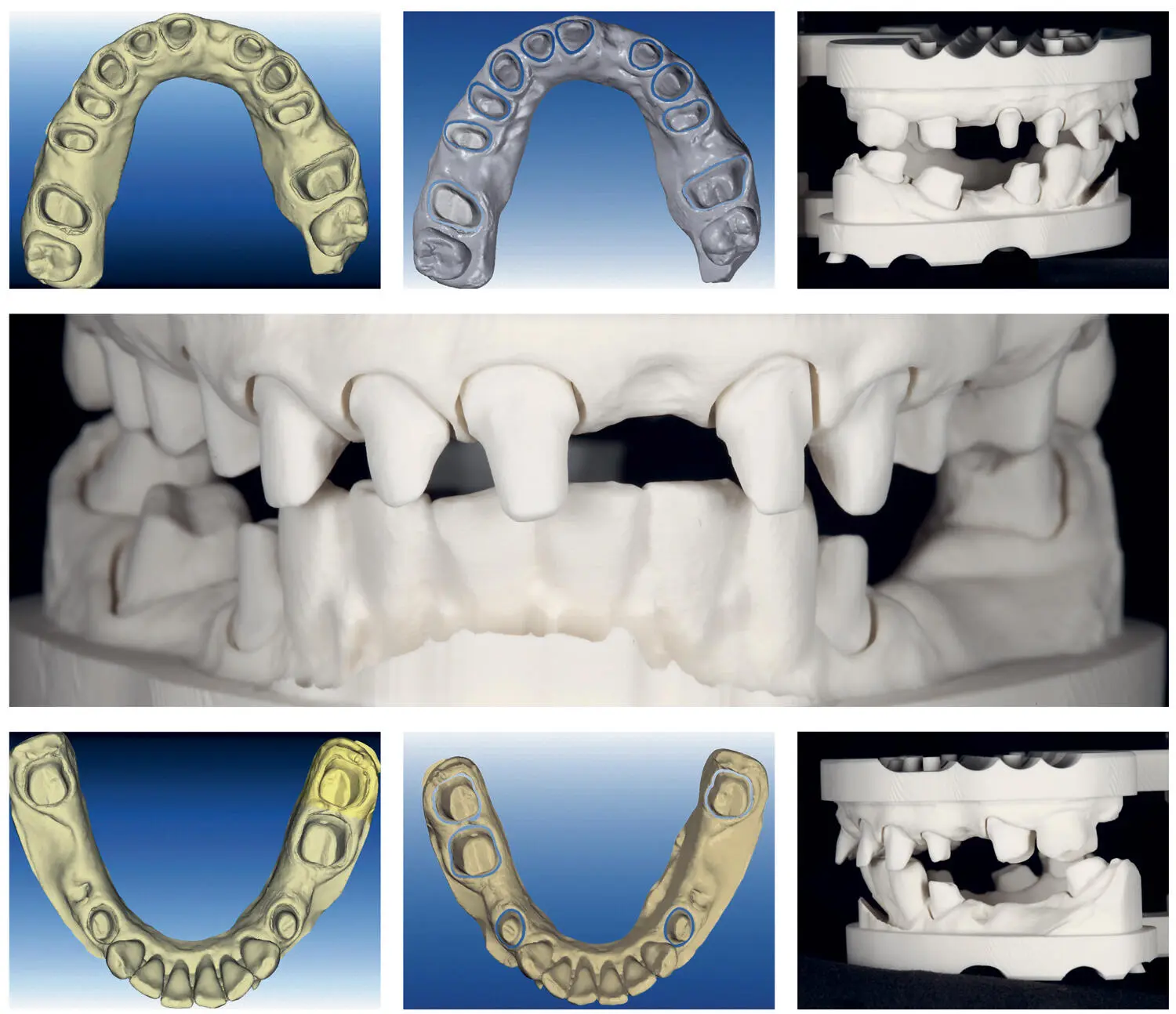
Fig 2.23Digital images of the scanned maxilla and mandible. The optical impressions were processed by the company to portray the preparation margins. Polyurethane casts were fabricated by using digital data sets from the scanning procedure.
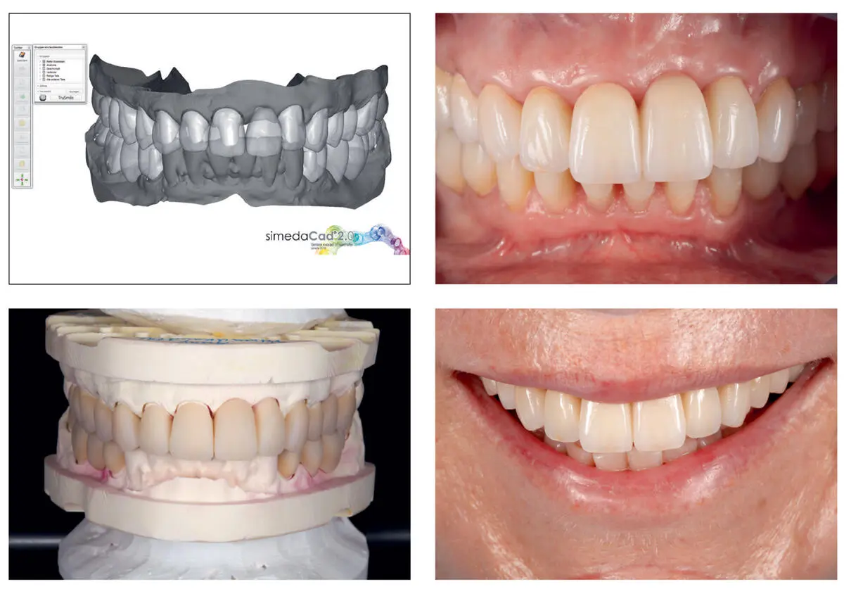
Fig 2.24The SimedaCad 2.0 software was used to design the restorations. A prototype of the restorations was fabricated and tried intraorally.
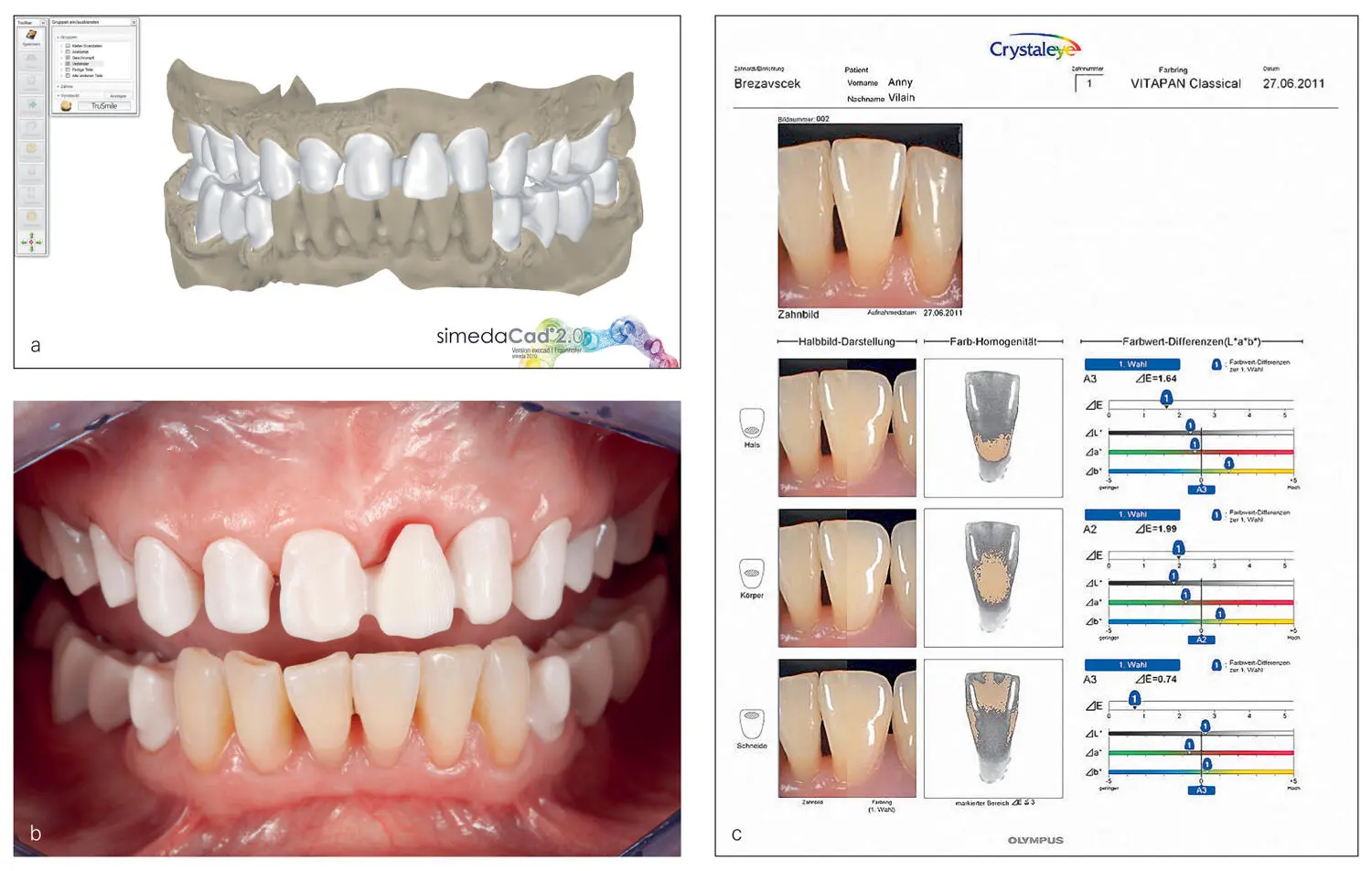
Fig 2.25By using a digital cutback, the framework design was created. The intraoral try-in of the zirconia frameworks showed proper fit and retention. The selection of the tooth shade was performed using a spectrophotometer.
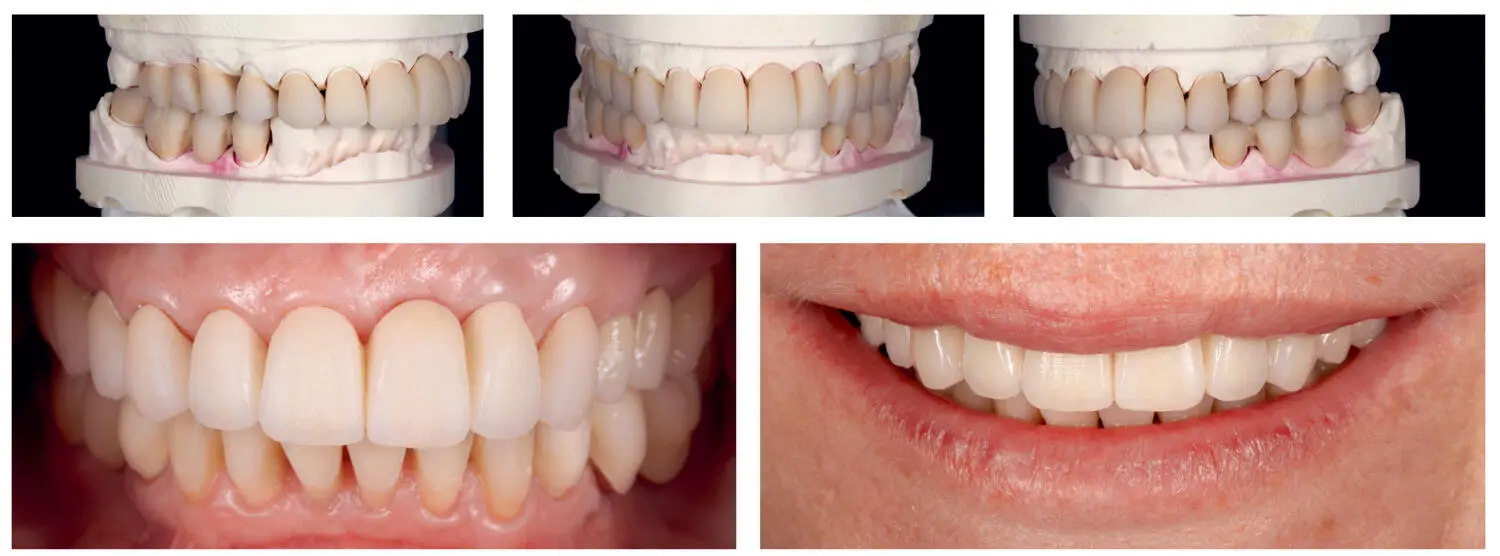
Fig 2.26Before finalization of the restorations, a biscuit try-in was performed to check esthetics, phonetics, and occlusion.
Before cementation of the final restorations, a radiographic examination and re-evaluation of the intraoral status were performed to check the vitality and the periodontal status of the abutment teeth. Furthermore, the inner zirconia surface of the final crowns was airborne-particle abraded with alumina oxide under a pressure of 0.5 bars and chemically treated with a monophosphate monomer (Clearfil Ceramic Primer, Kuraray Dental, Tokyo, Japan). Afterwards the crowns and FPDs were cemented with a dual curing adhesive cement (Panavia 21, Kuraray Dental; Figs 2.27– 2.29).
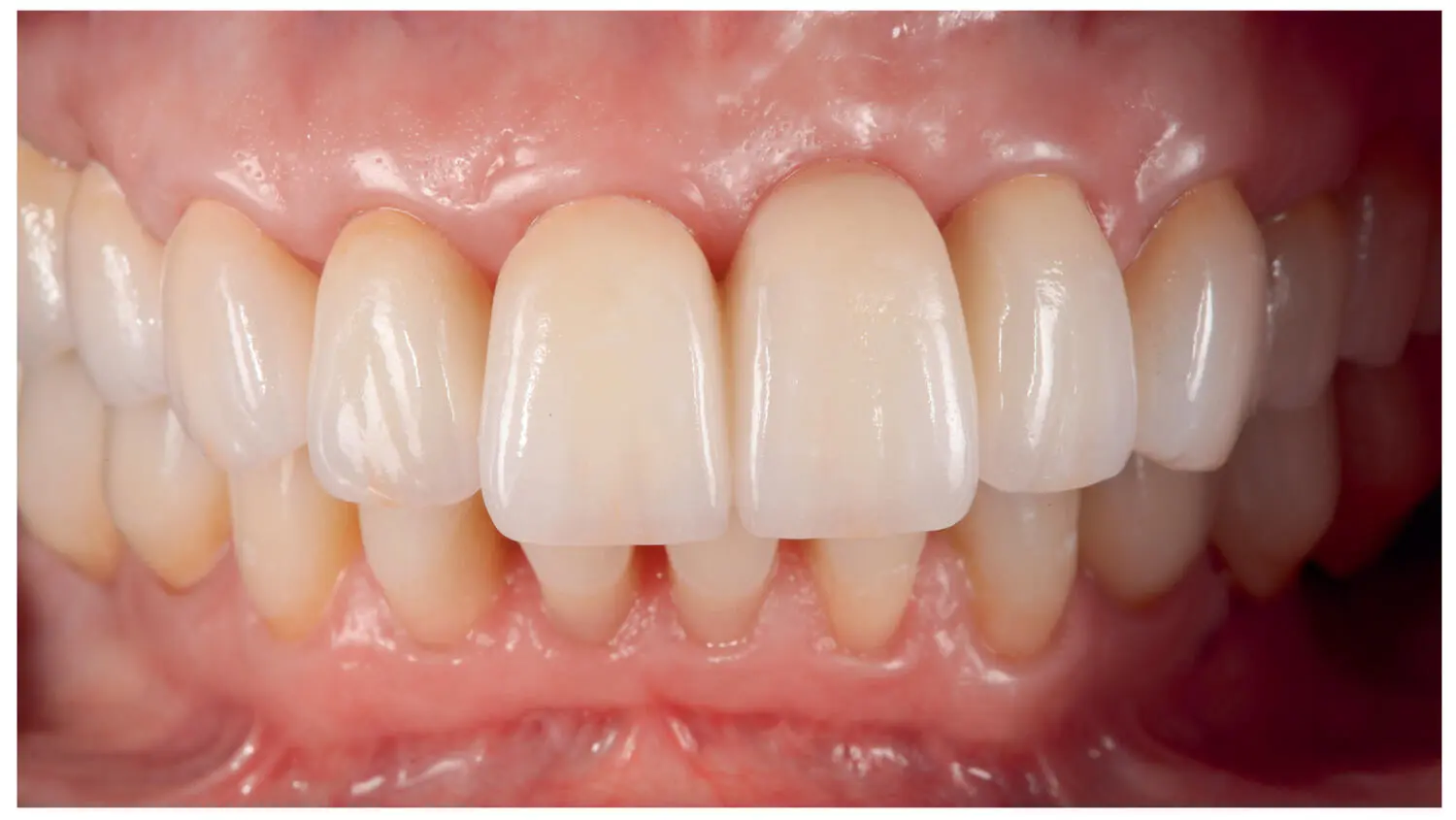
Fig 2.27Frontal view of the final crowns after cementation.
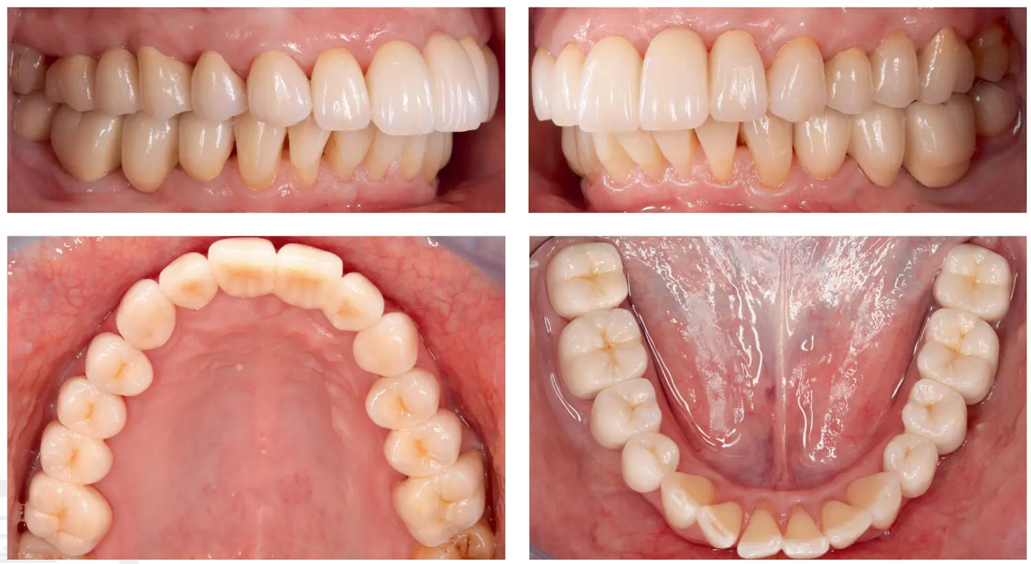
Fig 2.28Lateral and occlusal views of the cemented restorations.
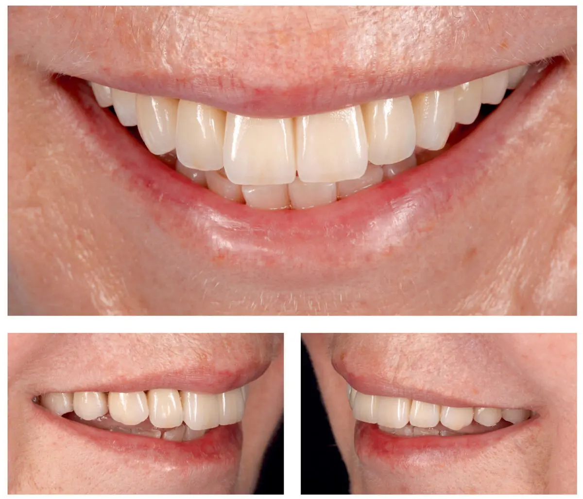
Fig 2.29Frontal lateral views of the patient’s smile showing a successful esthetic outcome of the prosthetic rehabilitation.
One week after the insertion, the final re-evaluation of the occlusal relationship was conducted. The patient was enrolled in a 6-month recall regimen.
References
1.Young JM, Altschuler BR. Laser holography in dentistry. J Prosthet Dent 1977;38:216–225.
2.Duret F, Termoz C. Method of and apparatus for making a prosthesis, especially a dental prosthesis. United States Patent and Trademark Office. G06F 15/00; A61C 9/00 ed. USA: Francois Duret, Christian Termoz, 1987.
3.Duret F, Preston JD. CAD/CAM imaging in dentistry. Curr Opin Dent 1991;1:150–154.
4.Liu PR, Essig ME. Panorama of dental CAD/CAM restorative systems. Compend Contin Educ Dent 2008;29:482, 484, 486–488.
5.Birnbaum NS, Aaronson HB, Stevens C, Cohen B. 3D digital scanners: a high-tech approach to more accurate dental impressions. Inside Dentistry 2009;5:70–74.
6.Duret F. The practical dental CAD/CAM in 1993. J Can Dent Assoc 1993;59:445–446, 448–453.
7.Liu PR. A panorama of dental CAD/CAM restorative systems. Compend Contin Educ Dent 2005;26:507–508.
8.Mormann WH, Brandestini M, Lutz F, Barbakow F. Chairside computer-aided direct ceramic inlays. Quintessence Int 1989;20:329–339.
9.Birnbaum NS, Aaronson HB. Dental impressions using 3D digital scanners: virtual becomes reality. Compend Contin Educ Dent 2008;29:498–505.
10.Andersson M, Oden A. A new all-ceramic crown. A dense-sintered, high-purity alumina coping with porcelain. Acta Odontol Scand 1993;51:59–64.
11.Andersson M, Carlsson L, Persson M, Bergman B. Accuracy of machine milling and spark erosion with a CAD/CAM system. J Prosthet Dent 1996;76:187–193.
12.Mormann WH. The evolution of the CEREC system. J Am Dent Assoc 2006;137 Suppl:7S–13S.
13.Christensen GJ. Will digital impressions eliminate the current problems with conventional impressions? J Am Dent Assoc 2008;139:761–763.
14.Stein JM. Stand-alone scanning systems simplify intraoral digital impressioning. Compend Contin Educ Dent 2011;32 Spec No 4:56, 58–59.
15.Christensen GJ. Impressions are changing: deciding on conventional, digital or digital plus in-office milling. J Am Dent Assoc 2009;140:1301–1304.
16.Christensen GJ. The state of fixed prosthodontic impressions: room for improvement. J Am Dent Assoc 2005;136:343–346.
17.Jokstad A. Computer-assisted technologies used in oral rehabilitation and the clinical documentation of alleged advantages – a systematic review. J Oral Rehabil 2017;44:261–290.
18.Henkel GL. A comparison of fixed prostheses generated from conventional vs digitally scanned dental impressions. Compend Contin Educ Dent 2007;28:422–424, 426–428, 430–421.
19.Zimmermann M, Mehl A, Mormann WH, Reich S. Intraoral scanning systems – a current overview. Int J Comput Dent 2015;18:101–129.
20.Kachalia PR, Geissberger MJ. Dentistry a la carte: in-office CAD/CAM technology. J Calif Dent Assoc 2010;38:323–330.
21.Steinbrenner H. The new Cerec AC Bluecam recording unit: a case report. Int J Comput Dent 2009;12:71–77.
22.Pieper R. Digital impressions – easier than ever. Int J Comput Dent 2009;12:47–52.
23.Yoshida M, Hanaoka Y, Tsuzuki T, et al. Collection of intraoral findings in corpse with small-scale color dental scanner system. Forensic Sci Int 2009;185:e25–e28.
24.Kutulakos K, Steger E. A theory of refractive and specular 3D shape by light-path triangulation. Int J Computer Vision 2008;76:13–29.
25.Kravitz ND, Groth C, Jones PE, Graham JW, Redmond WR. Intraoral digital scanners. J Clin Orthod 2014;48:337–347.
26.Mehl A, Ender A, Mormann W, Attin T. Accuracy testing of a new intraoral 3D camera. Int J Comput Dent 2009;12:11–28.
27.Wiedhahn K, Schenk O, Fritzsche G. Cerec Omnicam – Intraoralscan 2.0. Int J Comput Dent 2012;15:199–205.
28.Rohaly J. The development of the Lava chairside oral scanner C.O.S. technology – masterstroke of a legion of talented and committed people. Interview by Laslo Faith. Int J Comput Dent 2009;12:165–169.
29.Chiu HL, Lin SH, Chen CH, et al. Analysis of photostimulable phosphor plate image artifacts in an oral and maxillofacial radiology department. Oral Surg Oral Med Oral Pathol Oral Radiol Endod 2008;106:749–756.
30.Rohaly J, Hart DP, Brukilacchio TJ. Three-channel camera systems with non-collinear apertures. United States Patent and Trademark Office. G02B 9/08; H04N 15/00; H04N 5/225; G03B 35/00 (2006.01) ed. St. Paul, MN, USA: 3M Innovative Properties Company, 2008.
31.Reich S, Vollborn T, Mehl A, Zimmermann M. Intraoral optical impression systems –an overview. Int J Comput Dent 2013;16:143–162.
32.Decker JD, Bollen AM, Chen CS. A model for digital archiving of radiographs into a searchable database. Am J Orthod Dentofacial Orthop 2007;132:856–859.
33.Syrek A, Reich G, Ranftl D, Klein C, Cerny B, Brodesser J. Clinical evaluation of all-ceramic crowns fabricated from intraoral digital impressions based on the principle of active wavefront sampling. J Dent 2010;38:553–559.
Читать дальшеИнтервал:
Закладка:
Похожие книги на «Digital Workflow in Reconstructive Dentistry»
Представляем Вашему вниманию похожие книги на «Digital Workflow in Reconstructive Dentistry» списком для выбора. Мы отобрали схожую по названию и смыслу литературу в надежде предоставить читателям больше вариантов отыскать новые, интересные, ещё непрочитанные произведения.


Обсуждение, отзывы о книге «Digital Workflow in Reconstructive Dentistry» и просто собственные мнения читателей. Оставьте ваши комментарии, напишите, что Вы думаете о произведении, его смысле или главных героях. Укажите что конкретно понравилось, а что нет, и почему Вы так считаете.