Digital Workflow in Reconstructive Dentistry
Здесь есть возможность читать онлайн «Digital Workflow in Reconstructive Dentistry» — ознакомительный отрывок электронной книги совершенно бесплатно, а после прочтения отрывка купить полную версию. В некоторых случаях можно слушать аудио, скачать через торрент в формате fb2 и присутствует краткое содержание. Жанр: unrecognised, на английском языке. Описание произведения, (предисловие) а так же отзывы посетителей доступны на портале библиотеки ЛибКат.
- Название:Digital Workflow in Reconstructive Dentistry
- Автор:
- Жанр:
- Год:неизвестен
- ISBN:нет данных
- Рейтинг книги:4 / 5. Голосов: 1
-
Избранное:Добавить в избранное
- Отзывы:
-
Ваша оценка:
Digital Workflow in Reconstructive Dentistry: краткое содержание, описание и аннотация
Предлагаем к чтению аннотацию, описание, краткое содержание или предисловие (зависит от того, что написал сам автор книги «Digital Workflow in Reconstructive Dentistry»). Если вы не нашли необходимую информацию о книге — напишите в комментариях, мы постараемся отыскать её.
Contributors
Amirah M. R. Alammar • Abdulaziz Alsahaf • Wael Att • Maria Bateli • Jasmin Bernhart • Shaza Bishti • Sarah Blattner • Miha Brezavšček • Sandy Cepa • Nadine Emmanoulidi • Ahmed Fawzy • Manrique Fonseca • Michele Frapporti • Rumpa Ganguly • Yousef Al-Ghamdi • Petra Ch. Gierthmuehlen • Aiste Gintaute • Ulrich Lamott • Christos Lamprinos • Matthias Petsch • Udo Plaster • Aikaterini Ploumaki • Hanna Rauberger • Elisabeth Schwartzkopff • Christian F. Selz • Thamer Al-Sharif • Benedikt Spies • Frank A. Spitznagel • Jörg R. Strub • Michael Swain • Taskin Tuna • Alexander Vuck • Siegbert Witkowski
Digital Workflow in Reconstructive Dentistry — читать онлайн ознакомительный отрывок
Ниже представлен текст книги, разбитый по страницам. Система сохранения места последней прочитанной страницы, позволяет с удобством читать онлайн бесплатно книгу «Digital Workflow in Reconstructive Dentistry», без необходимости каждый раз заново искать на чём Вы остановились. Поставьте закладку, и сможете в любой момент перейти на страницу, на которой закончили чтение.
Интервал:
Закладка:
CASE EXAMPLE
Miha Brezavšček, Dr Med Dent/Wael Att DDS, Dr Med Dent, PhD
A 70-year-old woman presented at the Department of Prosthodontics, University Hospital of Freiburg, Germany. Her chief complaint was the unattractive appearance of the upper front teeth, due to defective veneering material and discoloration ( Fig 2.18). Because of the unesthetic appearance of the anterior teeth, the patient avoided showing her teeth while talking and smiling. Her wish was to renew all of the 20-year-old dental restorations in order gain a beautiful and harmonious smile ( Fig 2.18). The patient’s medical history revealed a noncontributory general condition and the patient was not under any treatment or medications.
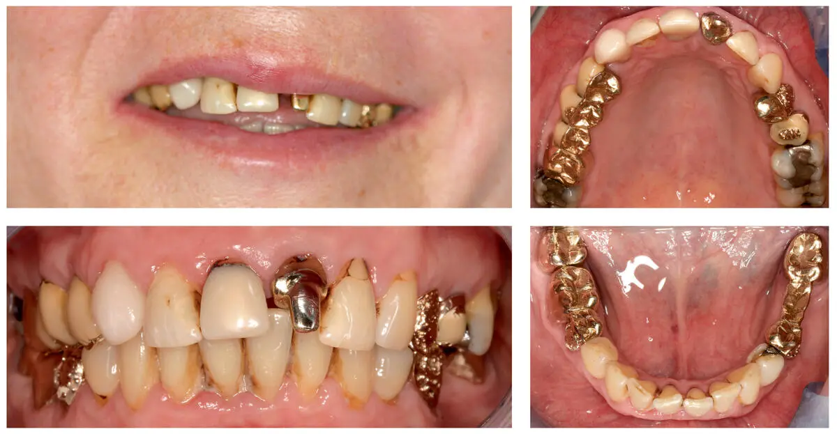
Fig 2.18A photo of the patient’s smile showing intraoral anterior and occlusal views of the old prosthetic rehabilitation.
A comprehensive examination revealed multiple insufficient restorations, secondary caries, and insufficient composite fillings ( Fig 2.18). The periodontal assessment depicted localized gingival inflammation. Functional examination revealed a stable occlusal scheme with canine-anterior-protected guidance during lateral and protrusive movements. The radiographic examination showed insufficient root canal fillings on teeth 45, 35, 25, 11, and an apical external resorption of tooth 11. The esthetic appearance of the existing fixed rehabilitation was deemed insufficient. All teeth were given a fair prognosis, except for teeth 25, 35, and 45, which were rated questionable, and tooth 21, which was deemed hopeless.
The final treatment plan comprised zirconia-based tooth-supported single crowns and FPDs in the maxilla and mandible.
Active Clinical Treatment
Preprosthetic Phase
Following a professional dental cleaning, alginate impressions (Alginat Super, Pluradent, Offenbach, Germany) were made. A full diagnostic wax-up was performed in order to evaluate the feasibility of the final esthetic and functional outcomes. To evaluate the esthetic and phonetic parameters, the wax-up was tried-in clinically via a direct mock-up ( Fig 2.19). Then, the old crowns and FPDs in the maxilla and mandible were removed. Direct provisional restorations (Luxatemp, DMG, Hamburg, Germany) were fabricated via silicone keys from the wax-up and delivered using a provisional cement (TempBond NE, Kerr, Orange, CA, USA) ( Fig 2.20). During the preprosthetic phase, tooth 21 was extracted because of external resorption and tooth 35 due to secondary caries. Teeth 25 and 45 were extracted after removal of the existing posts and cores, which revealed an extensive loss of the tooth structure. In addition, all insufficient fillings were replaced and a direct post-and-core buildup of tooth 13 was performed. Soft tissue enhancement via a connective tissue graft was performed in tooth 21. The shade of the mandibular anterior teeth was improved through external bleaching.
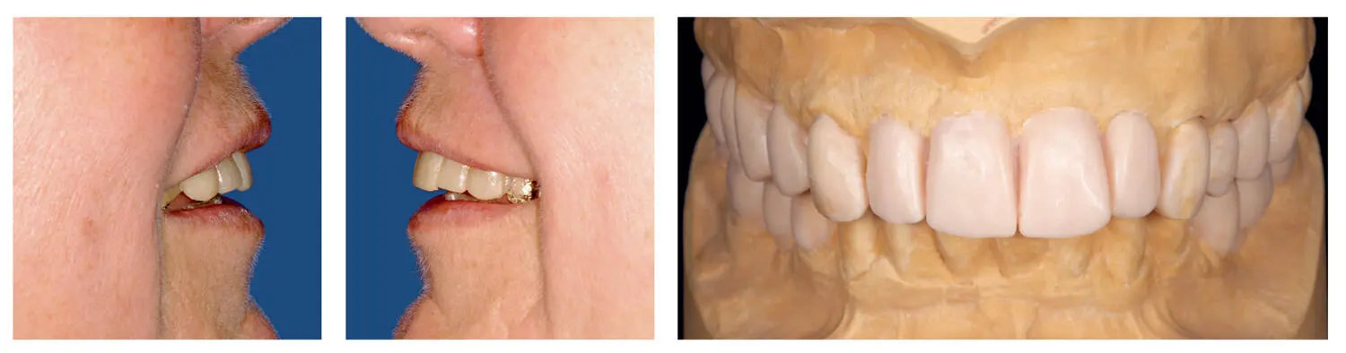
Fig 2.19A diagnostic wax-up was created to verify the predictability of the prosthetic rehabilitation. Esthetic and phonetic parameters were then evaluated via a mock-up try-in.
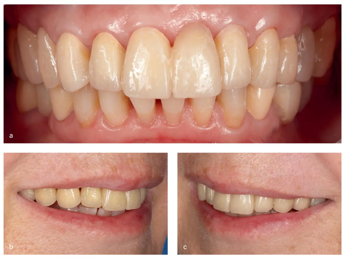
Fig 2.20An intraoral frontal view of the inserted provisionals (a). Side views of the patient’s smile after the delivery of the provisional restorations (b, c).
A re-evaluation of the preprosthetic phase followed after 4 months. The patient exhibited a stable periodontal status with probing depths ranging from 2 to 3 mm with no bleeding. The extraction sites healed successfully without any complications. The successful outcome of the preprosthetic phase allowed for the execution of the prosthodontic treatment plan.
Prosthetic Phase
After the definitive preparation of maxillary and mandibular teeth, retraction cords (Ultrapak #000 Retraction Cord, Ultradent, South Jordan, UT, USA) were placed ( Fig 2.21). The final impression was performed digitally with the help of an iTero intraoral scanner (iTero, Align Technology) ( Figs 2.21d, e). This scanner uses powder-free technology and produces images via parallel confocal microscopy (details about the scan technology can be found earlier in this chapter).
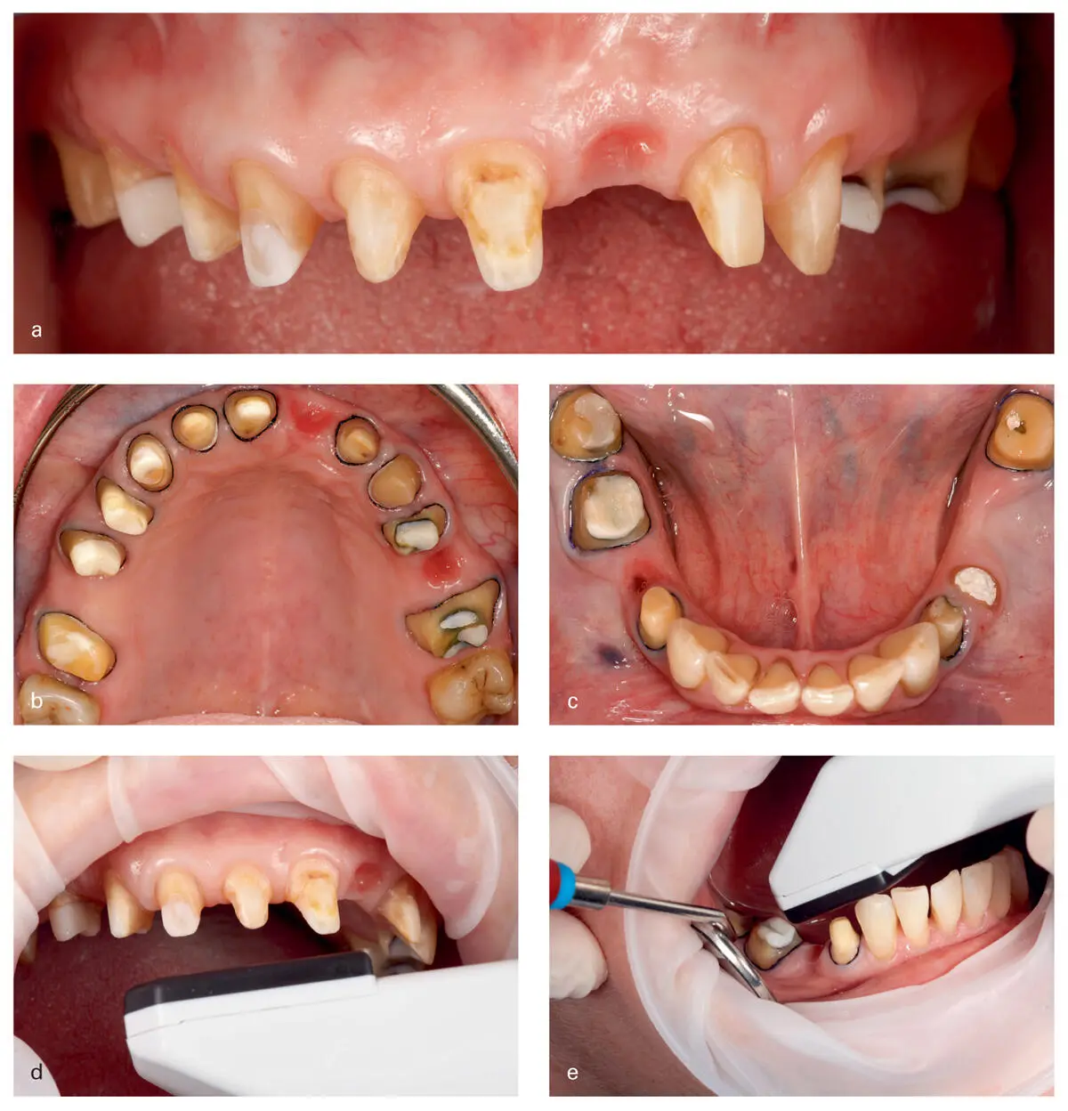
Fig 2.21Intraoral frontal and occlusal views of the prepared abutment teeth (a, b, c). The scanning procedure in the maxilla and mandible (d, e).
The scanning procedure began with the creation of the patient profile in the software interface. The digital impression was conducted first in the maxilla, starting with the most posterior tooth, and moving forwards through the whole arch. Scanning of each abutment tooth began at the occlusal surface, followed by the buccal surface and ending at the oral surface. The same scanning technique was also applied in the mandible. After taking the digital impression, a registration of the occlusal relationship was performed by scanning the three different areas of the arch (right, left, and anterior teeth). In order to assure a proper interocclusal relationship, the provisional restorations were used interchangeably in nonscanned areas as a bite reference during the scanning procedure ( Fig 2.22).
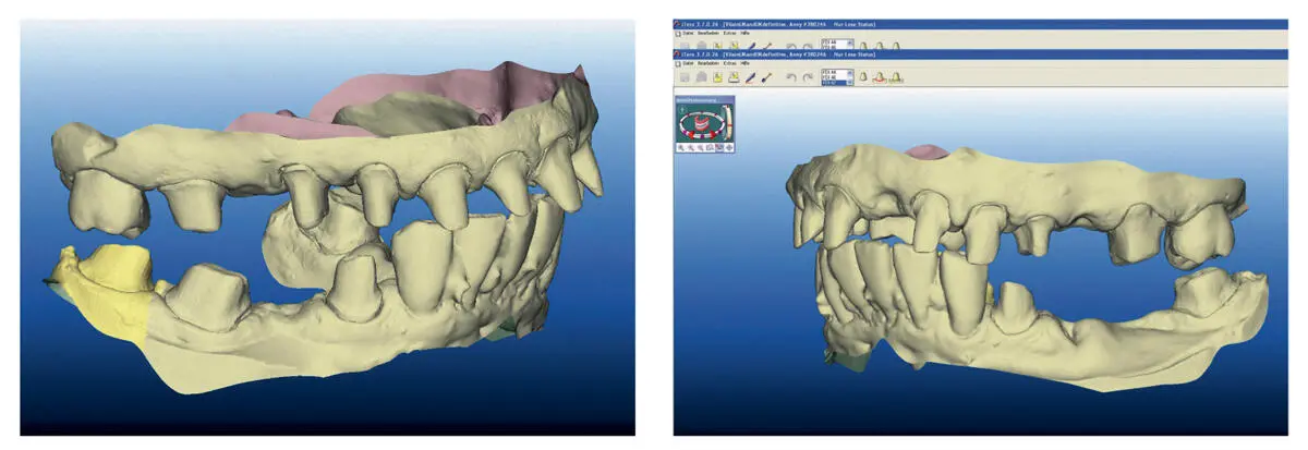
Fig 2.22Interocclusal relationship of the scanned images.
The obtained data sets were uploaded via a secure internet connection to the company server (Align Technology), where the initial digital file was cleaned and processed by the company computer software ( Fig 2.23). Afterwards the data sets were converted into STL data format and sent together with the CAD/CAM-fabricated polyurethane casts to the dental laboratory for restoration design ( Fig 2.23). By using CAD software (SimedaCad 2.0, Simeda Medical, Luxemburg), the STL files were imported and the restorations were digitally designed in their full anatomical form ( Fig 2.24). Before performing the necessary cutback for the fabrication of the final zirconia framework, a prototype made of composite was milled and tried intraorally ( Fig 2.24). Corrections of tooth length and form were performed either by adding flowable composite or by bur reshaping of the prototype. The modified prototype was scanned again with a laboratory scanner (3Shape) and the obtained data were matched with the previous restoration design, in order to assure an exact transfer of the performed corrections. Subsequently, anatomically shaped frameworks were designed using a digital cut back and milled from a zirconia block (SinaZ, Simeda Medical) ( Fig 2.25a). An intraoral try-in followed, showing proper fit and retention of the zirconia frameworks ( Fig 2.25b). At the same appointment, a spectrophotometer (Crystaleye, Olympus, Tokyo, Japan) was utilized to determine the appropriate tooth shade ( Fig 2.25c). The veneering of the zirconia frameworks was performed with feldspathic ceramic (HeraCeram Zirkonia, Heraus, Hanau, Germany). Before finalization of the restorations, a biscuit try-in was conducted to evaluate the occlusion, esthetics, and phonetics ( Fig 2.26). As the result was rated satisfactory by both the patient and the clinician, the restorations were finalized.
Читать дальшеИнтервал:
Закладка:
Похожие книги на «Digital Workflow in Reconstructive Dentistry»
Представляем Вашему вниманию похожие книги на «Digital Workflow in Reconstructive Dentistry» списком для выбора. Мы отобрали схожую по названию и смыслу литературу в надежде предоставить читателям больше вариантов отыскать новые, интересные, ещё непрочитанные произведения.


Обсуждение, отзывы о книге «Digital Workflow in Reconstructive Dentistry» и просто собственные мнения читателей. Оставьте ваши комментарии, напишите, что Вы думаете о произведении, его смысле или главных героях. Укажите что конкретно понравилось, а что нет, и почему Вы так считаете.