Richard J. Miron - Understanding Platelet-Rich Fibrin
Здесь есть возможность читать онлайн «Richard J. Miron - Understanding Platelet-Rich Fibrin» — ознакомительный отрывок электронной книги совершенно бесплатно, а после прочтения отрывка купить полную версию. В некоторых случаях можно слушать аудио, скачать через торрент в формате fb2 и присутствует краткое содержание. Жанр: unrecognised, на английском языке. Описание произведения, (предисловие) а так же отзывы посетителей доступны на портале библиотеки ЛибКат.
- Название:Understanding Platelet-Rich Fibrin
- Автор:
- Жанр:
- Год:неизвестен
- ISBN:нет данных
- Рейтинг книги:5 / 5. Голосов: 1
-
Избранное:Добавить в избранное
- Отзывы:
-
Ваша оценка:
Understanding Platelet-Rich Fibrin: краткое содержание, описание и аннотация
Предлагаем к чтению аннотацию, описание, краткое содержание или предисловие (зависит от того, что написал сам автор книги «Understanding Platelet-Rich Fibrin»). Если вы не нашли необходимую информацию о книге — напишите в комментариях, мы постараемся отыскать её.
Understanding Platelet-Rich Fibrin — читать онлайн ознакомительный отрывок
Ниже представлен текст книги, разбитый по страницам. Система сохранения места последней прочитанной страницы, позволяет с удобством читать онлайн бесплатно книгу «Understanding Platelet-Rich Fibrin», без необходимости каждый раз заново искать на чём Вы остановились. Поставьте закладку, и сможете в любой момент перейти на страницу, на которой закончили чтение.
Интервал:
Закладка:
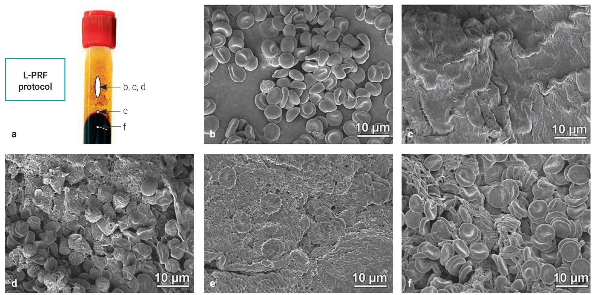
Fig 3-10SEM images of the distal surface of PRF clots prepared utilizing the L-PRF protocol. (a) The described areas observed by SEM. The L-PRF clot surfaces showed typically three types, as shown in b to d . (b) Clusters of RBCs were observed overlaying a smooth clot surface. (c) A wavy surface was observed, including RBCs. (d) The rough surface included leukocytes, platelets, and crushed RBCs. (e) The dense fibrin networks were observed, including RBCs at the border between the yellow plasma and RBC layers. (f) Many RBCs were observed within a fibrin network in the RBC layer. (Reprinted with permission from Fujioka-Kobayashi et al. 5)
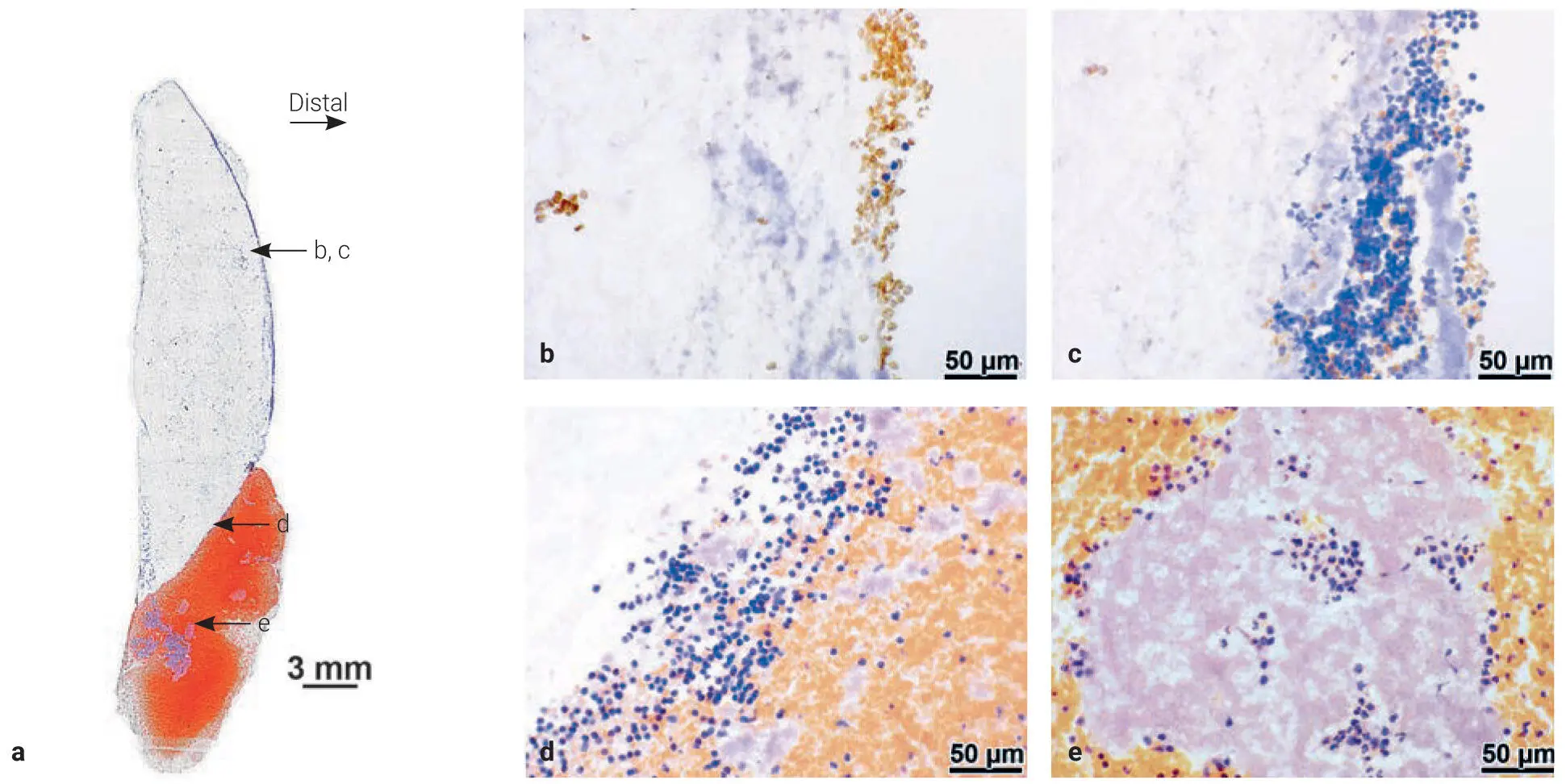
Fig 3-11Histologic observation of the frozen section of L-PRF sectioned transaxially. (a) The panoramic view of the sections from the whole PRF clot including the RBC layer stained with hematoxylin. The L-PRF clots and RBC layer were separated by a fixed-angle centrifuge. The distal wall showed two typical patterns, shown in b and c . (b) Clusters of RBCs with a few leukocytes were located on fibrin networks on the distal surface. (c) The aggregated cluster consisting of platelets, leukocytes, and RBCs occasionally observed. (d) Many leukocytes were located at the border between the PRF clot and the RBC layer. (e) The aggregated clusters of cells containing leukocytes were occasionally observed within the RBC layer within the red buffy coat zone. (Reprinted with permission from Fujioka-Kobayashi et al. 5)
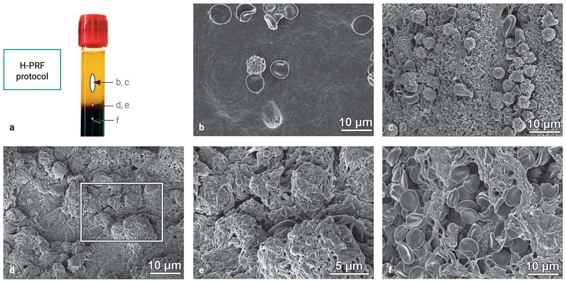
Fig 3-12SEM images of the distal surface of PRF clots prepared utilizing the H-PRF protocol. (a) The described areas observed by SEM. The H-PRF clot surfaces showed typically two types, as shown in b and c . (b) Few leukocytes and RBCs were observed on the smooth surfaces. (c) The rough surfaces included leukocytes, platelets, and RBCs. (d and e) The fibrin networks twisted around the leukocytes with platelets at the border between the yellow plasma and RBC layers. (f) Many RBCs were observed within a fibrin network in the RBC layer. (Reprinted with permission from Fujioka-Kobayashi et al. 5)
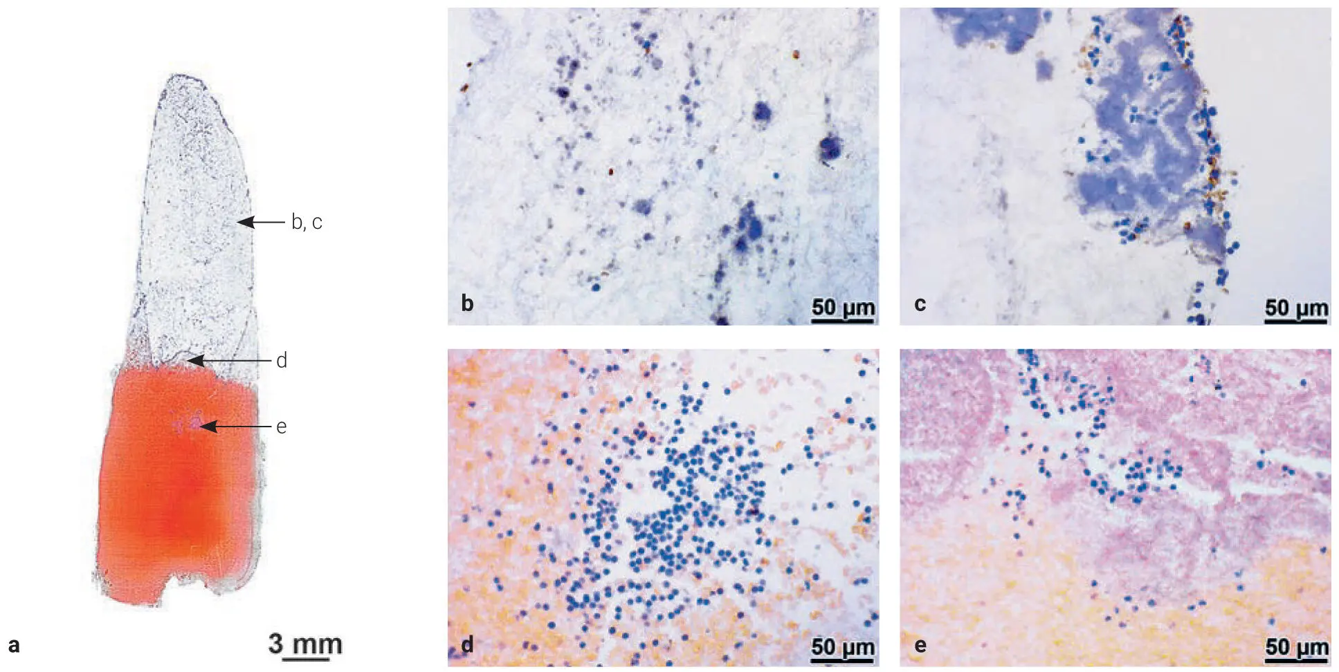
Fig 3-13Histologic observation of the frozen section of H-PRF sectioned transaxially. (a) The panoramic view of the sections from the whole PRF clot including the RBC layer stained with hematoxylin. The H-PRF clots and RBC layers were separated evenly and horizontally with no obvious accumulation of cells on the distal surface. The clots showed two typical patterns shown in b and c . (b) The fibrin networks were observed in the clots with many platelets and few leukocytes/RBCs. (c) Aggregated clusters consisting of leukocytes and a few RBCs were occasionally observed. (d) Many leukocytes were located at the border between the clots and RBC layer. (e) Aggregated clusters of cells containing leukocytes were occasionally observed in the RBC layer within the red buffy coat zone. (Reprinted with permission from Fujioka-Kobayashi et al. 5)
Within H-PRF clots, two typical patterns were observed on the surface, including fewer blood cells on the smooth clot surfaces, with more found located on the rough surfaces (see Fig 3-12). Abundant platelets were found within the clots, with a few clusters located on the actual clot surface (see Fig 3-13). The border between the plasma and RBC layers included a dense fibrin network with many leukocytes.
Interestingly, aggregated clusters of platelets with leukocytes were found in both L-PRF and H-PRF within the RBC layer, approximately 5 mm below the precise separation typically referred to as the buffy coat zone . A representative summary figure is provided in Fig 3-14.
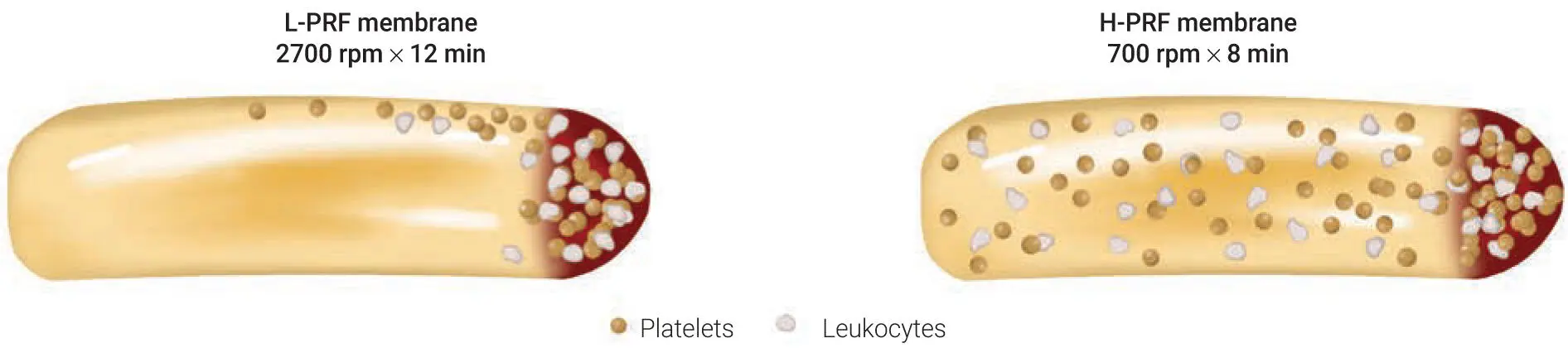
Fig 3-14Graphic demonstrating cell distribution within PRF when centrifugation was carried out either by fixed-angle or horizontal centrifugation. Note that the majority of cells following the L-PRF protocol are found along the back distal surface of PRF tubes as well as primarily contained within the buffy coat layer. A more even distribution of cells was observed when horizontal centrifugation was utilized. (Reprinted with permission from Fujioka-Kobayashi et al. 5)
Optimization of PRF Protocols
To this day, PRF has not been most efficiently optimized. Additionally, in the early 2000s, a variety of publications on the topic of PRF were utilized at different RCF/rpm parameters. As highlighted in chapter 4, many were actually using the same rpm values for devices with different rotor sizes (which completely changes the g-force) without understanding its pronounced impact on cell layer separation.
It is important to note that larger-radius centrifuges produce much greater g-force even at identical rpms.
One of the most common limitations to PRF is the fact that various protocols have never been investigated in studies. In 2014, Ghanaati et al discovered a way to further optimize the production of PRF using three different protocols and by gradually reducing RCF. By doing so, he discovered that more cells could be obtained in the upper PRF layers; this method has since been named the low-speed centrifugation concept (LSCC). 7
In 2019, drastically better results were obtained utilizing horizontal centrifugation. While 20% to 30% better results were obtained with the LSCC, the ability to simply shift from fixed-angle to horizontal centrifugation led to as much as a fourfold increase in cells. Our research team then investigated 24 different protocols (instead of the original 3) to better optimize PRF.
Evaluation of 24 protocols for the production of PRF
While in previous studies few protocols were compared (generally at most three), 7the desire and emphasis of our research group was to better investigate for the first time the effect of numerous centrifugation parameters on the final production of PRF. As such, 24 different protocols were evaluated ( Fig 3-15). 8All protocols were compared utilizing a recent method to quantify cells in PRF in 1-mL sequential layers pipetted from the upper layer downward until all 10 mL were harvested (see chapter 2for methodology). In total, 960 complete blood counts (CBCs) were investigated. Both solid- and liquid-based PRF protocols were investigated following 24 protocols involving six RCF values (100g, 200g, 400g, 700g, 1000g, and 1200g) at four centrifugation times (3, 5, 8, and 12 minutes).
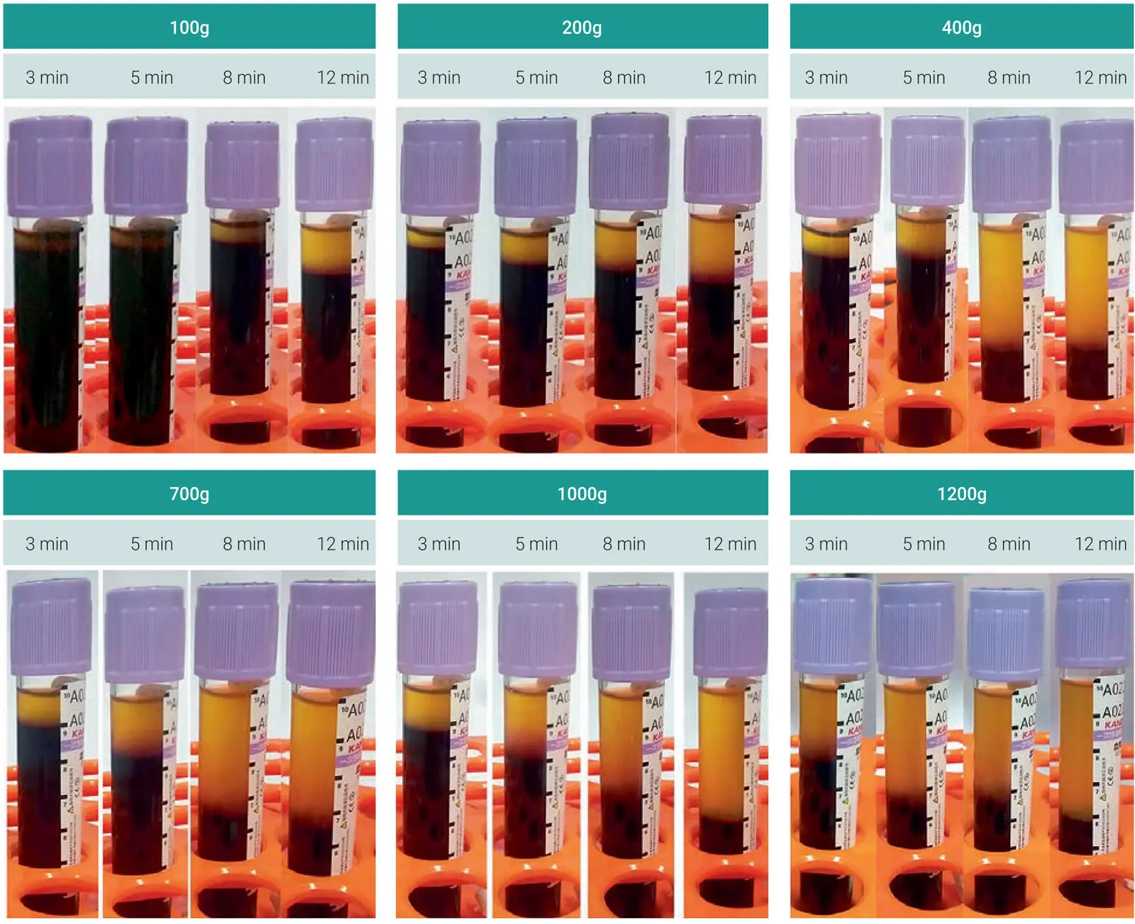
Fig 3-15Clinical image demonstrating the plasma layer separation for the 24 protocols investigated in this study. Note that while some protocols reveal roughly identical plasma layer separation, the underlying cellular content in the various protocols may be drastically different. (Reprinted with permission from Miron et al. 8)
Читать дальшеИнтервал:
Закладка:
Похожие книги на «Understanding Platelet-Rich Fibrin»
Представляем Вашему вниманию похожие книги на «Understanding Platelet-Rich Fibrin» списком для выбора. Мы отобрали схожую по названию и смыслу литературу в надежде предоставить читателям больше вариантов отыскать новые, интересные, ещё непрочитанные произведения.


Обсуждение, отзывы о книге «Understanding Platelet-Rich Fibrin» и просто собственные мнения читателей. Оставьте ваши комментарии, напишите, что Вы думаете о произведении, его смысле или главных героях. Укажите что конкретно понравилось, а что нет, и почему Вы так считаете.