Richard J. Miron - Understanding Platelet-Rich Fibrin
Здесь есть возможность читать онлайн «Richard J. Miron - Understanding Platelet-Rich Fibrin» — ознакомительный отрывок электронной книги совершенно бесплатно, а после прочтения отрывка купить полную версию. В некоторых случаях можно слушать аудио, скачать через торрент в формате fb2 и присутствует краткое содержание. Жанр: unrecognised, на английском языке. Описание произведения, (предисловие) а так же отзывы посетителей доступны на портале библиотеки ЛибКат.
- Название:Understanding Platelet-Rich Fibrin
- Автор:
- Жанр:
- Год:неизвестен
- ISBN:нет данных
- Рейтинг книги:5 / 5. Голосов: 1
-
Избранное:Добавить в избранное
- Отзывы:
-
Ваша оценка:
Understanding Platelet-Rich Fibrin: краткое содержание, описание и аннотация
Предлагаем к чтению аннотацию, описание, краткое содержание или предисловие (зависит от того, что написал сам автор книги «Understanding Platelet-Rich Fibrin»). Если вы не нашли необходимую информацию о книге — напишите в комментариях, мы постараемся отыскать её.
Understanding Platelet-Rich Fibrin — читать онлайн ознакомительный отрывок
Ниже представлен текст книги, разбитый по страницам. Система сохранения места последней прочитанной страницы, позволяет с удобством читать онлайн бесплатно книгу «Understanding Platelet-Rich Fibrin», без необходимости каждый раз заново искать на чём Вы остановились. Поставьте закладку, и сможете в любой момент перейти на страницу, на которой закончили чтение.
Интервал:
Закладка:
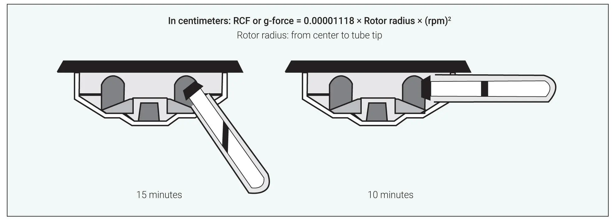
Fig 3-4Graphic from Drucker Diagnostics demonstrating that centrifugation carried out on a horizontal centrifuge requires only two-thirds the time required on a fixed-angle centrifuge. Therefore, a 12-minute protocol on a fixed-angle rotor would take only 8 minutes with a horizontal system. (Adapted from https://druckerdiagnostics.com/horizontal-vs-fixed-angle/.)
The time required for complete centrifugation on a horizontal rotor is only two-thirds of the time required on a fixed-angle centrifuge.
Accumulation of Cells in PRF Tubes
One thing commonly found following centrifugation is the angle produced after completion of the spin cycle ( Fig 3-5). 2In a study by Takahashi et al, the distribution of cells and growth factors (GFs) in PRF following centri-fugation using two different centrifugation devices was investigated. 3Blood samples were obtained in tubes and immediately centrifuged to prepare PRF using two protocols. Both matrices were compressed, embedded in paraffin, and subjected to immunohistochemical examination. 3
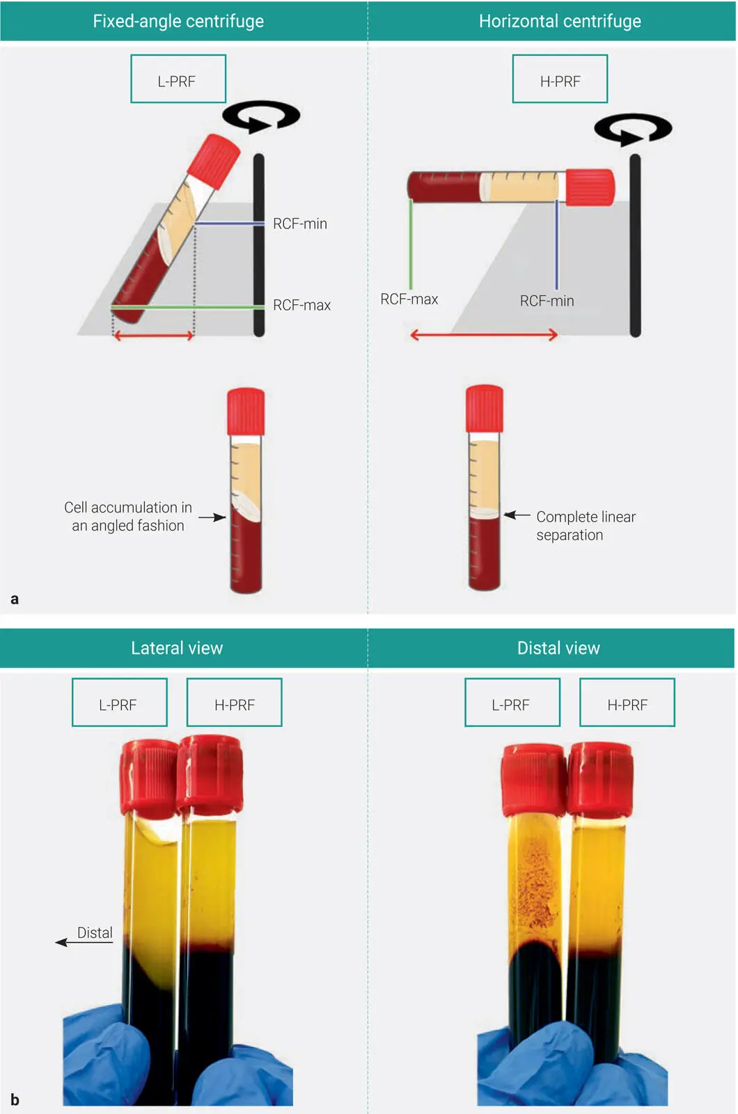
Fig 3-5 (a) Following centrifugation on fixed-angle centrifuges, blood layers do not separate evenly, and as a result, an angled blood separation is observed. In contrast, horizontal centrifugation produces an even separation. (Reprinted with permission from Miron et al. 1) (b) Layer separation following either L-PRF or H-PRF protocols. L-PRF clots are prepared with a sloped shape, and multiple red dots are often observed on the distal surface of PRF tubes; H-PRF, on the other hand, is prepared with a horizontal layer separation between the upper plasma and lower RBC layers.
Following histologic assessment, it was observed that leukocytes and plasma proteins were localized on the back walls of PRF tubes (referred to as distal surface ), including the interface corresponding to the buffy coat ( Figs 3-6and 3-7). 3These cells were being accumulated on the back distal surfaces only when utilizing fixed-angle centrifugation due to the gravitational pull (see Fig 3-8; see also Video 3-2).
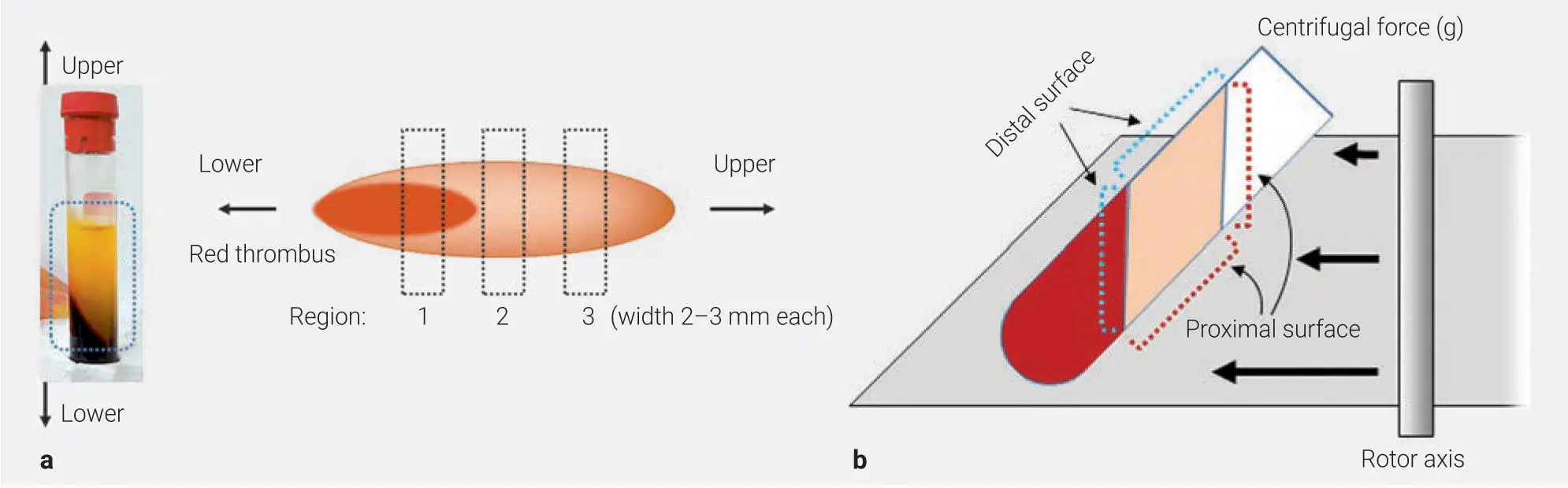
Fig 3-6Experimental setup describing the orientation of PRF membranes during histologic assessment. The proximal surface describes the inner tube wall (generally receiving the smallest g-force), whereas the distal surface is the outer tube wall, where cells generally accumulate during centrifugation at high g-force. (a) Regions in compressed A‐PRF or concentrated GF (CGF) matrix. This image is the proximal surface. (b) Centrifugal force and distal and proximal surfaces of A‐PRF or CGF matrix. (Reprinted with permission from Takahashi et al. 3)
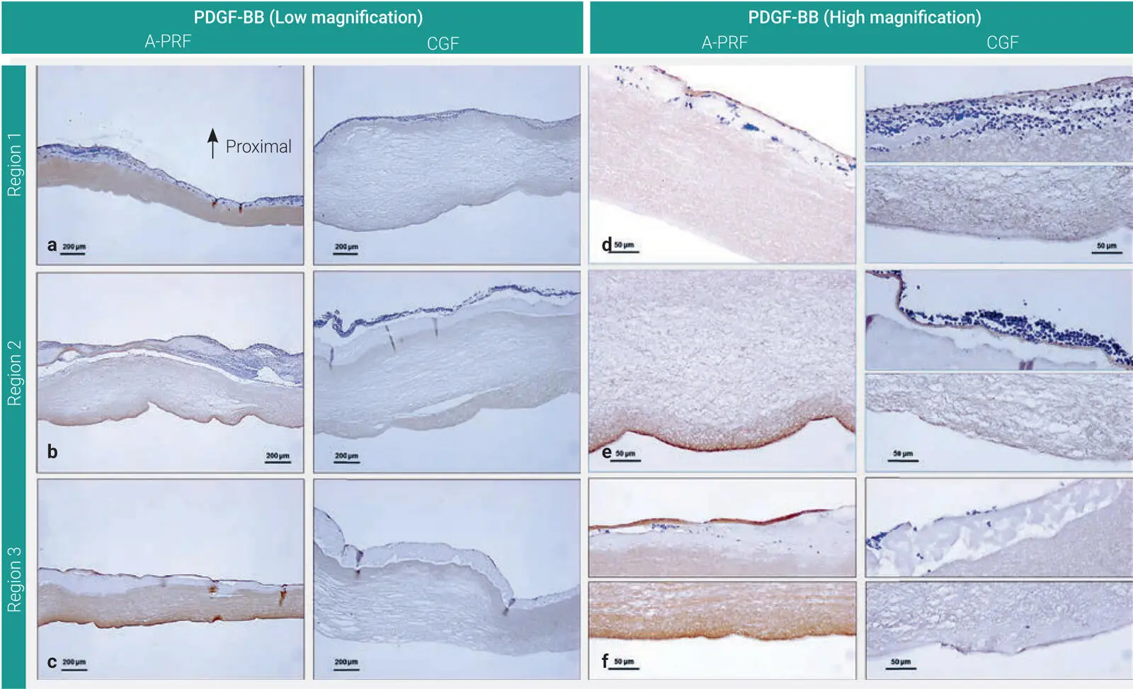
Fig 3-7Distribution of PDGF‐BB in A‐PRF and CGF matrices. (a and d) Region 1. (b and e) Region 2. (c and f) Region 3. Note that the majority of cells and GFs accumulated on the back distal surfaces of PRF tubes. (Reprinted with permission from Takahashi et al. 3)
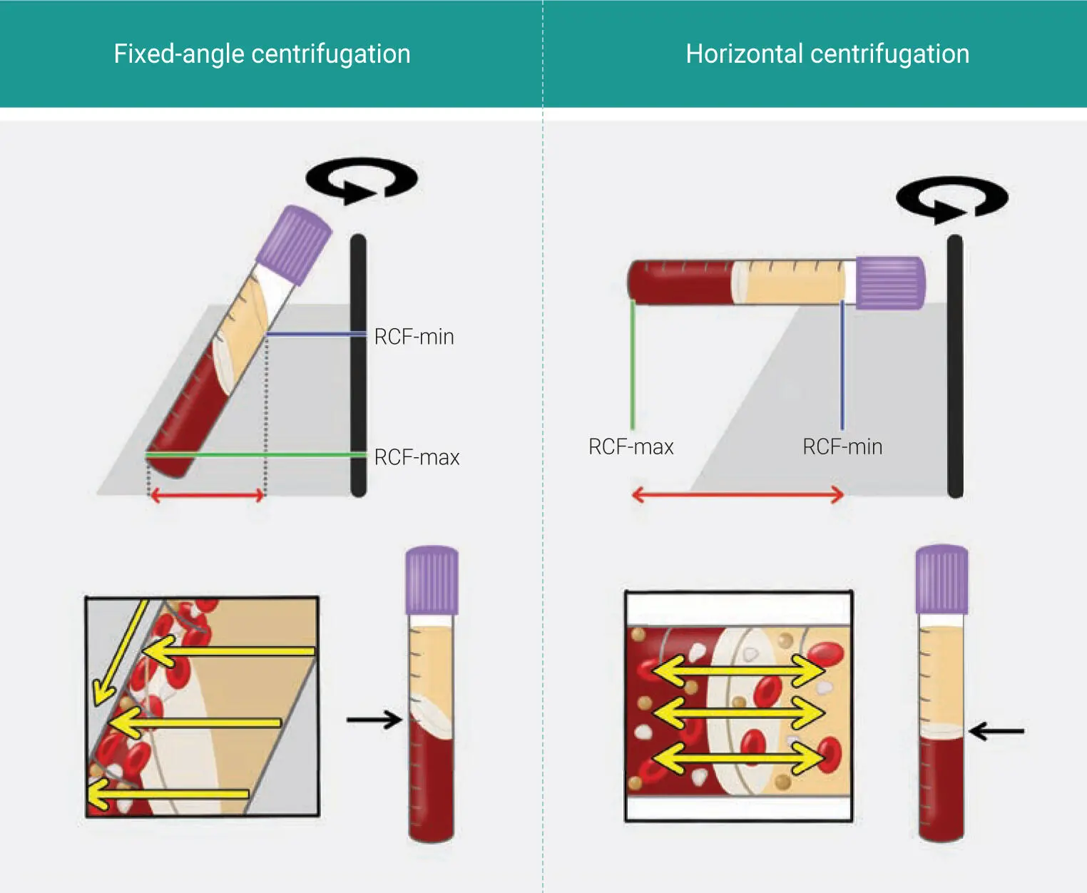
Fig 3-8Illustrations comparing fixed-angle and horizontal centrifuges. With horizontal centrifugation, increased separation of blood layers based on density is achieved due to the increased difference in RCF-min and RCF-max. Following centrifugation on fixed-angle centrifuges, blood layers do not separate evenly, and as a result, an angled blood separation is observed. In contrast, horizontal centrifugation produces even separation. Owing to the large RCF values (~200g–700g), the cells are pushed toward the outside and downward. On a fixed-angle centrifuge, cells are pushed toward the back of centrifugation tubes and then downward/upward based on cell density. These g-forces produce additional shear stress on cells as they separate based on density along the back walls of centrifugation tubes. In contrast, horizontal centrifugation allows for the free movement of cells to separate into their appropriate layers based on density, allowing for better cell separation and less trauma/shear stress on cells. (Adapted from Miron et al. 1)

 Video 3-2
More intriguing was the fact that the type of tube (plain glass versus silica-coated plastic) had a significant impact on the final distribution of cells found within PRF clots when the clots produced at different centrifugation speeds were investigated histologically. 4The specific role of tubes for the production of PRF has been such a hot topic as of late that an entire book chapter is dedicated solely to this topic (see chapter 5).
Cells were being accumulated on the back distal surfaces only when utilizing fixed-angle centrifugation.
Evaluation of PRF via Horizontal Centrifugation
As the field continues to progress, it has become clear that horizontal centrifugation offers numerous advantages. A simple evaluation of three protocols previously tested has demonstrated the very obvious improved blood cell layer separation observed with H-PRF when compared to L-PRF or A-PRF ( Fig 3-8; see chapter 2).
In a study titled “Histological comparison of platelet rich fibrin clots prepared by fixed-angle versus horizontal centrifugation,” Fujioka-Kobayashi et al compared L-PRF with H-PRF, observing the morphology of cells and their localization on the surface of PRF clots by SEM and histologically by transaxial frozen sections by means of a film method. 5It was consistently observed that L-PRF clots demonstrated a sloped separation between the upper plasma and the bottom RBC layers according to the angle of the rotor. Interestingly, red dots were often observed on the distal walls of the tubes in the upper layers, consisting of aggregations of RBCs, leukocytes, and platelets by SEM and histology 5( Fig 3-9). Clots produced on the horizontal centrifuge showed much smoother cell layer distribution and separation along the tube surfaces when compared to L-PRF.
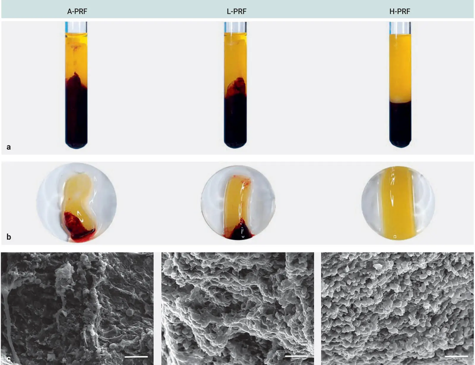
Fig 3-9Characterization of A-PRF, L-PRF, and H-PRF. (a and b) The morphology and size of A-PRF, L-PRF, and H-PRF after centrifugation using the manufacturer’s recommended centrifugation protocols and tubes. (c) SEM of A-PRF, L-PRF, and H-PRF clots. Scale bar = 20 μm. (Reprinted with permission from Zhang et al. 6)
Microscopic and histologic observation of L-PRF and H-PRF
PRF clots were further investigated by SEM and histologic assessment for cell distribution and surface configurations ( Figs 3-10to 3-13). Three distinct patterns were observed on the distal walls of L-PRF clots, including RBC clusters on the smooth or wavy fibrin clot surfaces as well as clusters of leukocytes, platelets, and crushed RBCs (see Fig 3-10). Histologic observation further confirmed these pattern types (see Fig 3-11). The border between the plasma and RBC layers included more dense fibrin networks covered with many blood cells (see Fig 3-10e). Many leukocytes were found at this layer (see Fig 3-11d).
Читать дальшеИнтервал:
Закладка:
Похожие книги на «Understanding Platelet-Rich Fibrin»
Представляем Вашему вниманию похожие книги на «Understanding Platelet-Rich Fibrin» списком для выбора. Мы отобрали схожую по названию и смыслу литературу в надежде предоставить читателям больше вариантов отыскать новые, интересные, ещё непрочитанные произведения.


Обсуждение, отзывы о книге «Understanding Platelet-Rich Fibrin» и просто собственные мнения читателей. Оставьте ваши комментарии, напишите, что Вы думаете о произведении, его смысле или главных героях. Укажите что конкретно понравилось, а что нет, и почему Вы так считаете.