Richard J. Miron - Understanding Platelet-Rich Fibrin
Здесь есть возможность читать онлайн «Richard J. Miron - Understanding Platelet-Rich Fibrin» — ознакомительный отрывок электронной книги совершенно бесплатно, а после прочтения отрывка купить полную версию. В некоторых случаях можно слушать аудио, скачать через торрент в формате fb2 и присутствует краткое содержание. Жанр: unrecognised, на английском языке. Описание произведения, (предисловие) а так же отзывы посетителей доступны на портале библиотеки ЛибКат.
- Название:Understanding Platelet-Rich Fibrin
- Автор:
- Жанр:
- Год:неизвестен
- ISBN:нет данных
- Рейтинг книги:5 / 5. Голосов: 1
-
Избранное:Добавить в избранное
- Отзывы:
-
Ваша оценка:
Understanding Platelet-Rich Fibrin: краткое содержание, описание и аннотация
Предлагаем к чтению аннотацию, описание, краткое содержание или предисловие (зависит от того, что написал сам автор книги «Understanding Platelet-Rich Fibrin»). Если вы не нашли необходимую информацию о книге — напишите в комментариях, мы постараемся отыскать её.
Understanding Platelet-Rich Fibrin — читать онлайн ознакомительный отрывок
Ниже представлен текст книги, разбитый по страницам. Система сохранения места последней прочитанной страницы, позволяет с удобством читать онлайн бесплатно книгу «Understanding Platelet-Rich Fibrin», без необходимости каждый раз заново искать на чём Вы остановились. Поставьте закладку, и сможете в любой момент перейти на страницу, на которой закончили чтение.
Интервал:
Закладка:
By utilizing this novel technique and investigating each 1 mL layer by layer, it was possible for the first time to investigate exactly each cell layer following centrifugation and determine the precise location of each blood cell type.
L-PRF protocol
Following centrifugation, 1-mL sequential layers were sent for CBC analysis according to Fig 2-14a. As Fig 2-14b shows, the original L-PRF protocol using the IntraSpin device (2700 rpm for 12 min; ~700g) with a 33-degree fixed-angle centrifuge revealed precisely that the number of leukocytes (control 6 × 10 9cells/L) and platelets was significantly concentrated in layer 5 (~17 × 10 9cells/L; arrows represent where the plasma and RBC layers separate). Interestingly, a threefold to fourfold increase in leukocyte number was observed specifically at this interface within the buffy coat. Notice, however, that no leukocytes were found in any of the upper 4 layers, displaying a very uneven PRF clot with respect to cell numbers. Almost all cells within the PRF clot were exclusively found within this 5th layer. Notice also that more leukocytes were found in the RBC layer below the PRF clot. A similar trend was also observed for lymphocytes, neutrophils, and monocytes (see Fig 2-14b). Naturally, all RBCs were found in layers 5 through 10 in the visually red layers. Platelets were accumulated once again precisely in layer 5 (six- to eightfold), within the buffy coat zone.
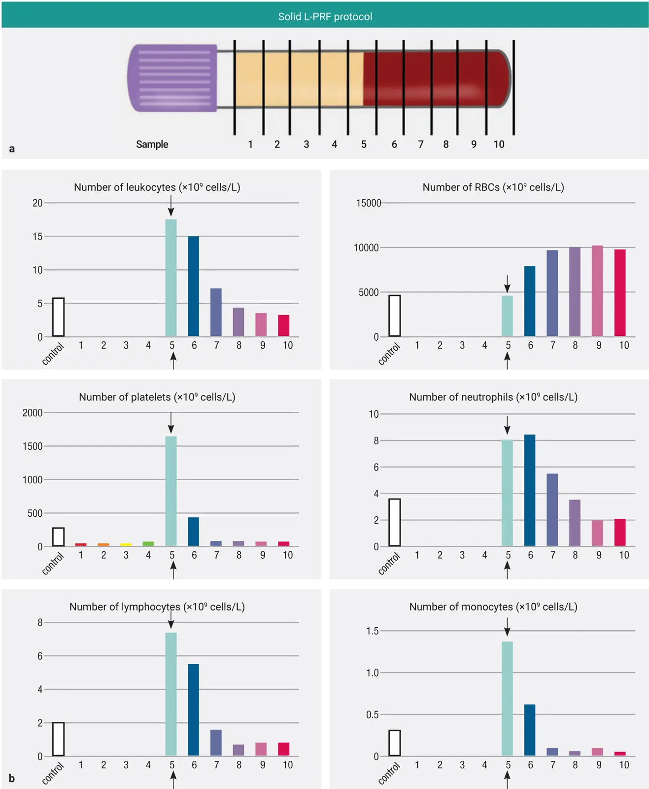
Fig 2-14 (a) Separation of the 10-mL tube into 10 1-ml layers for pipetting. (b) The concentration of cell types in each 1-mL layer utilizing the solid L-PRF protocol (2700 rpm for 12 minutes; ~700g). Notice that the majority of platelets accumulated directly within the 5th layer in the buffy coat. Furthermore, the highest concentration of leukocytes was also noted in this layer. The first 4 layers of this plasma layer were typically devoid of all cells. (Adapted with permission from Miron et al. 50)
Because the majority of cells were found in layer 5, we were interested to determine if these cells were specifically found within the yellow plasma layer (within the PRF clot) or within the RBC layer. For this, a second blood tube was utilized; 500 µL of blood volume was collected just above the RBC layer within the buffy coat, and 500 µL was taken from the RBC layer. It was revealed that the majority of platelets were found within the yellow plasma layer (> 80%), whereas the majority of leukocytes and other WBCs were found within the red blood layer ( Fig 2-15). This revealed that most leukocytes were in fact not found within the PRF layers utilizing the L-PRF protocol.
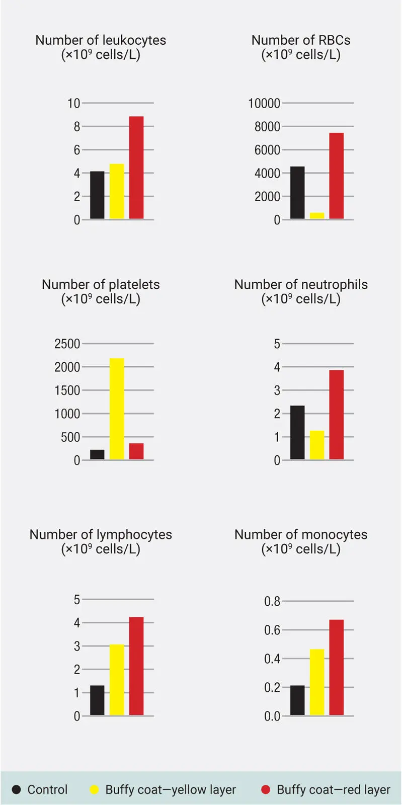
Fig 2-15Layer 5 (the zone that incorporates the buffy coat containing a plasma and RBC component) demonstrated the cell-rich zone. Analysis of this zone revealed that many of the cells were in fact located in the red zone (especially leukocytes). The cell-rich zone contains a yellow buffy coat zone, but the red portion of this buffy coat also contains many cells. Following these findings, it is generally recommended to harvest a small portion of the red zone, specifically when drawing liquid-PRF (i-PRF), because many cells are located within this region. (Adapted from Miron et al. 50)
Interestingly, the final concentration of leukocytes found using the L-PRF protocol was 4.13 × 10 9cells/L, whereas the control whole blood value from this patient was 6.125 × 10 9cells/L, representing a 33% reduction in leukocyte concentration when compared to control blood. Platelet numbers were increased 1.61-fold. The total leukocyte and platelet content represent 33% and 80% of the total blood cells found, respectively, within this 10-mL blood sample. This meant that roughly 20% of platelets and 66% of leukocytes were actually located within the RBC layer (similar to the observed histologic results by Ghanaati et al in 2014 6 ).
With the L-PRF protocol, the majority of leukocytes and platelets were not found within the plasma layer but rather in layer 5 within the buffy coat zone.
A-PRF protocol
Figure 2-16depicts centrifugation following A-PRF protocols (1300 rpm for 8 minutes on a Duo Quattro centrifuge). Interestingly, the number of platelets were concentrated throughout the first four to five layers, unlike the L-PRF protocol. Here, a twofold increase in platelets was observed compared to a 1.6-fold increase utilizing the L-PRF protocol. More importantly, however, the platelets were found evenly distributed throughout the A-PRF plasma layers. When investigating leukocyte number, however, a significantly lower concentration (33% original values) as well as total numbers (9.315 vs 20.65 × 10 9cells/L) were found in the A-PRF group when compared to L-PRF. Therefore, it was initially suspected that either the g-force or the total time was not sufficient to adequately accumulate or separate the leukocytes utilizing the A-PRF protocol. 50Once again, lower leukocytes in PRF were actually found when compared to whole blood.

Fig 2-16The concentration of cell types in each 1-mL layer utilizing the solid A-PRF protocol (1300 rpm for 8 minutes; ~200g). Notice that the platelets were more evenly distributed throughout the upper 5-mL plasma layer. Noteworthy, however, is that the majority of WBCs (leukocytes, neutrophils, lymphocytes, and monocytes) were not found in the upper plasma layer. (Adapted from Miron et al. 50)
While the A-PRF protocol with LSCC led to a higher concentration of platelets, it was not effectively capable of concentrating leukocytes.
i-PRF protocol
Liquid-PRF protocols were then investigated and compared ( Fig 2-17). The IntraSpin protocol (2700 rpm for 3 min; ~700g) is depicted in Fig 2-17a. Interestingly, this protocol accumulated platelets evenly throughout the PRF layer better than when utilizing the 12-minute protocol. Nevertheless, leukocytes were significantly lower once again when compared to whole blood, representing only 54% of the original control blood concentrations. This demonstrates that following centrifugation, lower numbers of leukocytes are found in L-PRF samples when compared to control blood in either L-PRF protocol. Platelet concentrates were increased 2.12-fold.
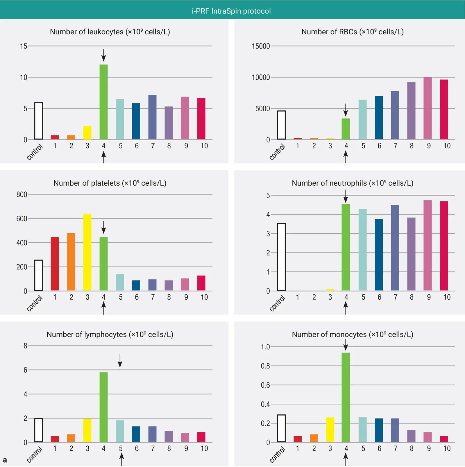
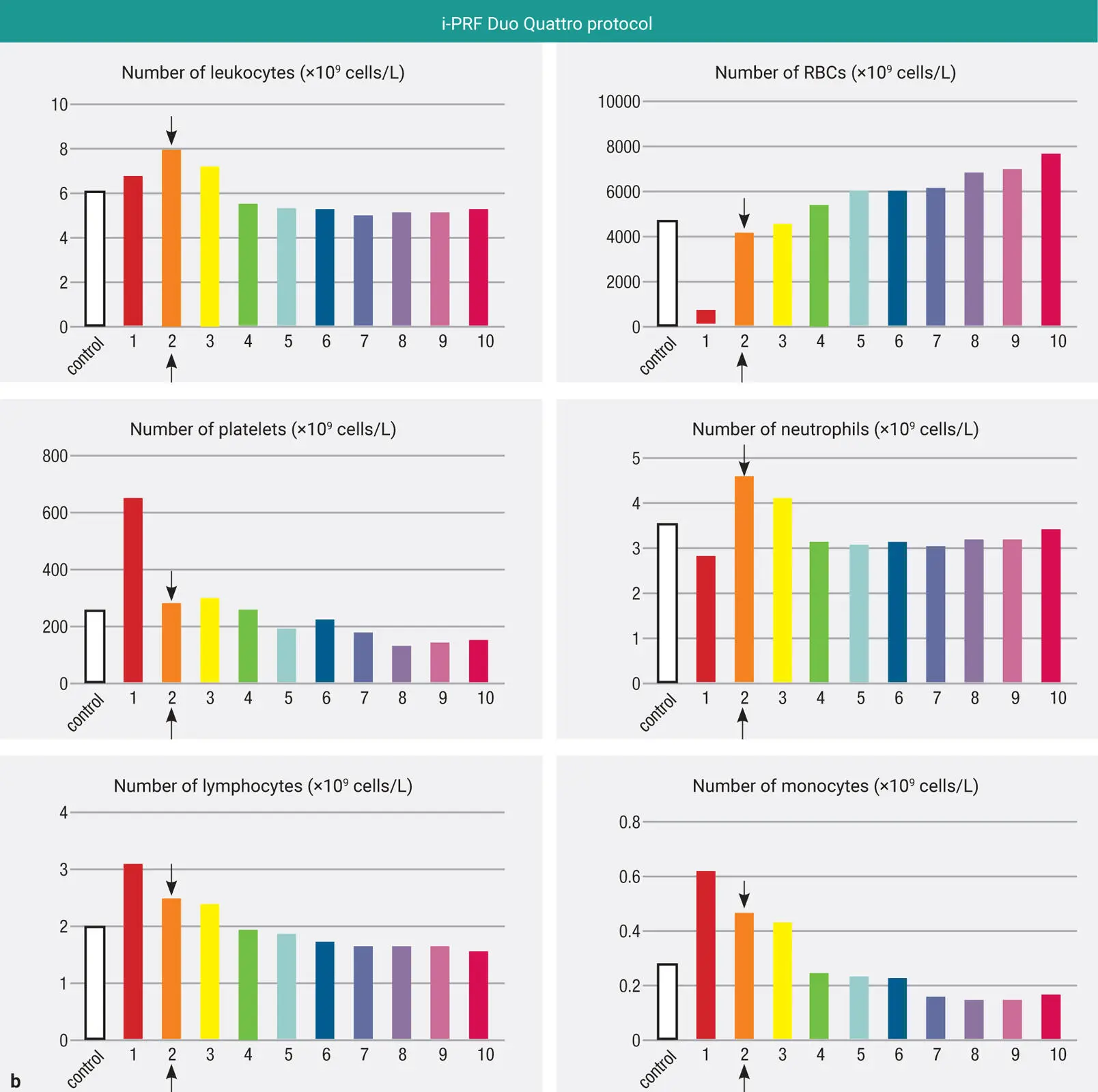
Fig 2-17 (a) The concentration of cell types in each 1-mL layer utilizing the i-PRF IntraSpin protocol (2700 rpm for 3 minutes; ~700g). Notice that most platelets are more evenly distributed utilizing this protocol when compared to the 12-minute solid-PRF IntraSpin protocol. (b) The concentration of cell types in each 1-mL layer utilizing the i-PRF Duo Quattro protocol (800 rpm for 3 minutes; ~60g). Notice that very little change in platelet or leukocyte accumulation is observed utilizing this centrifugation cycle. A slight increase in platelets and leukocytes is, however, observed when compared to the control. (Adapted from Miron et al. 50)
The i-PRF protocol recommended by Process for PRF (Duo Quattro centrifuge) produced a 1.23-fold increase in leukocyte concentration and a 2.07-fold increase in platelet concentration when compared to whole blood (see Fig 2-17b). The overall accumulation demonstrated an 18% total leukocyte content and a 31% total platelet count when compared to whole blood. This represented an extremely low platelet yield, as all other protocols produced at least 80% total yield. (Keep in mind here that this means 70% of platelets are found within the red layer following the use of this LSCC and not in the upper, liquid-PRF layer.) Most notably, the change in cell density layer by layer, as depicted in Fig 2-17b, was almost unnoticeable. These findings revealed that the i-PRF protocol displayed an inability to concentrate cells effectively, and it was clear that improvements were needed.
Читать дальшеИнтервал:
Закладка:
Похожие книги на «Understanding Platelet-Rich Fibrin»
Представляем Вашему вниманию похожие книги на «Understanding Platelet-Rich Fibrin» списком для выбора. Мы отобрали схожую по названию и смыслу литературу в надежде предоставить читателям больше вариантов отыскать новые, интересные, ещё непрочитанные произведения.


Обсуждение, отзывы о книге «Understanding Platelet-Rich Fibrin» и просто собственные мнения читателей. Оставьте ваши комментарии, напишите, что Вы думаете о произведении, его смысле или главных героях. Укажите что конкретно понравилось, а что нет, и почему Вы так считаете.