Irena Sailer - Fixed Restorations
Здесь есть возможность читать онлайн «Irena Sailer - Fixed Restorations» — ознакомительный отрывок электронной книги совершенно бесплатно, а после прочтения отрывка купить полную версию. В некоторых случаях можно слушать аудио, скачать через торрент в формате fb2 и присутствует краткое содержание. Жанр: unrecognised, на английском языке. Описание произведения, (предисловие) а так же отзывы посетителей доступны на портале библиотеки ЛибКат.
- Название:Fixed Restorations
- Автор:
- Жанр:
- Год:неизвестен
- ISBN:нет данных
- Рейтинг книги:3 / 5. Голосов: 1
-
Избранное:Добавить в избранное
- Отзывы:
-
Ваша оценка:
Fixed Restorations: краткое содержание, описание и аннотация
Предлагаем к чтению аннотацию, описание, краткое содержание или предисловие (зависит от того, что написал сам автор книги «Fixed Restorations»). Если вы не нашли необходимую информацию о книге — напишите в комментариях, мы постараемся отыскать её.
The book is divided into four parts: basic information regarding materials and production processes, step-by-step clinical procedures with extensive case presentations, long-term outcomes, and management of complications. With over 2000 clinical images and diagrams, backed up with the scientific evidence for recommendations, the best practice for tooth- and implant-supported fixed restorations is clearly described. The vast clinical and technical knowledge and experience of the authors has resulted in a unique textbook that will aid in decision making regarding material selection and procedures for all patients in need of fixed restorations.
Fixed Restorations — читать онлайн ознакомительный отрывок
Ниже представлен текст книги, разбитый по страницам. Система сохранения места последней прочитанной страницы, позволяет с удобством читать онлайн бесплатно книгу «Fixed Restorations», без необходимости каждый раз заново искать на чём Вы остановились. Поставьте закладку, и сможете в любой момент перейти на страницу, на которой закончили чтение.
Интервал:
Закладка:
It was assumed that for esthetic all-ceramic crowns less invasive tooth preparations were needed than for conventional metal-ceramic crowns, as the color of the ceramics already resembles the color of the tooth substance. Consequently, less loss of vitality of abutment teeth supporting all-ceramic crowns would be expected. This, however, does not apply for all dental ceramics. A recent review of the literature has shown that with weaker ceramics, like glass-ceramic, the incidence of loss of abutment tooth vitality was even higher than with metal-ceramics 9 , 10. Indeed, a laboratory study demonstrated that the amount of removed tooth substance for all-ceramic and metal-ceramic anterior and posterior crowns is rather similar. Both are the most invasive types of fixed restorations 16.
An overview of the different types of preparations for the different restorations is given in detail in Part I, Chapter 6.
Quality of tooth substance
The quality of the tooth substance influences the predictability of adhesive fixation of the restoration material to the abutment tooth substance. Materials for minimally invasive restorations like composites and ceramics depend on the adhesive fixation to the enamel and/or dentin in order to obtain sufficient stability for good clinical performance 17. Numerous studies have demonstrated that adhesively cemented ceramic crowns exhibited better clinical survival rates than conventionally cemented ceramic crowns 13. Veneers, onlays, and resin-bonded fixed/removable dental prostheses rely entirely on the adhesive fixation, as they have no or only little geometric retention to the abutment teeth. For good adhesion, the amount and the quality of enamel and/or dentin are crucial 18. In case of lack of enamel/dentin for predictable adhesive cementation, the conventional treatment protocols with conventionally cemented restorations shall still be considered.
1.2.5 Amount and quality of soft tissues
With tooth- and implant-supported restorations, the initial examination should include the evaluation of the patient- and site-specific soft tissues in addition to the previously discussed tooth-related factors.
The amount and the quality of the soft tissues play an important role for the selection of the restorative material. The thickness and the type of the soft tissues vary between patients. At approximately 80% of the population thin, delicate, and rather translucent soft tissues can be found, whereas at 20% of the population thick and resistant soft tissues are observed 19. This difference plays an important role at treatment planning as it influences the selection of the restorative material. The soft tissue color may be positively or negatively influenced by the restorative material, most specifically in the marginal area of tooth- or implant-supported restorations 20 – 22.
A recent study demonstrated that soft tissue color changes are perceived by dental professionals (dentist, dental technician) and laypeople to similar extent 23. This study tested the threshold value for the visibility of soft tissue color changes using photographs of ideal anterior dentitions with non-discolored soft tissues as test objects. The photographs were introduced into a specific software (Adobe Photoshop), and by means of this software the gingiva and the teeth were separated into two layers. Thereafter, the color parameters (Lab values) of the gingival layer were gradually changed to a 1–6% range of higher and lower Lab values, increasing or reducing the brightness and shifting the soft tissue color within the color spectrum (either to more red and yellow, or to more green and blue). The modified gingival layers were merged back with the tooth layers, resulting in 12 color-changed pictures of the respective clinical case and one original picture. In another software (Keynote), the changed and the unchanged pictures of each of the clinical cases were combined in the presentations in a way that half of each image was of original color and the other half was color-modified. These presentations were then separately examined by 3 groups of 10 observers each – 2 professional groups (dentists, technicians) and 1 group of laypeople. The observers had to determine whether or not they perceived a difference in soft tissue color between the unmodified and the modified sides at the 13 images per patient, and if yes, whether the color change was lighter or darker. With the aid of the color difference ∆E, calculated between the modified and unmodified soft tissues for each picture, and the evaluations of the different groups of observers, the respective threshold values for the perception of soft tissue color changes were assessed 23. The study showed that our human eyes are sensitive to soft color differences, and soft tissue discolorations are equally perceived by professionals and laypeople 23. Consequently, soft tissue discoloration caused by the restorative material can lead to the esthetic failure of the restoration and the restorative material has to be selected carefully.
The soft tissue color can be a critical factor for the esthetic outcome of a restoration both at non-vital discolored abutment teeth and at implant-supported restorations. In some situations, the restorative material is not capable of improving pre-existing discolorations. It has been shown that at non-vital abutment teeth, the soft tissue color was not related to the restoration material, ie, the post-and-core material or the prosthesis 24. The soft tissue color could not be influenced, neither positively by white root post nor negatively by dark root posts 24. Hence, in clinical situations with discolored non-vital abutment teeth, existing soft tissue discolorations have to be accepted to some extent as an esthetic compromise of the new restorations (Fig 1-2-4). An alternative solution is to augment the thickness of the buccal soft tissues with connective tissue grafts.
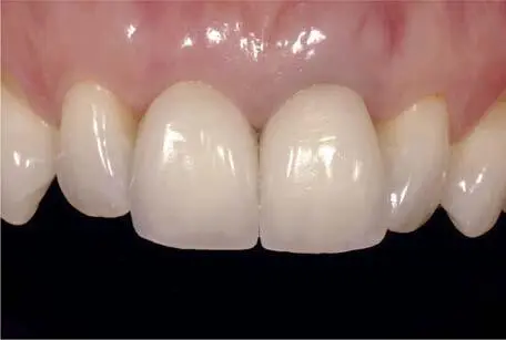
Fig 1-2-4 Clinical case example showing the different outcomes of two zirconia-ceramic crowns on the vital abutment of the maxillary right central incisor, and the non-vital, discolored abutment of the left central incisor (note the grayish shadowing at the gingiva).
With implant-supported restorations, the selection of the material of the prosthetic components exhibits an influence on the soft tissue color. Titanium implant abutments and metal-ceramic implant restorations can lead to dark, grayish peri-implant soft tissue discoloration, as has been shown in several clinical studies 21 , 22(Fig 1-2-5). The thickness of the soft tissues was identified as crucial parameter for the presence or absence of the soft tissue discoloration. A minimal soft tissue thickness of 2 mm was defined as the threshold value for the color influence 22.
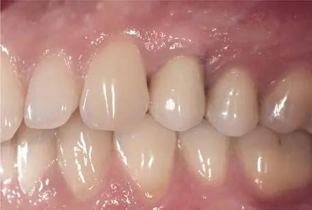
Fig 1-2-5 Grayish discoloration of metal-ceramic implant crown of the maxillary left first premolar, supported by a titanium abutment.
At a soft tissue thickness of >2 mm the material selection and, hence, color of the implant restoration is less critical for the esthetic outcome. Still, in patient cases with thin soft tissues and anterior implant-supported restorations, the use of ceramic abutments and all-ceramic restorations is recommended (Fig 1-2-6).
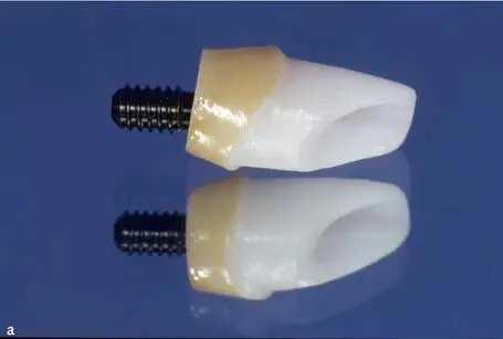
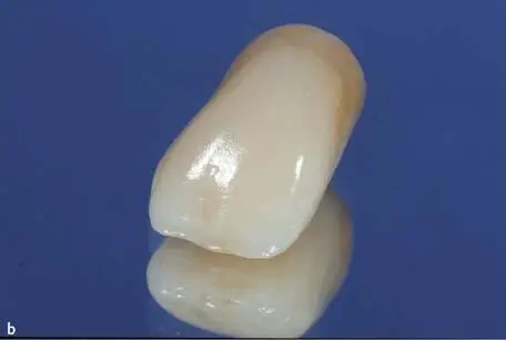
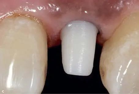
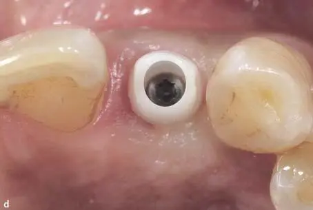
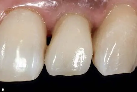
Fig 1-2-6a to 1-2-6e All-ceramic implant single crown supported by a veneered customized zirconia abutment for the replacement of the maxillary left lateral incisor.
Читать дальшеИнтервал:
Закладка:
Похожие книги на «Fixed Restorations»
Представляем Вашему вниманию похожие книги на «Fixed Restorations» списком для выбора. Мы отобрали схожую по названию и смыслу литературу в надежде предоставить читателям больше вариантов отыскать новые, интересные, ещё непрочитанные произведения.


Обсуждение, отзывы о книге «Fixed Restorations» и просто собственные мнения читателей. Оставьте ваши комментарии, напишите, что Вы думаете о произведении, его смысле или главных героях. Укажите что конкретно понравилось, а что нет, и почему Вы так считаете.