Salivary Gland Pathology
Здесь есть возможность читать онлайн «Salivary Gland Pathology» — ознакомительный отрывок электронной книги совершенно бесплатно, а после прочтения отрывка купить полную версию. В некоторых случаях можно слушать аудио, скачать через торрент в формате fb2 и присутствует краткое содержание. Жанр: unrecognised, на английском языке. Описание произведения, (предисловие) а так же отзывы посетителей доступны на портале библиотеки ЛибКат.

- Название:Salivary Gland Pathology
- Автор:
- Жанр:
- Год:неизвестен
- ISBN:нет данных
- Рейтинг книги:3 / 5. Голосов: 1
-
Избранное:Добавить в избранное
- Отзывы:
-
Ваша оценка:
Salivary Gland Pathology: краткое содержание, описание и аннотация
Предлагаем к чтению аннотацию, описание, краткое содержание или предисловие (зависит от того, что написал сам автор книги «Salivary Gland Pathology»). Если вы не нашли необходимую информацию о книге — напишите в комментариях, мы постараемся отыскать её.
now features case presentations to place the information in context, as well as additional treatment algorithms in each chapter to assist in clinical decision making. Written by highly respected clinicians, educators, and researchers with extensive expertise in oral and maxillofacial surgery, this authoritative resource:
Reviews the etiology, diagnosis, and treatment of all salivary gland pathologies, with detailed explanations and hundreds of full-color clinical images Incorporates new information on the taxonomy of salivary gland tumors, neoplastic and non-neoplastic entities, image guided biopsies of salivary gland lesions, and complications in traditional and non-traditional forms of salivary gland surgery Offers expanded coverage of histopathology, including classification, grading, and staging of salivary gland tumors Features up-to-date chapters on anatomy and physiology, imaging, cysts, systemic diseases, tumors, trauma, and innovative salivary gland surgical techniques Includes a discussion of meta-analyses and systematic reviews to support evidence-based practice remains the definitive resource for oral and maxillofacial surgeons, otolaryngologists, head and neck surgeons, and general surgeons as well as their residents providing care for patients with salivary gland pathology.
Salivary Gland Pathology — читать онлайн ознакомительный отрывок
Ниже представлен текст книги, разбитый по страницам. Система сохранения места последней прочитанной страницы, позволяет с удобством читать онлайн бесплатно книгу «Salivary Gland Pathology», без необходимости каждый раз заново искать на чём Вы остановились. Поставьте закладку, и сможете в любой момент перейти на страницу, на которой закончили чтение.
Интервал:
Закладка:
b Signal from calcium deposition is complex. Calcium concentrations of under 30% by weight have high T1 signal and intermediate T2 signal, but over 40% have decreasing signal on T1 and T2. The surface area of the calcium particle also has an effect, with large surface area resulting in increased T1 signal (Henkelman et al. 1991).
c Depends on the protein concentration (complex cysts, abscess).
d MRI signal of intracranial hemorrhage is quite complex and dependent on multiple factors with degrees of variability.
CSF = cerebrospinal fluid.
Gradient recalled echo imaging (GRE)
Gradient recalled echo imaging is the second most common type of imaging sequence after the spin‐echo. This sequence is very susceptible (more than spin‐echo T2) to magnetic field inhomogeneity and is commonly used in the brain to identify blood products, metal deposition such as iron, manganese, and nonmetals such as calcium. This sequence is very sensitive but not specific. The “flip angle” used in obtaining GRE can be altered, resulting in either T1 weighted (long flip angle) or T2 weighted (short flip angle) images ( Figure 2.10).
Short tau inversion recovery (STIR)
Short tau inversion recovery (STIR) is commonly acquired because of its very high sensitivity to fluid and readily detects subtle edema in tissues. When acquired in the conventional method, it also results in nulling the fat signal, thereby further increasing the signal of tissue fluid relative to background. This is the best sequence for edema, particularly when trying to determine bone invasion by tumors. It can also be useful in assessing skull base foramina ( Figures 2.11and 2.12).
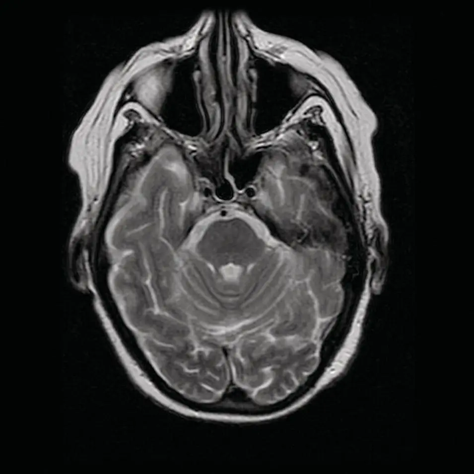
Figure 2.9. Axial MRI FSE T2 weighted image demonstrating the high signal of CSF and subcutaneous fat, intermediate signal of the brain and mucosa, and the low signal in the arteries.
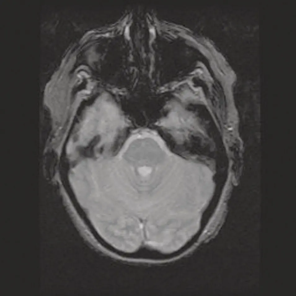
Figure 2.10. Axial MRI GRE image.
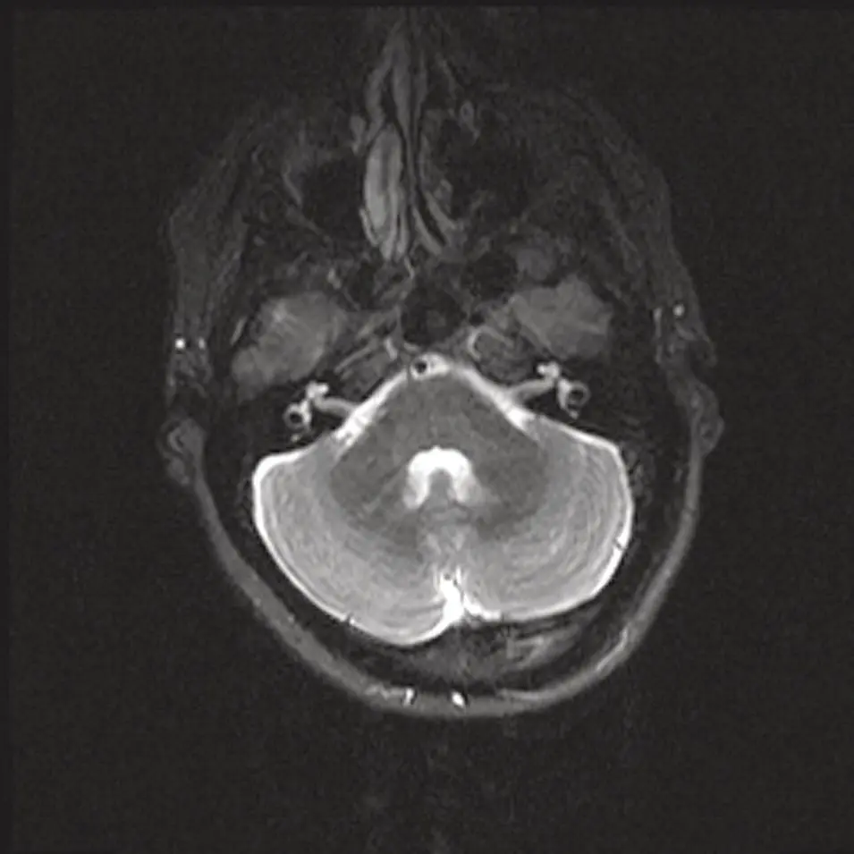
Figure 2.11. Axial MRI STIR image at the skull base demonstrating the high signal of CSF but suppression of subcutaneous fat signal.
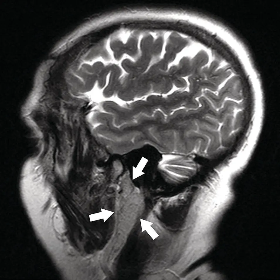
Figure 2.12. Sagittal MRI STIR image at the level of the parotid gland demonstrating the deep lobe seen through the stylomandibular tunnel (arrows). Note the parotid gland extending superiorly to the skull base.
Gadolinium (Gd) contrast
Intravenous contrast with gadolinium, a paramagnetic element, alters (shortens) T1 and T2 relaxation times, which results in a brighter signal. Its effect is greater on T1 than on T2 weighted images. Areas of tissue that accumulate Gd will have a higher or brighter signal and “enhance.” In the head and neck, post‐contrast T1 images should be produced with fat saturation to null the fat signal and therefore increase the signal of Gd accumulation ( Figure 2.13).
Fluid attenuation inversion recovery (FLAIR)
Fluid attenuation inversion recovery (FLAIR) is not as commonly used in the neck but is a necessity in brain imaging. By nulling the CSF signal, brain tissue edema from a variety of causes stands out and is easily identified. It is however not specific. FLAIR can be useful for assessing skull base or foraminal invasion by tumors. However, artifacts can result from CSF pulsation or high FiO 2administration and can mimic pathologic processes such as subarachnoid hemorrhage, or meningitis (bacterial, carcinomatous, viral, or aseptic) ( Figure 2.14).
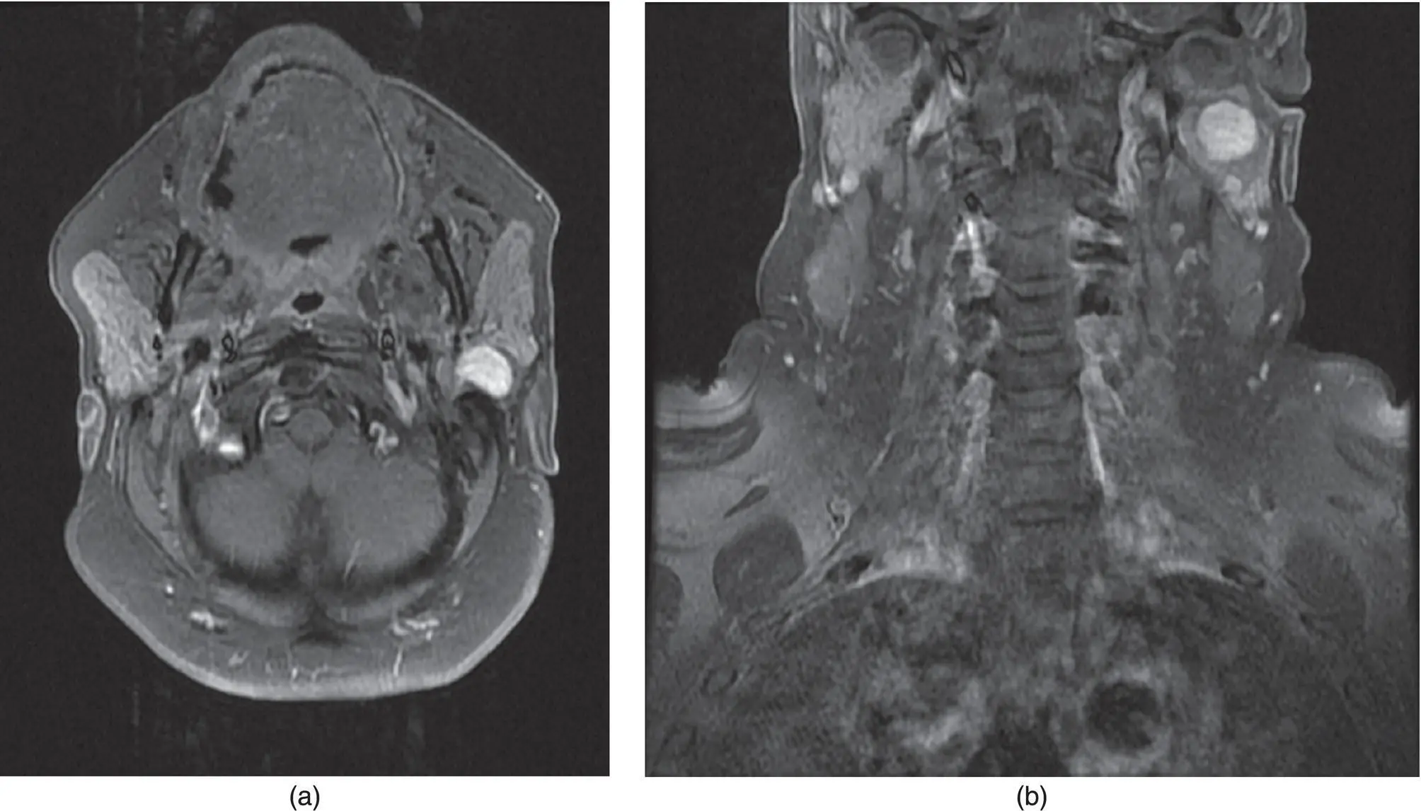
Figure 2.13. Axial (a) and coronal (b) MRI T1 post‐contrast fat saturated image demonstrating a mass in the left parotid gland. Note the mild vascular enhancement and suppression of fat high signal on T1 weighted image.
Diffusion weighted images (DWI)
Diffusion weighted images are not routinely clinically used in the neck or head but are indispensable in the brain. Typical intracranial application is for assessing acute stroke, but can be applied for the assessment of active multiple sclerosis (MS) plaques, and abscesses ( Figure 2.15). The concept of DWI is based on the molecular motion of water and the sensitivity of certain MRI sequences to detect the diffusion or movement of water in tissues at the cellular level.
The use of DWI and specifically apparent diffusion coefficient (ADC) values and maps for salivary gland imaging are under investigation and show promise in differentiating benign from malignant tissues (Shah et al. 2003; Abdel‐Razek et al. 2007; Eida et al. 2007; Habermann et al. 2007). The ADC values are affected by technical factors (b‐value setting, image resolution, choice of region of interest, susceptibility artifacts, and adequate shimming) as well as physiologic factors (biochemical composition of tumors, hemorrhage, perfusion, and salivary flow) (Eida et al. 2007). The ADC values of salivary glands change with gustatory stimulation. Although mixed results have been reported, there is generally an increase in the ADC value from pre‐stimulation to post‐stimulation measurements (Habermann et al. 2007). The normal parotid, submandibular, and sublingual glands have measured ADC values of 0.63 ± 0.11 × 10 −3mm 2/s, 0.97 ± 0.09 × 10 −3mm 2/s, and 0.87 ± 0.05 × 10 −3mm 2/s, respectively (Eida et al. 2007). In pleomorphic adenomas, the ADC maps demonstrate areas of cellular proliferation to have intermediate ADC levels and areas of myxomatous changes to have high ADC values (Eida et al. 2007). Warthin's tumor showed lymphoid tissue to have a very low ADC, necrosis with intermediate ADC, and low ADC in cysts among the lymphoid tissue (Eida et al. 2007). Among the malignant lesions, mucoepidermoid carcinoma shows low ADC in a more homogenous pattern whereas the adenoid cystic carcinomas demonstrated a more speckled pattern with areas of low and high ADC likely from multiple areas of cystic or necrotic change (Eida et al. 2007). Lymphoma in salivary glands has been demonstrated to have a diffuse extremely low ADC likely from the diffuse uniform cellularity of lymphoma (Eida et al. 2007). In general, cystic, necrotic, or myxomatous changes tend to have higher ADC and regions of cellularity, low ADC. Malignant tumors tend to show very low to intermediate ADC whereas benign lesions have higher ADC, but with heterogenous pattern. Overlaps do occur for example with Warthin's tumor demonstrating very low ADC regions and adenoid cystic carcinoma with areas of high ADC (Eida et al. 2007).
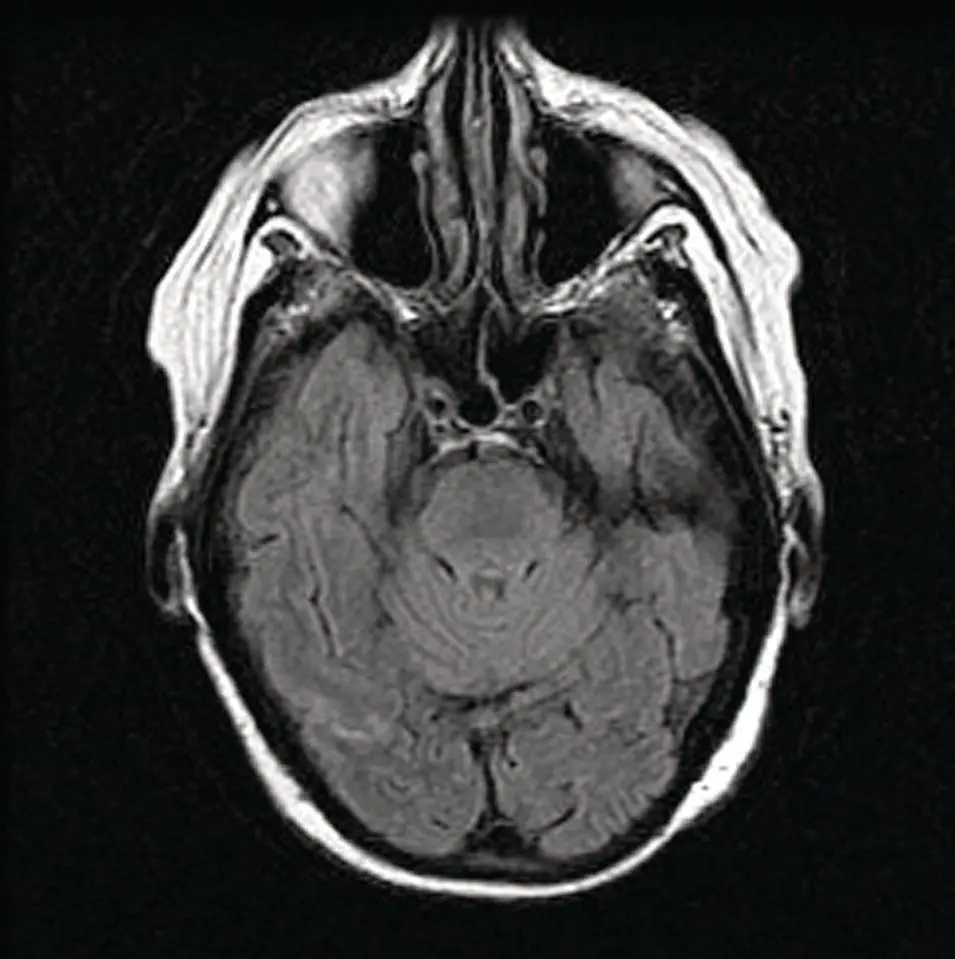
Figure 2.14. Axial MRI FLAIR image at the skull base demonstrating CSF flow‐related artifactual increased signal in the right prepontine cistern.
Читать дальшеИнтервал:
Закладка:
Похожие книги на «Salivary Gland Pathology»
Представляем Вашему вниманию похожие книги на «Salivary Gland Pathology» списком для выбора. Мы отобрали схожую по названию и смыслу литературу в надежде предоставить читателям больше вариантов отыскать новые, интересные, ещё непрочитанные произведения.


Обсуждение, отзывы о книге «Salivary Gland Pathology» и просто собственные мнения читателей. Оставьте ваши комментарии, напишите, что Вы думаете о произведении, его смысле или главных героях. Укажите что конкретно понравилось, а что нет, и почему Вы так считаете.