Salivary Gland Pathology
Здесь есть возможность читать онлайн «Salivary Gland Pathology» — ознакомительный отрывок электронной книги совершенно бесплатно, а после прочтения отрывка купить полную версию. В некоторых случаях можно слушать аудио, скачать через торрент в формате fb2 и присутствует краткое содержание. Жанр: unrecognised, на английском языке. Описание произведения, (предисловие) а так же отзывы посетителей доступны на портале библиотеки ЛибКат.

- Название:Salivary Gland Pathology
- Автор:
- Жанр:
- Год:неизвестен
- ISBN:нет данных
- Рейтинг книги:3 / 5. Голосов: 1
-
Избранное:Добавить в избранное
- Отзывы:
-
Ваша оценка:
Salivary Gland Pathology: краткое содержание, описание и аннотация
Предлагаем к чтению аннотацию, описание, краткое содержание или предисловие (зависит от того, что написал сам автор книги «Salivary Gland Pathology»). Если вы не нашли необходимую информацию о книге — напишите в комментариях, мы постараемся отыскать её.
now features case presentations to place the information in context, as well as additional treatment algorithms in each chapter to assist in clinical decision making. Written by highly respected clinicians, educators, and researchers with extensive expertise in oral and maxillofacial surgery, this authoritative resource:
Reviews the etiology, diagnosis, and treatment of all salivary gland pathologies, with detailed explanations and hundreds of full-color clinical images Incorporates new information on the taxonomy of salivary gland tumors, neoplastic and non-neoplastic entities, image guided biopsies of salivary gland lesions, and complications in traditional and non-traditional forms of salivary gland surgery Offers expanded coverage of histopathology, including classification, grading, and staging of salivary gland tumors Features up-to-date chapters on anatomy and physiology, imaging, cysts, systemic diseases, tumors, trauma, and innovative salivary gland surgical techniques Includes a discussion of meta-analyses and systematic reviews to support evidence-based practice remains the definitive resource for oral and maxillofacial surgeons, otolaryngologists, head and neck surgeons, and general surgeons as well as their residents providing care for patients with salivary gland pathology.
Salivary Gland Pathology — читать онлайн ознакомительный отрывок
Ниже представлен текст книги, разбитый по страницам. Система сохранения места последней прочитанной страницы, позволяет с удобством читать онлайн бесплатно книгу «Salivary Gland Pathology», без необходимости каждый раз заново искать на чём Вы остановились. Поставьте закладку, и сможете в любой момент перейти на страницу, на которой закончили чтение.
Интервал:
Закладка:
The Hounsfield unit (H) (named for Godfrey Hounsfield, inventor of the CT scanner) is the unit of density measurement for CT. These units are assigned based on the degree of attenuation of the X‐ray beam by tissue in a given voxel (volume ele ment) and are assigned relative to water (0H) ( Table 2.1). The scale ranges from −1024H for air, to +4000H for very dense bone. The images are created based on a gray scale from black (−1024 H) to white (+4000 H) and shades of gray. Despite the wide range of units, majority of tissues in the human body are between −100 and +100 H. Soft tissues and parenchymal organs are in a range between 20 and 80 H, whereas fat is approximately −100 H. Simple fluid is 0 H, but proteinaceous fluid can be upward of 25 H. Unclotted and clotted blood varies depending on the hemoglobin concentration and hematocrit but average measurements are 50 and 80 H, respectively. CT images are displayed using a combination of “window widths” (WW, range of CT numbers from black to white), and “window levels” (WL, position of the window on the scale), which are based on the attenuation characteristics of tissues. Typically, head and neck images are interpreted using “soft‐tissue windows” (WW 500 H, WL 30 H), “bone windows” (WW 2000, WL 500), or “lung windows” (WW 1500, WL 500). The “soft‐tissue windows” demonstrate the slight density differences of soft tissues, whereas “bone windows” demonstrated cortical and medullary features of bones with sharp detail. “Lung windows” demonstrate the sharp interface of air and the fine soft‐tissue components of lung parenchyma.
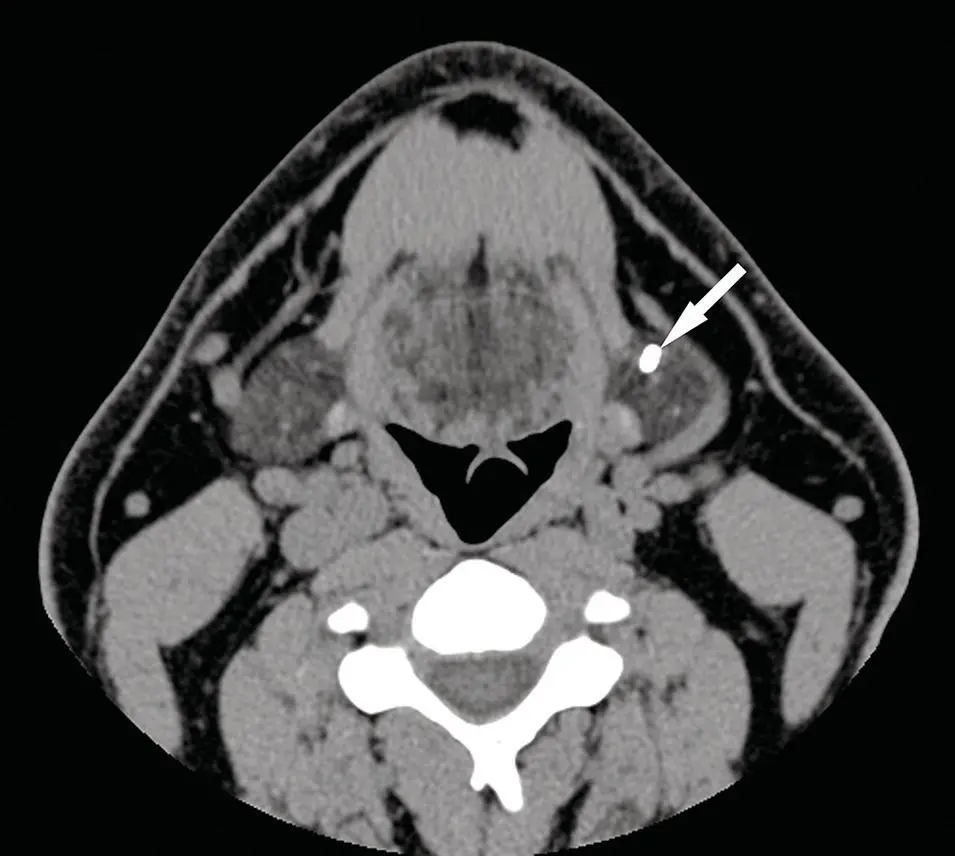
Figure 2.1. Axial CT of the neck in soft‐tissue window without contrast demonstrating poor definition between soft‐tissue structures. The blood vessels are unopacified and cannot be easily distinguished from lymph nodes. Note the sialolith (arrow) in the hilum of the left submandibular gland.
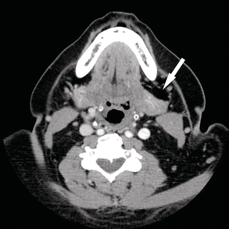
Figure 2.2. Axial CT of the neck in soft‐tissue window with IV contrast demonstrates improved visualization of structures with enhancement of tissues and vasculature. Note the small lipoma (arrow) anterior to the left submandibular gland, which distorts the anterior aspect of the gland with slight posterior displacement.
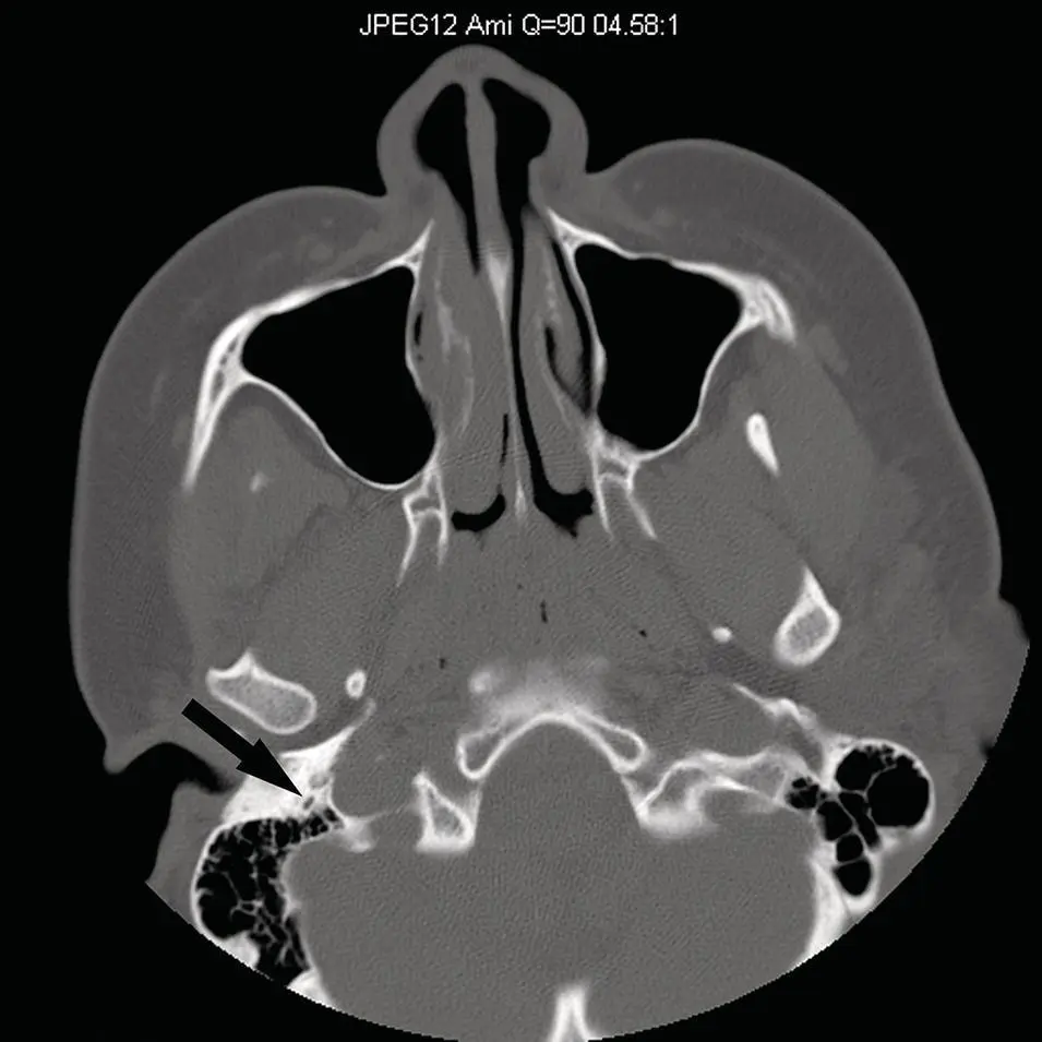
Figure 2.3. Axial CT of the skull base reconstructed in a sharp algorithm and in bone window and level display demonstrating sharp bone detail. Note the sharply defined normal right stylomastoid foramen (arrow).
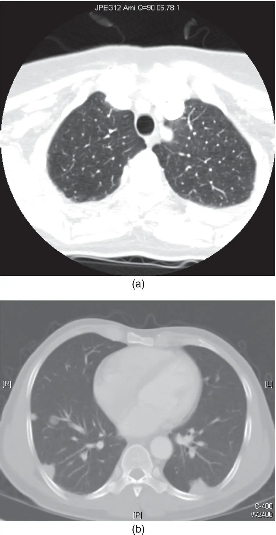
Figure 2.4. Axial CT of the neck at the thoracic inlet in lung windows demonstrating lung parenchyma (a). Axial image of dedicated CT of chest demonstrating cannon ball lesions in a patient previously treated for adenoid cystic carcinoma of the palate (b). These lesions are representative of diffuse metastatic disease of the lungs, but not pathognomonic of adenoid cystic carcinoma.
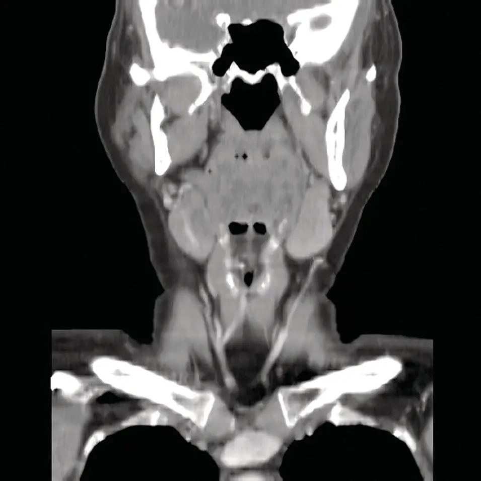
Figure 2.5. Coronal CT reformation of the neck in soft‐tissue window at the level of the submandibular glands. Orthogonal images with MDCT offer very good soft‐tissue detail in virtually any plane of interest to assess anatomic and pathologic relationships.
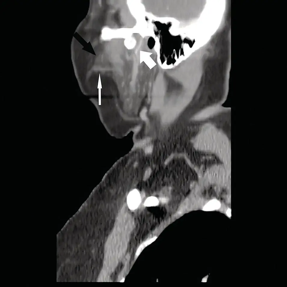
Figure 2.6. Sagittal CT reformation of the neck in soft‐tissue window at the level of the parotid gland. Note the accessory parotid gland (black arrow) sitting atop the parotid (Stensen) duct (thin white arrow). Also, note the retromandibular vein (large white arrow) and external auditory canal.
Table 2.1. CT density in Hounsfield units (H).
| Tissue or structure | Hounsfield unit (H) |
|---|---|
| Water or CSF | 0 |
| Fat | −30 to −100 |
| Soft tissue, muscle a | 50–60 |
| Unclotted blood b | 35–50 |
| Clotted blood b | 50–75 |
| Parotid gland c | −10 to +30 |
| Submandibular gland c | 30–60 |
| Sublingual gland d | 60–90 |
| Bone | 1000 |
| Lung | −850 |
| Air | −1000 |
| Calcification | 150–200 |
| Gray matter | 35–40 |
| White matter | 25–35 |
aDepends on degree of fat deposition.
bDepends on the hemoglobin concentration and hematocrit.
cDepends on age and fat deposition.
dVery limited evaluation secondary to partial volume effect.
CSF = cerebrospinal fluid.
Although the density of the salivary glands is variable, the parotid glands tend to be slightly lower in density relative to muscle, secondary to a higher fat content and become progressively more fat replaced over time. The CT density of parotid glands varies from −10 to +30 H. The submandibular glands are denser than parotid glands and are equivalent in density to muscle. The submandibular glands vary in density from +30 to +60 H.
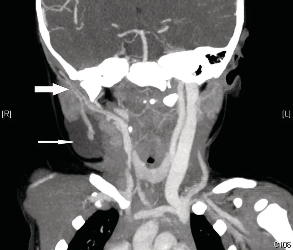
Figure 2.7. CT angiogram (CTA) of the neck at the level of the parotid gland demonstrating the retromandibular vein and adjacent external carotid artery (large white arrow). Note the right cervical lymphangioma (thin white arrow) associated with the tail of the right parotid gland.
CT angiography (CTA) is a powerful method, which allows visualization of arterial vasculature, demonstrating the vascular anatomy of arteries and veins. CTA can be critical in preoperative evaluation to determine the degree of vascularity of lesions and plan an appropriate surgical approach to minimize blood loss or perform preoperative embolization. CTA is obtained with fast image acquisition over a defined region of interest while administering a rapid IV contrast bolus timed to arrive in the region of interest during image acquisition. CTA images may be rendered in 3D data sets and rotated in any plane ( Figure 2.7). CTA is not only useful for preoperative planning, but it can also be quite useful in diagnosis of salivary gland vascular pathology such as aneurysms, or arteriovenous fistulas (AVFs) (Wong et al. 2004).
CT scanning, as with all imaging modalities, is prone to artifacts. Artifacts can be caused by motion, very dense or metallic implants (dental amalgam), and volume averaging. Motion artifact is common and may result from breathing, swallowing, coughing, or sneezing during the image acquisition or from an unaware or uncooperative patient. Metallic implants cause complete attenuation of X‐rays in the beam and result in focal loss of data and bright and dark steaks in the image. Because the image is created from a three‐dimensional section of tissue averaged to form a two‐dimensional image, the partial volume or volume averaging artifact results from partial inclusion of structures in adjacent images. Finally, the beam hardening artifact is produced by attenuation of low‐energy X‐rays, by dense objects, from the energy spectrum of the X‐ray beam, resulting in a residual average high‐energy beam (or hard X‐rays), which results in loss of data and dark lines on the image. This phenomenon is often seen in the posterior fossa of head CT scans caused by the very dense petrous bones. Multi‐detector row CT scanner can help reduce metallic artifacts using advanced algorithms, and reduce motion artifacts secondary to faster scanning speeds.
Читать дальшеИнтервал:
Закладка:
Похожие книги на «Salivary Gland Pathology»
Представляем Вашему вниманию похожие книги на «Salivary Gland Pathology» списком для выбора. Мы отобрали схожую по названию и смыслу литературу в надежде предоставить читателям больше вариантов отыскать новые, интересные, ещё непрочитанные произведения.


Обсуждение, отзывы о книге «Salivary Gland Pathology» и просто собственные мнения читателей. Оставьте ваши комментарии, напишите, что Вы думаете о произведении, его смысле или главных героях. Укажите что конкретно понравилось, а что нет, и почему Вы так считаете.