Interpretation Basics of Cone Beam Computed Tomography
Здесь есть возможность читать онлайн «Interpretation Basics of Cone Beam Computed Tomography» — ознакомительный отрывок электронной книги совершенно бесплатно, а после прочтения отрывка купить полную версию. В некоторых случаях можно слушать аудио, скачать через торрент в формате fb2 и присутствует краткое содержание. Жанр: unrecognised, на английском языке. Описание произведения, (предисловие) а так же отзывы посетителей доступны на портале библиотеки ЛибКат.
- Название:Interpretation Basics of Cone Beam Computed Tomography
- Автор:
- Жанр:
- Год:неизвестен
- ISBN:нет данных
- Рейтинг книги:4 / 5. Голосов: 1
-
Избранное:Добавить в избранное
- Отзывы:
-
Ваша оценка:
Interpretation Basics of Cone Beam Computed Tomography: краткое содержание, описание и аннотация
Предлагаем к чтению аннотацию, описание, краткое содержание или предисловие (зависит от того, что написал сам автор книги «Interpretation Basics of Cone Beam Computed Tomography»). Если вы не нашли необходимую информацию о книге — напишите в комментариях, мы постараемся отыскать её.
Enables rapid reference to common CBCT findings, with multiple images for each finding Features a streamlined framework that makes relevant information easier to find and apply in dental practice Offers hundreds of new images to aid in correctly identifying findings Contains new and updated content, including expanded coverage of CBCT and implants Provides sample reports and explains how they are used in day-to-day clinical practice
remains a must-have resource for all dental practitioner and specialists who use CBCT, dental students in radiology interpretation courses, and residents beginning to use CBCT in their specialty.
Interpretation Basics of Cone Beam Computed Tomography — читать онлайн ознакомительный отрывок
Ниже представлен текст книги, разбитый по страницам. Система сохранения места последней прочитанной страницы, позволяет с удобством читать онлайн бесплатно книгу «Interpretation Basics of Cone Beam Computed Tomography», без необходимости каждый раз заново искать на чём Вы остановились. Поставьте закладку, и сможете в любой момент перейти на страницу, на которой закончили чтение.
Интервал:
Закладка:
Periodontics
The American Academy of Periodontology (AAP) created a set of questions regarding CBCT use based on published research in 2017. They asked three main questions when reviewing CBCT research, covering goals, benefits, potential risks, and the bottom line.
Implants
“Should CBCT imaging replace two‐dimensional (2D) radiographic analysis of regional anatomy in the surgical management of patients requiring dental implants?”
Goals
Diagnosis and treatment outcome assessment.
Implant treatment planning.
Anatomic characterization.
Benefits
CBCT helped to identify incidental findings that may influence treatment including but not limited to anatomic variants, pathologies and fractures. CBCT supports minimally invasive therapy for dental implants and provides a method to educate patients.
Quantity of bone, alveolar ridge morphology, maxillary sinus location, and mandibular canal location are important information prior to placing an implant. Standard intraoral radiographs provide the height of bone available but do not show whether there are ridge defects or concavities ( Figures 2.17and 2.18). CBCT imaging provides information on these things to ensure implant placement within the bone and not surrounding soft tissues. Cross‐sectional views are recommended to view the facial‐lingual width and morphology of the alveolar ridge. The recommended voxel size is 0.3 mm to reduce the overall radiation exposure.
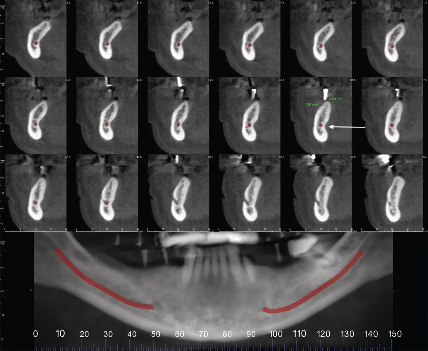
Figure 2.17. Reconstructed pantomograph and cross‐sectional slices showing lingual concavity in posterior mandible (white arrow) and mandibular canal noted in red.
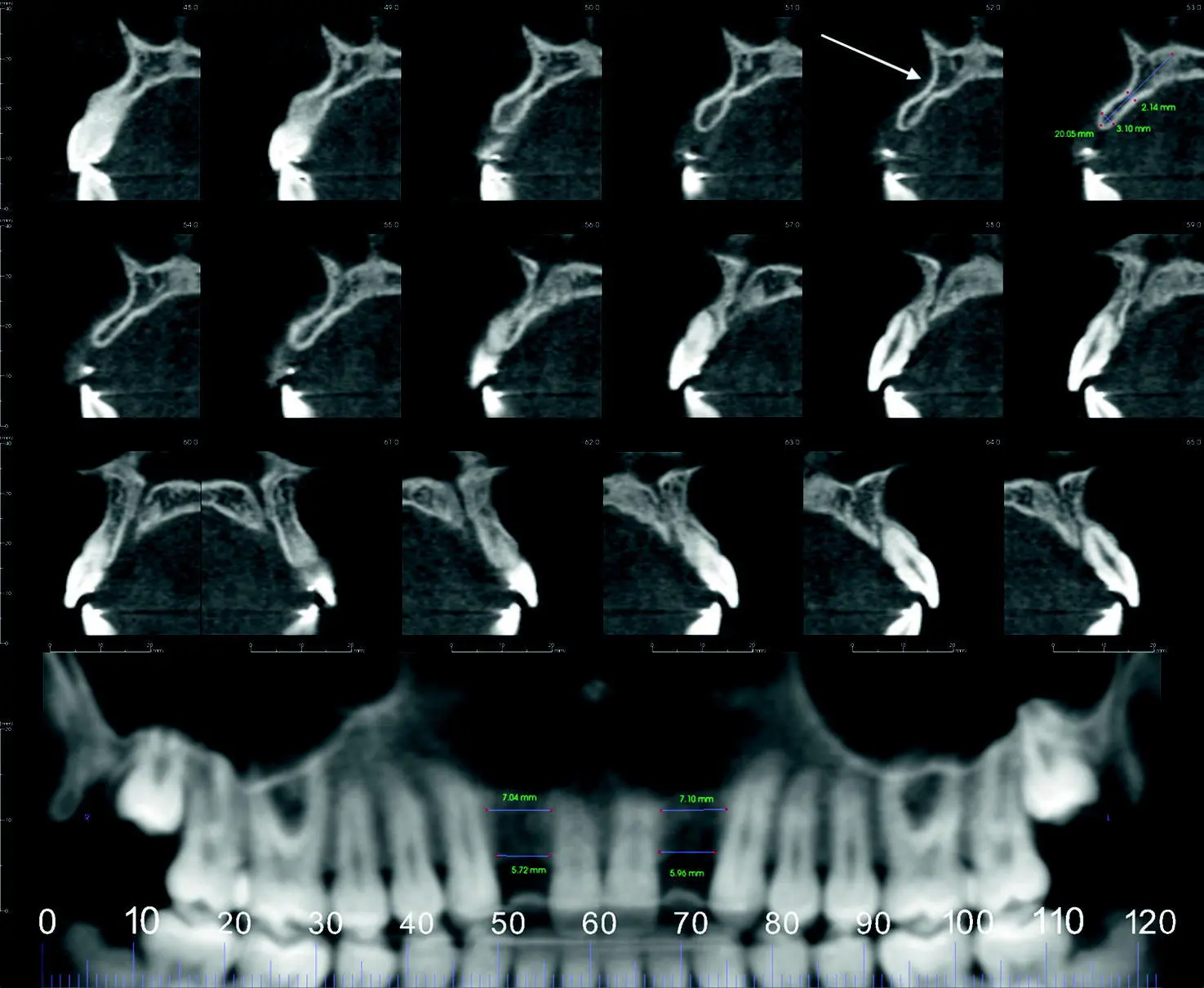
Figure 2.18. Reconstructed pantomograph and cross‐sectional slices showing facial concavity in anterior maxilla (white arrow).
Potential Risks
Long‐term radiation hazards of effective dose accumulation are still unknown, so the practitioner should minimize exposure when possible.
Bottom Line
CBCT should be used as an adjunct to 2D imaging when the benefits outweigh the risks.
Tooth Movement
“Is CBCT imaging useful in determining risk to periodontal structures in patients requiring tooth movement?”
Goals
Evaluate changes in alveolar bone thickness and height around natural teeth.
Benefits
CBCT aids in identifying patients undergoing orthodontic treatment who are at risk for alveolar bone/soft‐tissue deficiencies.
Potential Risks
Long‐term radiation hazards of effective dose accumulation are still unknown, so the practitioner should minimize exposure when possible.
Bottom Line
CBCT can assist in planning orthodontic therapy and aid in identifying those with thin bone.
Periodontitis
“Does CBCT imaging add clinical value in diagnostic assessment and treatment planning for the management of periodontitis?”
Benefits
Current evidence does not support routine use of CBCT in managing periodontitis.
Potential Risks
Long‐term radiation hazards of effective dose accumulation are still unknown, so the practitioner should minimize exposure when possible.
Bottom Line
CBCT provides little benefit managing periodontal disease.
References
Endodontics
1 AAE and AAOMR Joint Position Statement; use of cone beam computed tomography in endodontics—2015/2016 update. ( https://www.aae.org/specialty/clinical‐resources/guidelines‐position‐statements/).
2 Chakravarthy, P. V. K., Telang, L. A., Nerali, J., et al. (2012). Cracked tooth: A report of two cases and role of cone beam computed tomography in diagnosis. Case Reports in Dentistry, 2012, 525364.
3 Durack, C., and Patel, S. (2012). Cone beam computed tomography in endodontics. Braz Dent J, 23 (3), 179–91.
4 Joint Position Paper AAE and AAOMR; use of cone‐beam computed tomography in endodontics ( http://c.ymcdn.com/sites/www.aaomr.org/resource/resmgr/Docs/AAOMR‐AAE_postition_paper_CB.pdf).
Orthodontics
1 Clinical recommendations regarding use of cone beam computed tomography in orthodontics. Position statement by the American Academy of Oral and Maxillofacial Radiology. ( https://www.aaomr.org/assets/Journal_Publications/Position_Papers/1.%20clinical%20recommendations%20%20regarding%20use%20of%20cbct%20in%20orthodontics.%20position%20statement%20by%20the%20american%20%20academy%20of%20oral%20and%20maxillofaci.pdf).
2 Fisher, J. (2015). Take only CBCTs on these types of orthodontic cases. New Advances in Digital Dentistry, 1, 1.
3 Kapila, S., Conley, R. S., Harrell Jr, W. E. (2011). The current status of cone beam computed tomography imaging in orthodontics. Dentomaxillofac Radiol, 40 (1), 24–34.
4 Rossini, G., Cavallini, C., Cassetta, M., et al. (2012). Localization of impacted maxillary canines using cone beam computed tomography. Review of the literature. Ann Stomatol (Roma), 3 (1), 14–8.
Periodontics
1 Mandelaris, G. A., Scheyer, E. T., Evans, M., et. al. (2017). American Academy of Periodontology best evidence consensus statement on selected oral applications for cone‐beam computed tomography. J Periodontol, 88, 939–45.
2 Quereshy, F. A., Barnum, G., Demko, C., et al. (2012). Use of cone beam computed tomography to volumetrically assess alveolar cleft defects—preliminary results. J Oral Maxillofac Surg, 70 (1), 188–91.
3 Tsai, P., Torabinejad, M., Rice, D., et al. (2012). Accuracy of cone‐beam computed tomography and periapical radiography in detecting small periapical lesions. J Endod, 38, 965–70.
Конец ознакомительного фрагмента.
Текст предоставлен ООО «ЛитРес».
Прочитайте эту книгу целиком, купив полную легальную версию на ЛитРес.
Безопасно оплатить книгу можно банковской картой Visa, MasterCard, Maestro, со счета мобильного телефона, с платежного терминала, в салоне МТС или Связной, через PayPal, WebMoney, Яндекс.Деньги, QIWI Кошелек, бонусными картами или другим удобным Вам способом.
Интервал:
Закладка:
Похожие книги на «Interpretation Basics of Cone Beam Computed Tomography»
Представляем Вашему вниманию похожие книги на «Interpretation Basics of Cone Beam Computed Tomography» списком для выбора. Мы отобрали схожую по названию и смыслу литературу в надежде предоставить читателям больше вариантов отыскать новые, интересные, ещё непрочитанные произведения.


Обсуждение, отзывы о книге «Interpretation Basics of Cone Beam Computed Tomography» и просто собственные мнения читателей. Оставьте ваши комментарии, напишите, что Вы думаете о произведении, его смысле или главных героях. Укажите что конкретно понравилось, а что нет, и почему Вы так считаете.