Interpretation Basics of Cone Beam Computed Tomography
Здесь есть возможность читать онлайн «Interpretation Basics of Cone Beam Computed Tomography» — ознакомительный отрывок электронной книги совершенно бесплатно, а после прочтения отрывка купить полную версию. В некоторых случаях можно слушать аудио, скачать через торрент в формате fb2 и присутствует краткое содержание. Жанр: unrecognised, на английском языке. Описание произведения, (предисловие) а так же отзывы посетителей доступны на портале библиотеки ЛибКат.
- Название:Interpretation Basics of Cone Beam Computed Tomography
- Автор:
- Жанр:
- Год:неизвестен
- ISBN:нет данных
- Рейтинг книги:4 / 5. Голосов: 1
-
Избранное:Добавить в избранное
- Отзывы:
-
Ваша оценка:
Interpretation Basics of Cone Beam Computed Tomography: краткое содержание, описание и аннотация
Предлагаем к чтению аннотацию, описание, краткое содержание или предисловие (зависит от того, что написал сам автор книги «Interpretation Basics of Cone Beam Computed Tomography»). Если вы не нашли необходимую информацию о книге — напишите в комментариях, мы постараемся отыскать её.
Enables rapid reference to common CBCT findings, with multiple images for each finding Features a streamlined framework that makes relevant information easier to find and apply in dental practice Offers hundreds of new images to aid in correctly identifying findings Contains new and updated content, including expanded coverage of CBCT and implants Provides sample reports and explains how they are used in day-to-day clinical practice
remains a must-have resource for all dental practitioner and specialists who use CBCT, dental students in radiology interpretation courses, and residents beginning to use CBCT in their specialty.
Interpretation Basics of Cone Beam Computed Tomography — читать онлайн ознакомительный отрывок
Ниже представлен текст книги, разбитый по страницам. Система сохранения места последней прочитанной страницы, позволяет с удобством читать онлайн бесплатно книгу «Interpretation Basics of Cone Beam Computed Tomography», без необходимости каждый раз заново искать на чём Вы остановились. Поставьте закладку, и сможете в любой момент перейти на страницу, на которой закончили чтение.
Интервал:
Закладка:
The appendix shows example written reports of CBCT scans to view and consider when writing radiology interpretations.
Acknowledgments
I’d like to thank Gayle Reardon for her contributions to this book and sharing her knowledge of the paranasal sinuses, nasal cavity and airway, temporomandibular joints, and implants. Thanks to the staff at Wiley for guidance and support in the creation of this book. Last, a giant thanks to my family (Max, Max, and Rugen) for their love, support, and encouragement throughout this entire process. I would not have been able to complete this without them.
About the Companion Website
Do not forget to visit the companion website for this book:
www.wiley.com/go/gonzalez/interpretation 
Sample cases with videos
Scan this QR code to visit the companion website:

1 Introduction to Cone Beam Computed Tomography
Shawneen M. Gonzalez
Introduction
This chapter covers basics of cone beam computed tomography including comparison to traditional computed tomography, artifacts frequently seen, and views created with a cone beam computed tomography dataset.
Conventional Computed Tomography
General Information
Computed tomography (CT) is credited to Godfrey Hounsfield, who in 1967 wrote about the technology and then created a unit in 1972. He was awarded the Nobel Prize in Physiology/Medicine in 1979. Conventional CT units are both hard‐tissue and soft‐tissue imaging modalities. The first CT, first generation, had a scan time of 10+ minutes depending on how much of the body was being imaged. The processing time would take 2½ hours or longer. All first‐generation CT units were only a single slice. This means that one fan of radiation exposed the patient and would have to circle around the patient several times to cover the area of concern. Current CT units are fifth generation, or helical/spiral. The scan times have gone down to 20–60 seconds with a processing time of 2–20 minutes. The number of slices available is up to 64, 128, 256, and 512+. The more slices available makes it possible to scan more of the patient in one circle, hence the shorter scan times. Conventional CT units work with the patient lying down on a table while being scanned. The table moves in and out of the bore to cover the area of concern. Once all the data are received, they are compiled to create a dataset. This dataset can be manipulated to look at the information in many different angles.
Cone Beam Computed Tomography
General Information
Cone beam computed tomography (CBCT) was developed in Italy in 1997. The first unit created was the NewTom. The NewTom was similar to conventional CT in having the patient lying down with an open bore where the radiation exposes the patient. Instead of a fan of radiation (used in conventional CT units), a cone of radiation is used to expose the patient, hence the name cone beam computed tomography. As new CBCT units were created, companies started using seated or standing options. With continued updates to the units, the sizes have become smaller, with many needing only as much space as a pantomograph machine.
Conventional CT versus Cone Beam CT
Voxels
Voxels are 3D data blocks representing a specific x‐ray absorption. CBCT units capture isotropic voxels. An isotropic voxel is equal in all three dimensions (x, y, and z planes), producing higher‐resolution images compared to conventional CT units. Conventional CT unit voxels are non‐isotropic with two sides equal, but the third side (z‐plane) different. The voxel sizes currently available in CBCT units range from 0.076 mm to 0.6 mm. The voxel sizes currently available in conventional CT units range from 1.25 mm to 5.0 mm. Resolution of the final image is determined by the unit’s voxel size. The smaller the voxel size the higher the resolution.
Field of View
Field of view (FOV) is the area of the patient captured. CBCT units vary in FOV options, with some units having a fixed FOV and some having variable FOVs. The ranges of FOVs are from 5 cm × 3.8 cm, commonly referred to as a small/quadrant FOV, to 23 cm × 26 cm, commonly referred to as a large FOV ( Figures 1.1– 1.6).
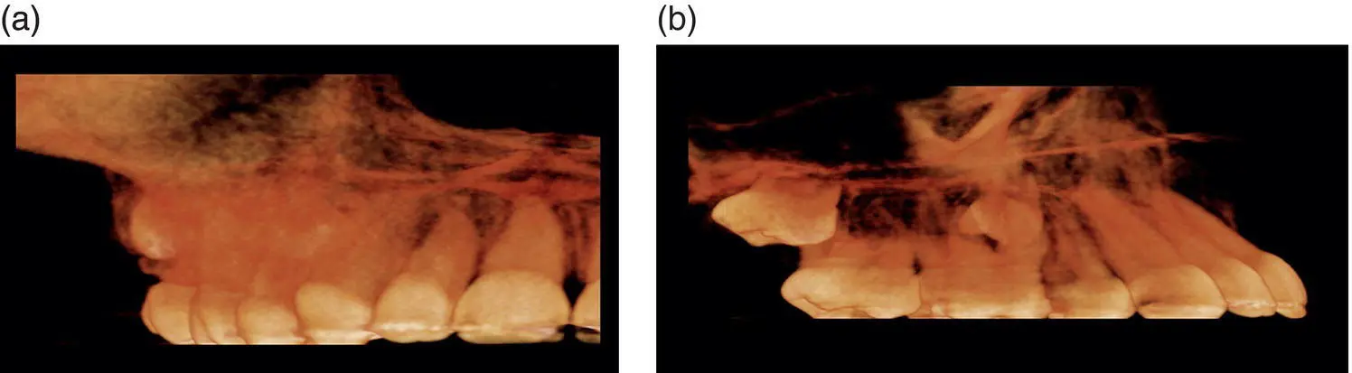
Figure 1.1. (a) 3D rendering of a small FOV of 5 cm × 8 cm from an anteroposterior (AP) view. (b) 3D rendering of a small FOV of 5 cm × 8 cm from a lateral view.
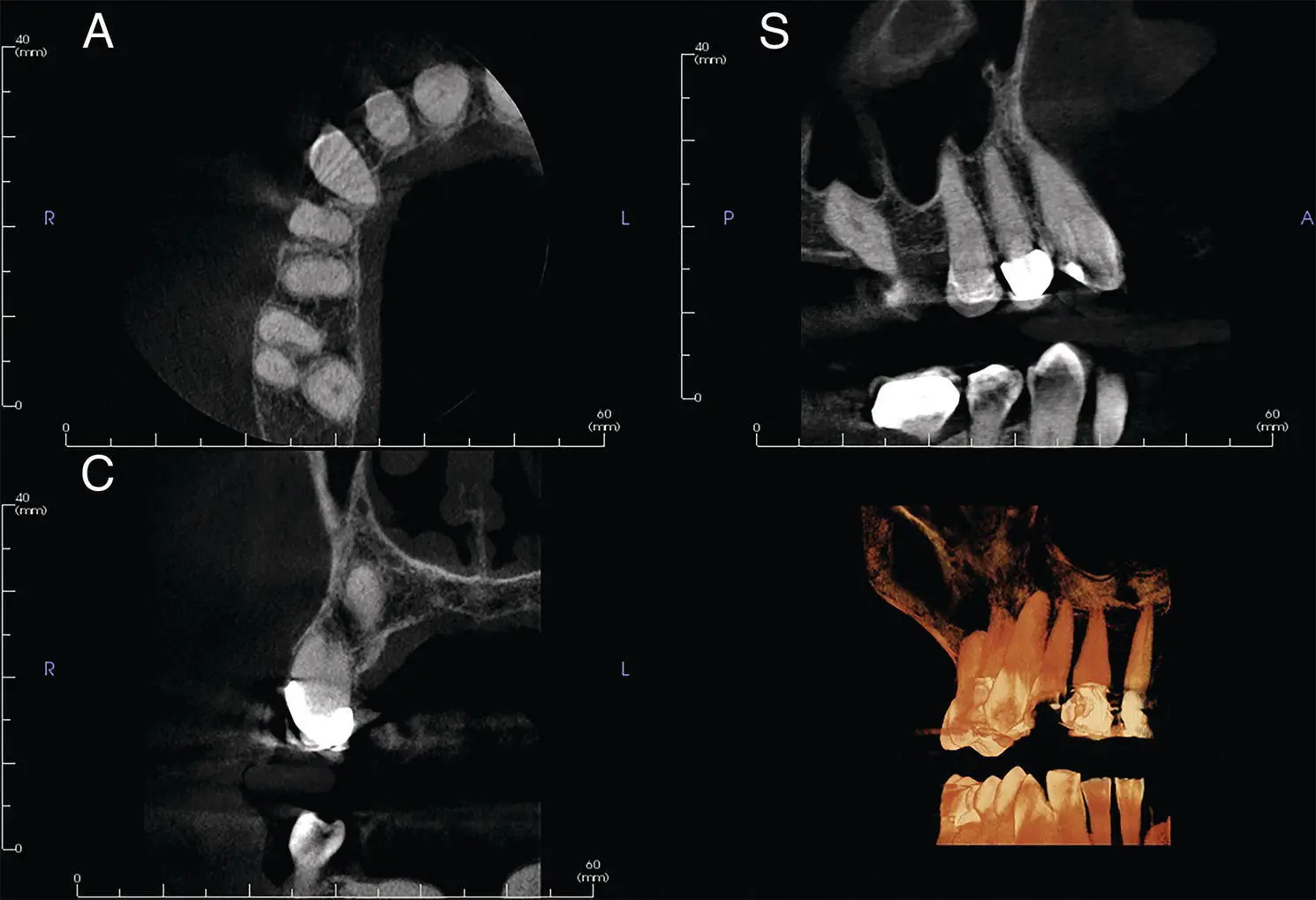
Figure 1.2.Axial (A), coronal (C), sagittal (S), and reconstructed 3D views from a small FOV.
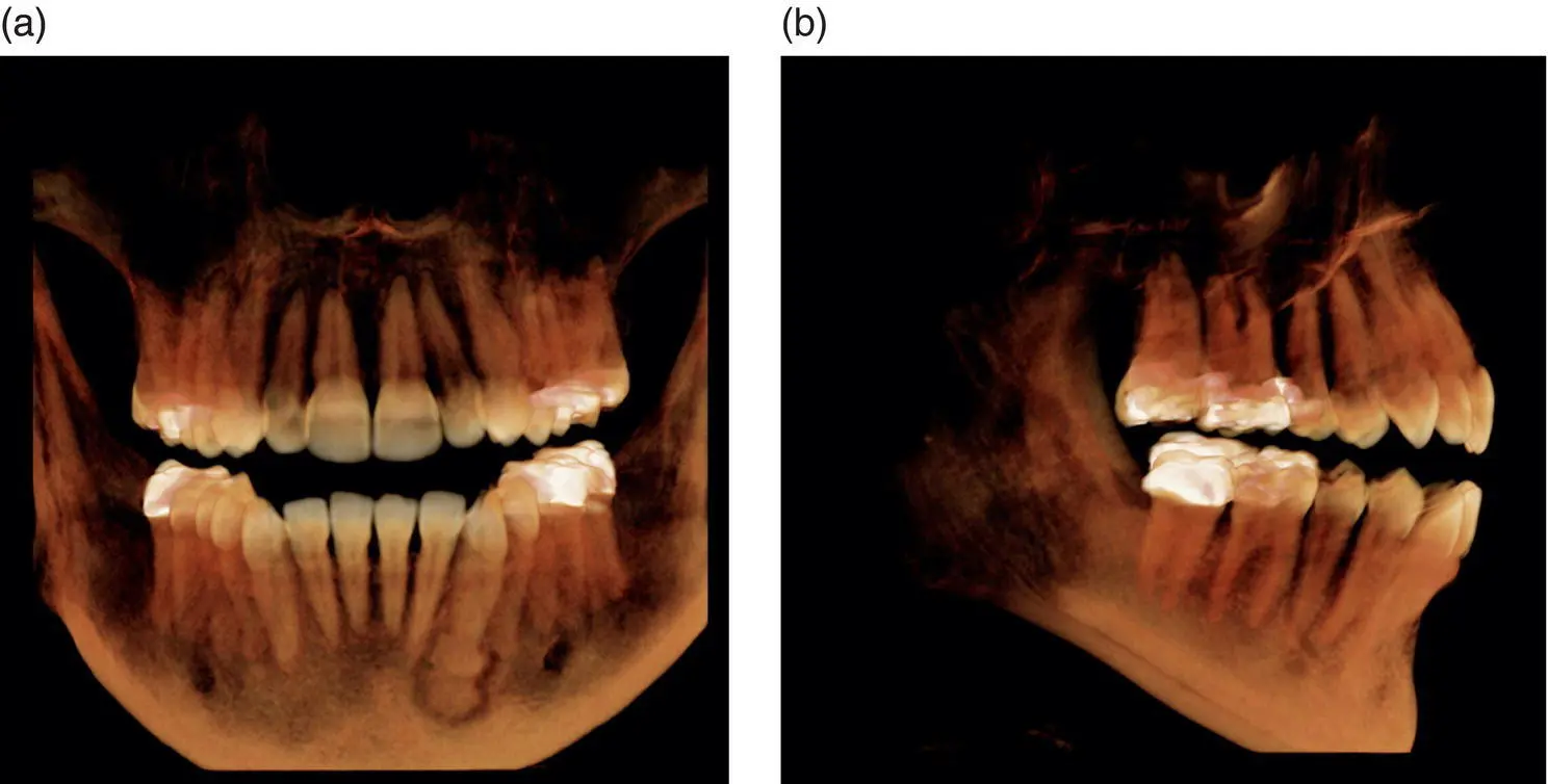
Figure 1.3.(a) 3D rendering of a medium FOV of 8 cm × 8 cm from an anteroposterior (AP) view. (b) 3D rendering of a medium FOV of 8 cm × 8 cm from a lateral view.
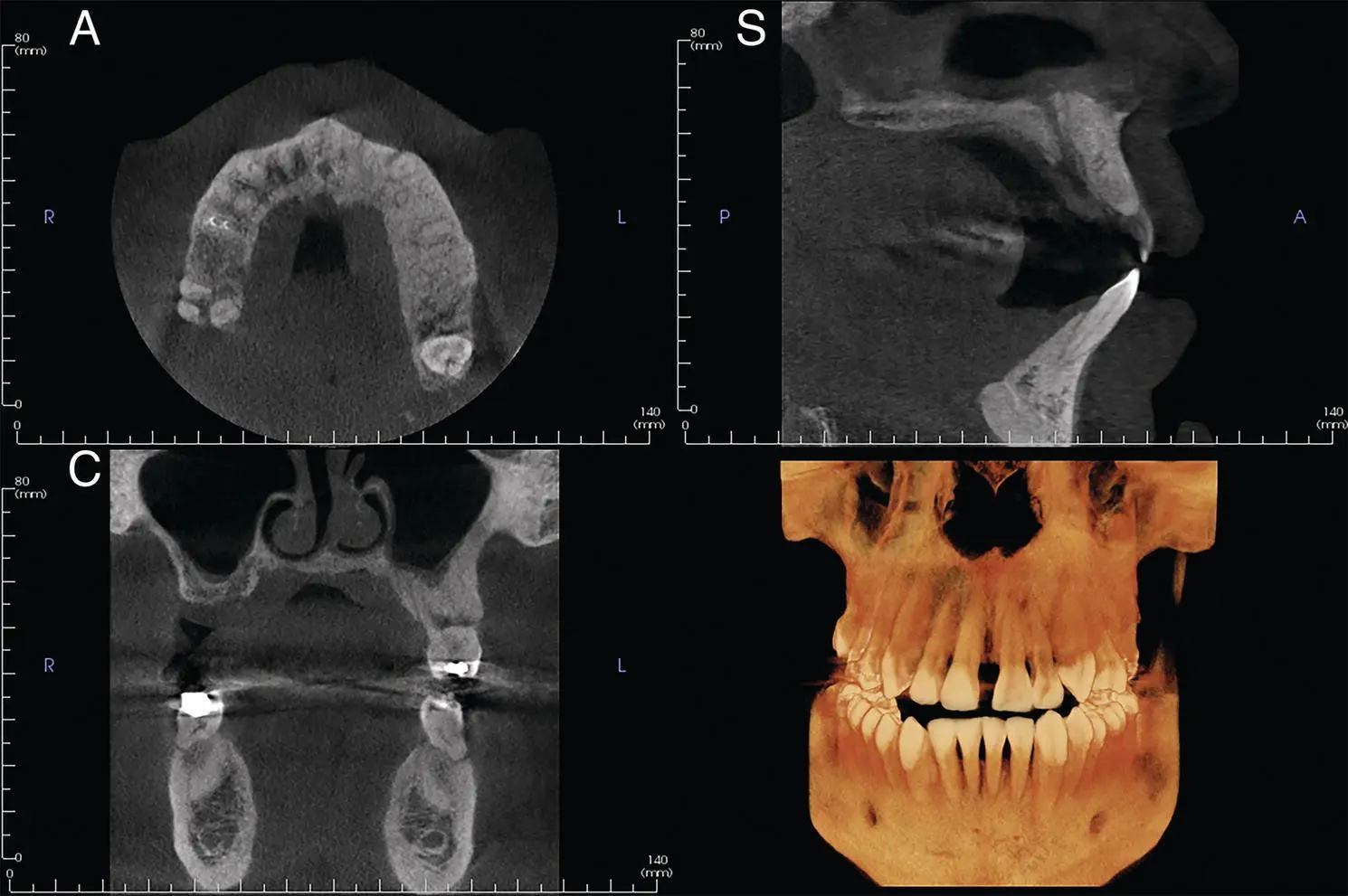
Figure 1.4.Axial (A), coronal (C), sagittal (S), and reconstructed 3D views from medium FOV.
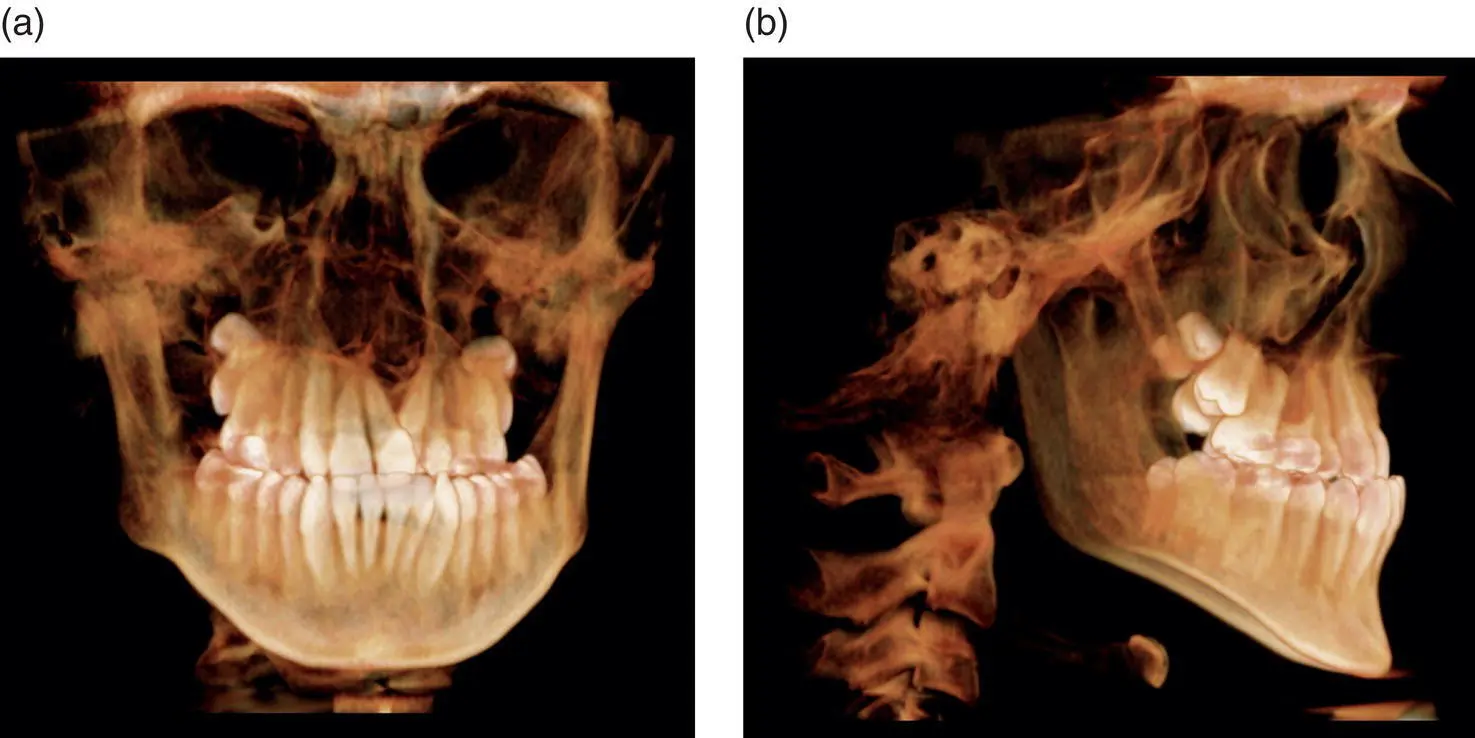
Figure 1.5.(a) 3D rendering of a large FOV of 16 cm × 16 cm from an anteroposterior (AP) view. (b) 3D rendering of a large FOV of 16 cm × 16 cm from a lateral view.
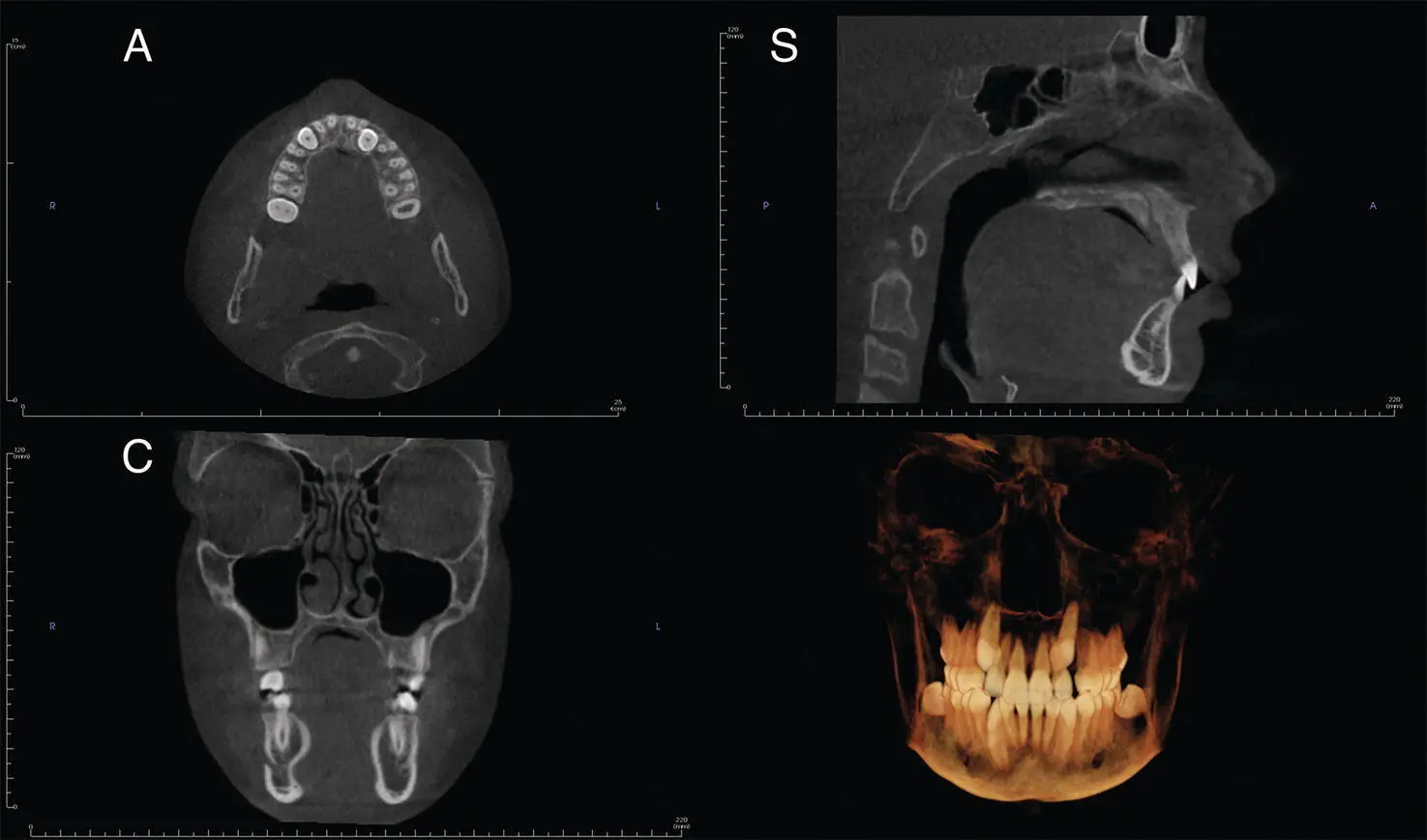
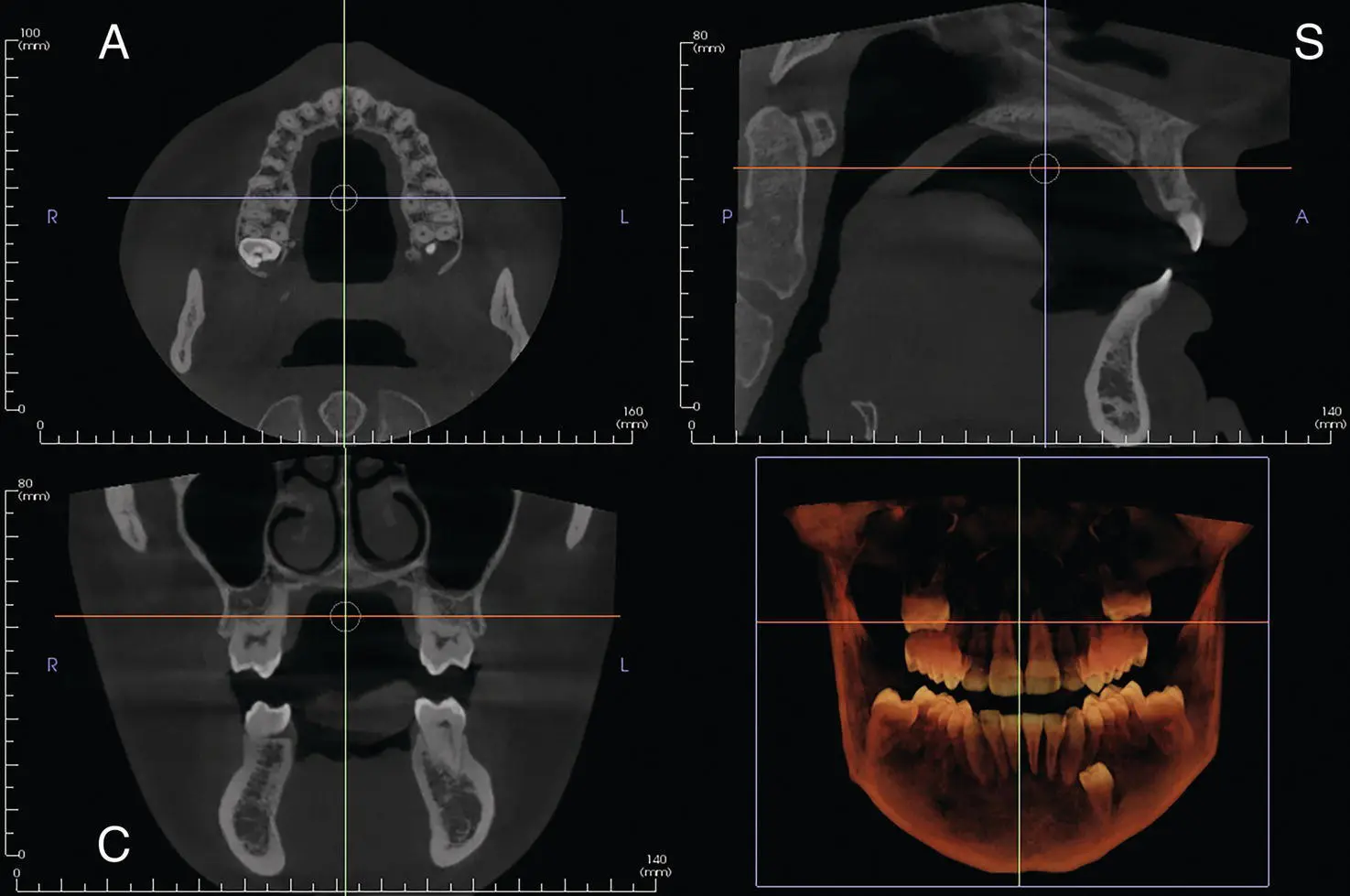
Figure 1.6. Axial (A), coronal (C), sagittal (S), and reconstructed 3D views from large FOV.
Radiation Doses
Radiation doses with CBCT units are as varied as the FOV options. CBCT units have approximate radiation dose ranges of 5 microSieverts to 1073 microSieverts. Conventional CT units have much higher radiation doses due to their soft tissue capabilities, with doses of 1200 microSieverts and higher per each scan, depending on the selected scan field.
Viewing CBCT Data
Multiplanar Reformation
Multiplanar reformation, or MPR, is a view of three different directional 2D images (axial, coronal, and sagittal planes) ( Figure 1.7). Within this view, the images may be manipulated in the thickness of data, and direction of viewing can be altered. Reconstructed pantomographs and lateral cephalometric skulls ( Figures 1.8and 1.9) are possible without distortion from standard 2D radiography. The dataset may also be manipulated to create cross‐sectional (orthogonal) views of the jaws and condyles ( Figures 1.10and 1.11).
Интервал:
Закладка:
Похожие книги на «Interpretation Basics of Cone Beam Computed Tomography»
Представляем Вашему вниманию похожие книги на «Interpretation Basics of Cone Beam Computed Tomography» списком для выбора. Мы отобрали схожую по названию и смыслу литературу в надежде предоставить читателям больше вариантов отыскать новые, интересные, ещё непрочитанные произведения.


Обсуждение, отзывы о книге «Interpretation Basics of Cone Beam Computed Tomography» и просто собственные мнения читателей. Оставьте ваши комментарии, напишите, что Вы думаете о произведении, его смысле или главных героях. Укажите что конкретно понравилось, а что нет, и почему Вы так считаете.