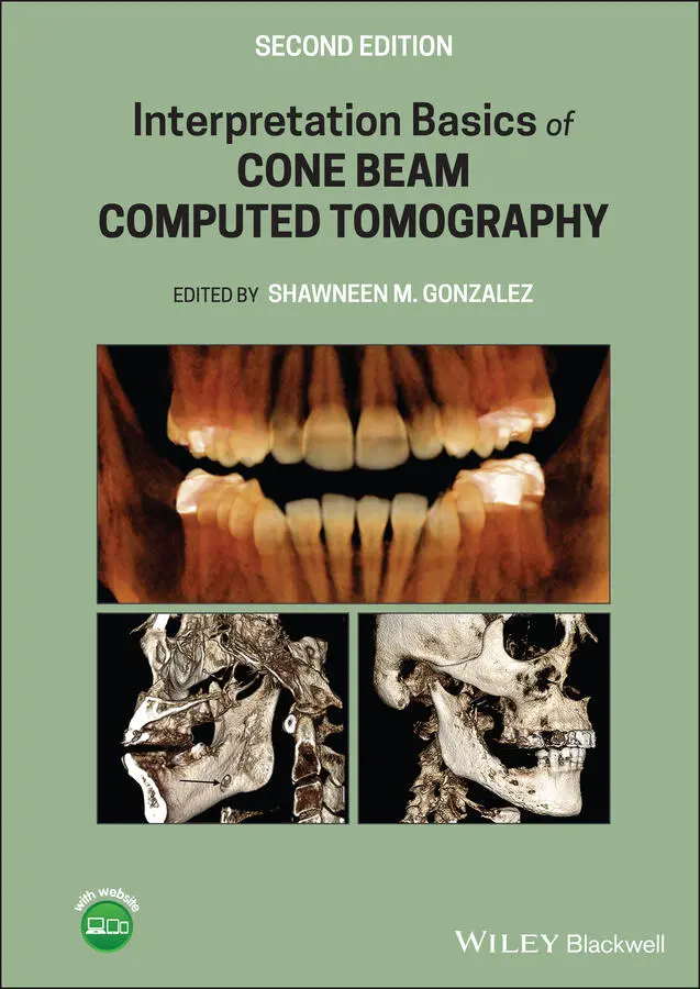
Interpretation Basics of Cone Beam Computed Tomography
Здесь есть возможность читать онлайн «Interpretation Basics of Cone Beam Computed Tomography» — ознакомительный отрывок электронной книги совершенно бесплатно, а после прочтения отрывка купить полную версию. В некоторых случаях можно слушать аудио, скачать через торрент в формате fb2 и присутствует краткое содержание. Жанр: unrecognised, на английском языке. Описание произведения, (предисловие) а так же отзывы посетителей доступны на портале библиотеки ЛибКат.
- Название:Interpretation Basics of Cone Beam Computed Tomography
- Автор:
- Жанр:
- Год:неизвестен
- ISBN:нет данных
- Рейтинг книги:4 / 5. Голосов: 1
-
Избранное:Добавить в избранное
- Отзывы:
-
Ваша оценка:
Interpretation Basics of Cone Beam Computed Tomography: краткое содержание, описание и аннотация
Предлагаем к чтению аннотацию, описание, краткое содержание или предисловие (зависит от того, что написал сам автор книги «Interpretation Basics of Cone Beam Computed Tomography»). Если вы не нашли необходимую информацию о книге — напишите в комментариях, мы постараемся отыскать её.
Enables rapid reference to common CBCT findings, with multiple images for each finding Features a streamlined framework that makes relevant information easier to find and apply in dental practice Offers hundreds of new images to aid in correctly identifying findings Contains new and updated content, including expanded coverage of CBCT and implants Provides sample reports and explains how they are used in day-to-day clinical practice
remains a must-have resource for all dental practitioner and specialists who use CBCT, dental students in radiology interpretation courses, and residents beginning to use CBCT in their specialty.
Interpretation Basics of Cone Beam Computed Tomography — читать онлайн ознакомительный отрывок
Ниже представлен текст книги, разбитый по страницам. Система сохранения места последней прочитанной страницы, позволяет с удобством читать онлайн бесплатно книгу «Interpretation Basics of Cone Beam Computed Tomography», без необходимости каждый раз заново искать на чём Вы остановились. Поставьте закладку, и сможете в любой момент перейти на страницу, на которой закончили чтение.
Интервал:
Закладка:
8 Chapter 9Figure 9.1. Axial view showing the nasopalatine canal (NPC). Yellow line sho...Figure 9.2. Axial view showing the incisive foramen (IF) and mandibular fora...Figure 9.3. Axial view showing the inferior alveolar nerve canal (IAN). Yell...Figure 9.4. Axial view showing the mental foramen (MF) and genial tubercles ...Figure 9.5. Coronal view showing the nasopalatine canal (NPC) with the incis...Figure 9.6. Coronal view showing the mental foramen (MF). Yellow line showin...Figure 9.7. Coronal view showing the inferior alveolar nerve canal (IAN). Ye...Figure 9.8. Coronal view showing the mandibular foramen (MNF). Yellow line s...Figure 9.9. Sagittal view showing the mental foramen (MF) with the inferior ...Figure 9.10. Sagittal view showing the nasopalatine canal (NPC), incisive fo...Figure 9.11. Axial (A), coronal (C), and sagittal (S) views showing a well‐d...Figure 9.12. Axial (A), coronal (C), and sagittal (S) views showing a well‐d...Figure 9.13. Rotated sagittal views showing a well‐defined, radiopaque area ...Figure 9.14. (a) Axial view showing a corticated lingual indentation consist...Figure 9.15. (a) Reconstructed pantomograph from a CBCT scan showing a well‐...Figure 9.16. (a) Axial view showing a corticated lingual indentation consist...Figure 9.17. (a) Sagittal view showing a well‐defined, round radiolucent are...Figure 9.18. (a) Sagittal view showing well‐defined, radiolucent areas (whit...Figure 9.19. (a) Axial (A), coronal (C), and sagittal (S) views showing a we...Figure 9.20. (a) Reconstructed pantomograph slice showing a diffuse radiopaq...Figure 9.21. (a) Reconstructed pantomograph showing irregular bone loss and ...Figure 9.22. (a) Rotated axial (A), coronal (C), and sagittal (S) views show...Figure 9.23. (a) Reconstructed pantomograph showing irregular bone loss in t...Figure 9.24. Coronal view showing multiple well‐defined, mixed radiopaque/ra...Figure 9.25. Sagittal view showing a well‐defined, mixed radiopaque/radioluc...Figure 9.26. Reconstructed pantomograph slice from a quadrant scan showing m...Figure 9.27. (a) 2D pantomograph showing a well‐defined, mixed radiopaque/ra...Figure 9.28. Axial (A), coronal (C), and sagittal (S) views showing multiple...
9 Chapter 10Figure 10.1. Sagittal view showing the glenoid fossa (GF), mandibular condyl...Figure 10.2. Coronal view showing the glenoid fossa (GF) and mandibular cond...Figure 10.3. Axial view showing the zygomatic process of the temporal bone (...Figure 10.4. (a) Coronal view showing various shapes of the condyle acceptab...Figure 10.5. Reconstructed pantomograph from a CBCT scan showing left condyl...Figure 10.6. Rotated sagittal cross‐sectional slices of the right and left c...Figure 10.7. Reconstructed pantomograph from a CBCT scan showing right condy...Figure 10.8. (a) Coronal view showing right condylar hyperplasia (white arro...Figure 10.9. (a) Axial view showing notching of the right condyle (white arr...Figure 10.10. Coronal view showing notching on superior aspect of right cond...Figure 10.11. (a) Reconstructed pantomograph from a CBCT scan showing superi...Figure 10.12. Sagittal and coronal slices showing normal condylar morphology...Figure 10.13. Sagittal and coronal slices showing mild flattening consistent...Figure 10.14. Sagittal and coronal slices showing flattening with anterior o...Figure 10.15. Sagittal and coronal slices showing flattening with loss of co...Figure 10.16. Coronal view showing flattening of the superior surface of the...Figure 10.17. (a) Rotated sagittal cross‐sectional slices showing anterior o...Figure 10.18. (a) Coronal view showing subchondral cyst formation (white arr...Figure 10.19. Reconstructed pantomograph from a CBCT scan of patient with rh...Figure 10.20. Rotated sagittal cross‐sectional slices showing severe bony de...Figure 10.21. (a) Coronal view showing erosions on the superior aspect of th...Figure 10.22. Rotated sagittal cross‐sectional slices showing erosions of th...Figure 10.23. (a) Axial view showing increased radiopacity of the right cond...
10 Chapter 11Figure 11.1. (a) Coronal view showing sample bone area of bone selected (whi...Figure 11.2. (a) Cross‐sectional slices through anterior maxilla with 1.0 mm...Figure 11.3. Reconstructed pantomograph from a CBCT scan showing the anterio...Figure 11.4. Reconstructed pantomograph from a CBCT scan showing right and l...Figure 11.5. Cross‐sectional views showing mandibular canal noted with red c...Figure 11.6. Rotated sagittal view showing mandibular canal as radiolucent b...Figure 11.7. (a) Implant screen using InVivo software with corresponding axi...
Guide
1 Cover Page
2 Title Page
3 Copyright Page
4 Preface
5 Acknowledgments
6 About the Companion Website
7 Table of Contents
8 Begin Reading
9 Appendix: Sample Reports
10 Index
11 Wiley End User License Agreement
Pages
1 iii
2 iv
3 ix
4 x
5 xi
6 xiii
7 1
8 2
9 3
10 4
11 5
12 6
13 7
14 8
15 9
16 10
17 11
18 13
19 14
20 15
21 16
22 17
23 18
24 19
25 20
26 21
27 22
28 23
29 24
30 25
31 26
32 27
33 28
34 29
35 30
36 31
37 33
38 34
39 35
40 36
41 37
42 39
43 40
44 41
45 42
46 43
47 44
48 45
49 46
50 47
51 48
52 49
53 50
54 51
55 52
56 53
57 54
58 55
59 56
60 57
61 58
62 59
63 60
64 61
65 62
66 63
67 64
68 65
69 66
70 67
71 68
72 69
73 70
74 71
75 73
76 74
77 75
78 76
79 77
80 78
81 79
82 80
83 81
84 82
85 83
86 84
87 85
88 86
89 87
90 88
91 89
92 90
93 91
94 92
95 93
96 94
97 95
98 96
99 97
100 98
101 99
102 100
103 101
104 102
105 103
106 104
107 105
108 106
109 107
110 108
111 109
112 110
113 111
114 112
115 113
116 115
117 116
118 117
119 118
120 119
121 120
122 121
123 122
124 123
125 124
126 125
127 126
128 127
129 128
130 129
131 130
132 131
133 132
134 133
135 134
136 135
137 136
138 137
139 138
140 139
141 141
142 142
143 143
144 144
145 145
146 146
147 147
148 148
149 149
150 150
151 151
152 152
153 153
154 154
155 155
156 156
157 157
158 158
159 159
160 161
161 162
162 163
163 164
164 165
165 166
166 167
167 168
168 169
169 170
170 171
171 172
172 173
173 174
174 175
175 176
176 177
177 178
178 179
179 180
180 181
181 182
182 183
183 184
184 185
185 186
186 187
187 188
188 189
189 190
190 191
191 192
192 193
193 194
194 195
195 196
196 197
197 198
198 199
199 200
200 201
201 202
202 203
203 204
204 205
205 206
206 207
207 209
208 210
209 211
210 212
211 213
212 214
213 215
214 216
215 217
216 218
217 219
218 220
219 221
220 223
221 224
222 225
223 226
224 227
225 228
226 229
227 230
228 231
229 232
Читать дальшеИнтервал:
Закладка:
Похожие книги на «Interpretation Basics of Cone Beam Computed Tomography»
Представляем Вашему вниманию похожие книги на «Interpretation Basics of Cone Beam Computed Tomography» списком для выбора. Мы отобрали схожую по названию и смыслу литературу в надежде предоставить читателям больше вариантов отыскать новые, интересные, ещё непрочитанные произведения.


Обсуждение, отзывы о книге «Interpretation Basics of Cone Beam Computed Tomography» и просто собственные мнения читателей. Оставьте ваши комментарии, напишите, что Вы думаете о произведении, его смысле или главных героях. Укажите что конкретно понравилось, а что нет, и почему Вы так считаете.