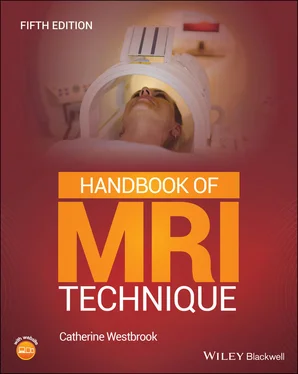
Catherine Westbrook - Handbook of MRI Technique
Здесь есть возможность читать онлайн «Catherine Westbrook - Handbook of MRI Technique» — ознакомительный отрывок электронной книги совершенно бесплатно, а после прочтения отрывка купить полную версию. В некоторых случаях можно слушать аудио, скачать через торрент в формате fb2 и присутствует краткое содержание. Жанр: unrecognised, на английском языке. Описание произведения, (предисловие) а так же отзывы посетителей доступны на портале библиотеки ЛибКат.
- Название:Handbook of MRI Technique
- Автор:
- Жанр:
- Год:неизвестен
- ISBN:нет данных
- Рейтинг книги:3 / 5. Голосов: 1
-
Избранное:Добавить в избранное
- Отзывы:
-
Ваша оценка:
- 60
- 1
- 2
- 3
- 4
- 5
Handbook of MRI Technique: краткое содержание, описание и аннотация
Предлагаем к чтению аннотацию, описание, краткое содержание или предисловие (зависит от того, что написал сам автор книги «Handbook of MRI Technique»). Если вы не нашли необходимую информацию о книге — напишите в комментариях, мы постараемся отыскать её.
FIFTH EDITION Handbook of MRI Technique.
Handbook of MRI Technique
Handbook of MRI Technique — читать онлайн ознакомительный отрывок
Ниже представлен текст книги, разбитый по страницам. Система сохранения места последней прочитанной страницы, позволяет с удобством читать онлайн бесплатно книгу «Handbook of MRI Technique», без необходимости каждый раз заново искать на чём Вы остановились. Поставьте закладку, и сможете в любой момент перейти на страницу, на которой закончили чтение.
Интервал:
Закладка:
Dr Catherine Westbrook
Acknowledgements
My heart‐felt thanks to the contributing authors, Anne Bright, Vera Kimbrell, Elizabeth Lorusso, Bac Nguyen and Sara Sullivan, without whom this book could never have been updated. As usual, I am extremely impressed with their professional and thoughtful contributions and I am very grateful for their valued opinions and support.
I would also like to thank Dr John Talbot for his beautifully drawn anatomical diagrams and Christos Tsiotsios from the Ygia Polytechnic Private Hospital, Cyprus for supplying many of the MR images used in this book. A big thank you to Matthew Hayes and Andrea Graziano of ScanLabMR for supplying the new localizer images. This has been a truly international collaboration.
And thank you, finally, to my wife Amabel, my children Adam, Ben and Maddie, my mother Maggie and my sister Francesca. I have no intention of embarrassing you too much, so I will just say, I love you all very much!
Dr Catherine Westbrook
About the Companion Website
The book is accompanied by a website:
www.wiley.com/go/westbrook/mritechnique5e 
The website features:
Flashcards
Multiple choice questions
Scan this QR code to visit the companion website.

1 How to Use This Book
Introduction
Common indications
Basic anatomy
Equipment
Patient positioning
Slice prescription
Suggested protocol
Protocol optimization
Patient considerations
Contrast usage
Summary
Terms and abbreviations used in Part 2
Conclusion
INTRODUCTION
This book has been written with the intention of providing a step‐by‐step explanation of the most common examinations currently carried out using magnetic resonance imaging (MRI). It is divided into two parts.
Part 1contains reviews or summaries of those theoretical and practical concepts that are frequently discussed in Part 2. These are:
protocol parameters and trade‐offs
pulse sequences
flow phenomena and artefacts
gating and respiratory compensation (RC) techniques
patient care and safety
contrast agents.
These summaries are not intended to be comprehensive but contain only a brief description of definitions and uses. For a more detailed discussion of these and other concepts, the reader is referred to MRI physics books. MRI in Practice by C. Westbrook and J. Talbot (Wiley Blackwell, 2019, fifth edition) is a particularly useful companion to this book.
Part 2is divided into the following examination areas:
head and neck
spine
chest
abdomen
pelvis
upper limb
lower limb
paediatric imaging.
Each anatomical region is subdivided into separate examinations. For example, the section entitled Head and neck includes explanations on imaging the brain, temporal lobes, pituitary fossa, and so on. Under each examination, the following categories are described:
common indications
basic anatomy
equipment
patient positioning
slice prescription
suggested protocol
protocol optimization
patient considerations
contrast usage.
COMMON INDICATIONS
These are the most usual reasons for scanning each area, although occasionally some rarer indications are included.
BASIC ANATOMY
Simple anatomical diagrams are provided for most examination areas to assist the reader.
EQUIPMENT
This contains a list of the equipment required for each examination and includes coil type, gating leads, bellows and immobilization devices. The correct use of gating and RC is discussed in Part 1(see Gating and respiratory compensation techniques ). The coil types described are the most common currently available. These are as follows.
Volume coils that both transmit and receive radiofrequency (RF) pulses and are specifically called transceivers. Most of these coils are quadrature in design, which means that they generate two RF fields perpendicular to each other. This maximises coupling between the coils and the spin population, thus improving image quality factors such as the signal‐to‐noise ratio (SNR). Volume coils often encompass large areas of anatomy and yield a uniform signal across the whole field of view (FOV). The head and body coil are examples of this type of coil.
Linear phased array coils consist of multiple coils and receivers. The signal from the receiver of each coil is combined to form one image. The image has the advantages of both a small coil (improved SNR) and those of the larger volume coils (increased coverage). Therefore, linear phased array coils can be used either to examine large areas such as the entire length of the spine, or to improve signal uniformity and intensity in small areas such as the breast. Figure 1.1 Correct placement of a flat surface receive coil.
Volume phased array (parallel imaging) uses the data from multiple coils or channels arranged around the area under examination to decrease scan time, increase phase resolution or a combination of both. Additional software and hardware are required. The hardware includes several coils perpendicular to each other or one coil with several channels. The number of possible coils/channels varies but currently ranges from 2 to 32 for routine imaging and up to 128 for cardiac imaging. During data acquisition, each coil fills its own lines of k‐space (e.g., if two coils are used together, one coil fills the even lines of k‐space and the other the odd lines. k‐space is therefore filled either twice as quickly or with twice the phase resolution in the same scan time). The number of selected coils/channels is generically called the reduction factor and is similar in principle to the turbo factor (TF)/echo train length (ETL) in fast or turbo spin echo (FSE/TSE) (see Pulse sequences). Every coil/channel produces a separate image that often displays aliasing artefact (see Flow phenomena and artefacts). Software called sensitivity encoding and additional algorithms remove this artefact and combine the images from each coil/channel to produce a single unwrapped image. This technology can be used in most examination areas and with any pulse sequence. It is often discussed in the Protocol optimization section in Part 2.
Surface/local coils are traditionally used to improve the SNR when imaging structures near to the skin surface. They are often specially designed to fit a certain area and in general they only receive signal. Surface coils increase SNR compared with volume coils. This is because they are placed close to the region under examination increasing signal amplitude that is generated in the coil, while noise is only received in the vicinity of the coil. However, surface coils only receive signal up to the edges of the coil and to a depth equal to the diameter of the conductive loop of the coil. To visualize structures deep within the patient, either a volume, linear or volume phased array coil or, more rarely, a local coil inserted into an orifice must be utilized (e.g., a rectal coil).
The choice of coil is one of the most important factors that determines the SNR. When using any type of coil remember to:
Check that the cables are intact and undamaged.
Check that the coil is plugged in properly and that the correct connector box is used.
Читать дальшеИнтервал:
Закладка:
Похожие книги на «Handbook of MRI Technique»
Представляем Вашему вниманию похожие книги на «Handbook of MRI Technique» списком для выбора. Мы отобрали схожую по названию и смыслу литературу в надежде предоставить читателям больше вариантов отыскать новые, интересные, ещё непрочитанные произведения.
Обсуждение, отзывы о книге «Handbook of MRI Technique» и просто собственные мнения читателей. Оставьте ваши комментарии, напишите, что Вы думаете о произведении, его смысле или главных героях. Укажите что конкретно понравилось, а что нет, и почему Вы так считаете.


