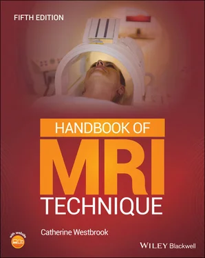
Catherine Westbrook - Handbook of MRI Technique
Здесь есть возможность читать онлайн «Catherine Westbrook - Handbook of MRI Technique» — ознакомительный отрывок электронной книги совершенно бесплатно, а после прочтения отрывка купить полную версию. В некоторых случаях можно слушать аудио, скачать через торрент в формате fb2 и присутствует краткое содержание. Жанр: unrecognised, на английском языке. Описание произведения, (предисловие) а так же отзывы посетителей доступны на портале библиотеки ЛибКат.
- Название:Handbook of MRI Technique
- Автор:
- Жанр:
- Год:неизвестен
- ISBN:нет данных
- Рейтинг книги:3 / 5. Голосов: 1
-
Избранное:Добавить в избранное
- Отзывы:
-
Ваша оценка:
- 60
- 1
- 2
- 3
- 4
- 5
Handbook of MRI Technique: краткое содержание, описание и аннотация
Предлагаем к чтению аннотацию, описание, краткое содержание или предисловие (зависит от того, что написал сам автор книги «Handbook of MRI Technique»). Если вы не нашли необходимую информацию о книге — напишите в комментариях, мы постараемся отыскать её.
FIFTH EDITION Handbook of MRI Technique.
Handbook of MRI Technique
Handbook of MRI Technique — читать онлайн ознакомительный отрывок
Ниже представлен текст книги, разбитый по страницам. Система сохранения места последней прочитанной страницы, позволяет с удобством читать онлайн бесплатно книгу «Handbook of MRI Technique», без необходимости каждый раз заново искать на чём Вы остановились. Поставьте закладку, и сможете в любой момент перейти на страницу, на которой закончили чтение.
Интервал:
Закладка:
13 Chapter 14Figure 14.1 Anterior view of the right hip demonstrating bony components and...Figure 14.2 Coronal localizer showing slice prescription for axial imaging o...Figure 14.3 Coronal localizer showing slice prescription and angulation for ...Figure 14.4 Coronal FSE/TSE T2‐weighted image of the right hip with fat supp...Figure 14.5 Sagittal FSE/TSE T2‐weighted image of the right hip with fat sup...Figure 14.6 Coronal arthrogram of the left hip.Figure 14.7 Anterior view of the right femur.Figure 14.8 Sagittal FSE/TSE T2‐weighted image with fat suppression and radi... Figure 14.9 Axial FSE/TSE PD‐weighted image of the femur with fat suppressio...Figure 14.10 Anterior view of the right knee showing joint structures and li...Figure 14.11 Sagittal localizer showing slice prescription boundaries and an...Figure 14.12 Axial localizer showing slice prescription boundaries and angul...Figure 14.13 Sagittal rewound GRE T2*‐weighted image of the knee with fat su...Figure 14.14 Coronal STIR of the knee.Figure 14.15 Axial 3D rewound GRE T2* with fat suppression.Figure 14.16 Sagittal spoiled GRE T1‐weighted image of a flexed knee during ...Figure 14.17 Anterior view of the right tibia and fibula.Figure 14.18 Coronal STIR with radial k ‐space of the tibiae demonstrating a ...Figure 14.19 Axial STIR with radial k ‐space of the right tibia demonstrating...Figure 14.20 Sagittal view of the foot ankle showing ligaments on the latera...Figure 14.21 Sagittal localizer showing slice prescription boundaries and an...Figure 14.22 Sagittal localizer showing slice prescription boundaries and an...Figure 14.23 Sagittal spoiled GRE T1‐weighted image of the ankle.Figure 14.24 Sagittal FSE/TSE T2‐weighted image of the ankle with fat suppre...Figure 14.25 Coronal FSE/TSE T1‐weighted image of the ankle.Figure 14.26 Coronal localizer showing slice prescription boundaries and ang...Figure 14.27 Sagittal localizer showing slice prescription boundaries and an...Figure 14.28 Sagittal FSE/TSE T1‐weighted image of the foot.Figure 14.29 Sagittal FSE/TSE T2‐weighted image of the foot with fat suppres...Figure 14.30 Vascular supply to the right leg.Figure 14.31 Venous drainage of the right leg.Figure 14.32 Sequential CE‐MRA of the iliac vessels showing an AVM (first pa...Figure 14.33 Sequential CE‐MRA of the iliac vessels showing an AVM (second p...Figure 14.34 Sequential CE‐MRA of the iliac vessels showing an AVM (third pa...
14 Chapter 15Figure 15.1 Axial FSE/TSE T2‐weighted image in a 4‐month‐old child. Polymicr...Figure 15.2 Coronal FSE/TSE T2‐weighted image demonstrating a double cortex ...Figure 15.3 Axial FLAIR image demonstrating a highly malignant sarcoma.Figure 15.4 3D spoiled GRE reformatted in the axial plane demonstrating tran...Figure 15.5 Axial SW image of the brain demonstrating metastases from a high...Figure 15.6 Coronal T2 FSE/TSE (left) and GRE‐EPI (right) showing subtle ear...Figure 15.7 Axial SE‐EPI demonstrating chronic haemorrhage.Figure 15.8 Coronal FSE/TSE T2‐weighted image demonstrating mesial temporal ...Figure 15.9 Sagittal (left) and coronal (right) FSE/TSE T1‐weighted images a...Figure 15.10 Axial FSE/TSE T2‐weighted imaging demonstrating a neonatal glio...Figure 15.11 3D TOF‐MRA in a 4‐year‐old child demonstrating normal appearanc...Figure 15.12 Sagittal FSE/TSE T2‐weighted image demonstrating a vein of Gale...Figure 15.13 Sagittal (left) and coronal (right) FSE/TSE T1‐weighted images ...Figure 15.14 Sagittal FSE/TSE T2‐weighted image showing a cyst (arrow) that ...Figure 15.15 Sagittal FSE/TSE T1‐weighted (left) and T2‐weighted with fat su...Figure 15.16 Sagittal FSE/TSE T1‐weighted images post‐contrast enhancement o...Figure 15.17 Sagittal FSE/TSE T2‐weighted image of the cervical spine demons...Figure 15.18 Sagittal FSE/TSE T2‐weighted image with fat suppression demonst...Figure 15.19 Coronal FSE/TSE T2‐weighted images. Neurofibromatosis lesions a...Figure 15.20 Sagittal FSE/TSE T2 weighted‐image demonstrating a large terato...Figure 15.21 Coronal FSE/TSE T1‐weighted image through the ankle joint demon...Figure 15.22 Coronal FSE/TSE T1‐weighted image (top) and rewound GRE T2*‐wei...Figure 15.23 Axial FSE/TSE T1‐weighted image of the knee demonstrating osteo...Figure 15.24 Coronal image FSE/TSE T2‐weighted image showing Ewing’s sarcoma...Figure 15.25 Axial FSE/TSE T2‐weighted image with fat suppression demonstrat...Figure 15.26 Coronal FSE/TSE T2‐weighted image. Same patient as in Figure 15...Figure 15.27 Coronal FSE/TSE T2‐weighted image demonstrating a large left re...Figure 15.28 Sagittal FSE/TSE T2‐weighted image with fat suppression of the ...Figure 15.29 Whole body STIR image.Figure 15.30 Sagittal/oblique balanced GRE T2*‐weighted ciné image through t...Figure 15.31 Short axis phase sensitive delay image.Figure 15.32 3D balanced GRE T2*‐weighted image demonstrating the left coron...Figure 15.33 Coronal FSE/TSE T2‐weighted image showing a foetal lymphatic le...
Guide
1 Cover Page
2 Title Page
3 Copyright Page
4 Contributors
5 Preface
6 Acknowledgements
7 About the Companion Website
8 Table of Contents
9 Begin Reading
10 Index
11 Wiley End User License Agreement
Pages
1 iii
2 iv
3 ix
4 x
5 xi
6 xiii
7 xiii
8 1
9 2
10 3
11 4
12 5
13 6
14 7
15 8
16 9
17 10
18 11
19 12
20 13
21 14
22 15
23 16
24 17
25 19
26 21
27 22
28 23
29 24
30 25
31 26
32 27
33 28
34 29
35 30
36 31
37 32
38 33
39 34
40 35
41 36
42 37
43 38
44 39
45 40
46 41
47 42
48 43
49 44
50 45
51 46
52 47
53 48
54 49
55 50
56 51
57 53
58 54
59 55
60 56
61 57
62 58
63 59
64 60
65 61
66 62
67 63
68 64
69 65
70 66
71 67
72 68
73 69
74 70
75 71
76 73
77 74
78 75
79 76
80 77
81 78
82 79
83 80
84 81
85 82
86 83
87 84
88 85
89 86
90 87
91 88
92 89
93 90
94 91
95 92
96 93
97 94
98 95
99 96
100 97
101 98
102 99
103 100
104 101
105 102
106 103
107 104
108 105
109 106
110 107
111 108
112 109
113 110
114 111
115 112
116 113
117 114
118 115
119 116
120 117
121 118
122 119
123 120
124 121
125 122
126 123
127 124
128 125
129 126
130 127
131 128
132 129
133 130
134 131
135 132
136 133
137 134
138 135
139 136
140 137
141 138
142 139
143 140
144 141
145 142
146 143
147 144
148 145
149 146
150 147
151 148
152 149
153 150
154 151
155 152
156 153
157 154
158 155
159 156
160 157
161 158
162 159
163 160
164 161
165 162
166 163
167 164
168 165
169 166
170 167
171 168
172 169
173 170
174 171
175 172
176 173
177 174
178 175
179 176
180 177
181 178
182 179
183 180
184 181
185 182
186 183
187 184
188 185
189 186
190 187
191 188
192 189
193 190
194 191
195 192
196 193
197 194
198 195
199 196
Читать дальшеИнтервал:
Закладка:
Похожие книги на «Handbook of MRI Technique»
Представляем Вашему вниманию похожие книги на «Handbook of MRI Technique» списком для выбора. Мы отобрали схожую по названию и смыслу литературу в надежде предоставить читателям больше вариантов отыскать новые, интересные, ещё непрочитанные произведения.
Обсуждение, отзывы о книге «Handbook of MRI Technique» и просто собственные мнения читателей. Оставьте ваши комментарии, напишите, что Вы думаете о произведении, его смысле или главных героях. Укажите что конкретно понравилось, а что нет, и почему Вы так считаете.


