Catherine Westbrook - Handbook of MRI Technique
Здесь есть возможность читать онлайн «Catherine Westbrook - Handbook of MRI Technique» — ознакомительный отрывок электронной книги совершенно бесплатно, а после прочтения отрывка купить полную версию. В некоторых случаях можно слушать аудио, скачать через торрент в формате fb2 и присутствует краткое содержание. Жанр: unrecognised, на английском языке. Описание произведения, (предисловие) а так же отзывы посетителей доступны на портале библиотеки ЛибКат.

- Название:Handbook of MRI Technique
- Автор:
- Жанр:
- Год:неизвестен
- ISBN:нет данных
- Рейтинг книги:3 / 5. Голосов: 1
-
Избранное:Добавить в избранное
- Отзывы:
-
Ваша оценка:
Handbook of MRI Technique: краткое содержание, описание и аннотация
Предлагаем к чтению аннотацию, описание, краткое содержание или предисловие (зависит от того, что написал сам автор книги «Handbook of MRI Technique»). Если вы не нашли необходимую информацию о книге — напишите в комментариях, мы постараемся отыскать её.
FIFTH EDITION Handbook of MRI Technique.
Handbook of MRI Technique
Handbook of MRI Technique — читать онлайн ознакомительный отрывок
Ниже представлен текст книги, разбитый по страницам. Система сохранения места последней прочитанной страницы, позволяет с удобством читать онлайн бесплатно книгу «Handbook of MRI Technique», без необходимости каждый раз заново искать на чём Вы остановились. Поставьте закладку, и сможете в любой момент перейти на страницу, на которой закончили чтение.
Интервал:
Закладка:
Ensure that the receiving side of the coil faces the patient. This is usually labelled on the coil itself. Note that both sides of the coil usually receive signal, but coils are designed so that one side receives optimum signal. This is especially true of shaped coils that fit a certain anatomical area. If the wrong side of the coil faces the patient, signal is lost, and image quality suffers.
Place the coil as close as possible to the area under examination.
The coil should not directly touch the patient’s skin as it may become warm during the examination and cause discomfort. A small foam pad placed between the skin surface and the coil is usually sufficient insulation.
Ensure that the coil does not move when placed on the patient. A moving coil during data acquisition always produces a moving image.
Ensure that the receiving surface of the coil is parallel to the z (long) axis of the magnet. This guarantees that the transverse component of magnetization is perpendicular to the coil and that maximum signal is induced. Placing the coil at an angle to this axis, or parallel to the x or y axis, results in a loss of signal ( Figure 1.1). Note that the axes discussed here relate to a superconducting magnet. Open systems may label their axes differently, but the principle remains the same. To generate signal in a receiver coil on any type of system, the receiver coil must be located perpendicular to the transverse component of magnetization.
PATIENT POSITIONING
This contains a description of the correct patient position, placement of the patient within the coil and proper immobilization techniques. Centring and landmarking are described relative to the laser light system on a superconducting system as follows ( Figure 1.2):
The longitudinal alignment light refers to the light running parallel to the bore of the magnet in the z axis.
The horizontal alignment light refers to the light that runs from left to right of the bore of the magnet in the x axis.
The vertical alignment light refers to the light that runs from the top to the bottom of the magnet in the y axis.
It is assumed in Part 2that the following areas are examined with the patient placed head‐first in the magnet:
head and neck (all areas)
cervical, thoracic and whole spine
chest (all areas)
abdomen (for areas superior to the iliac crests)
shoulders and upper limb (except where specified).
The remaining anatomical regions are examined with the patient placed feet‐first in the magnet. These are:
pelvis
hips
lower limbs.
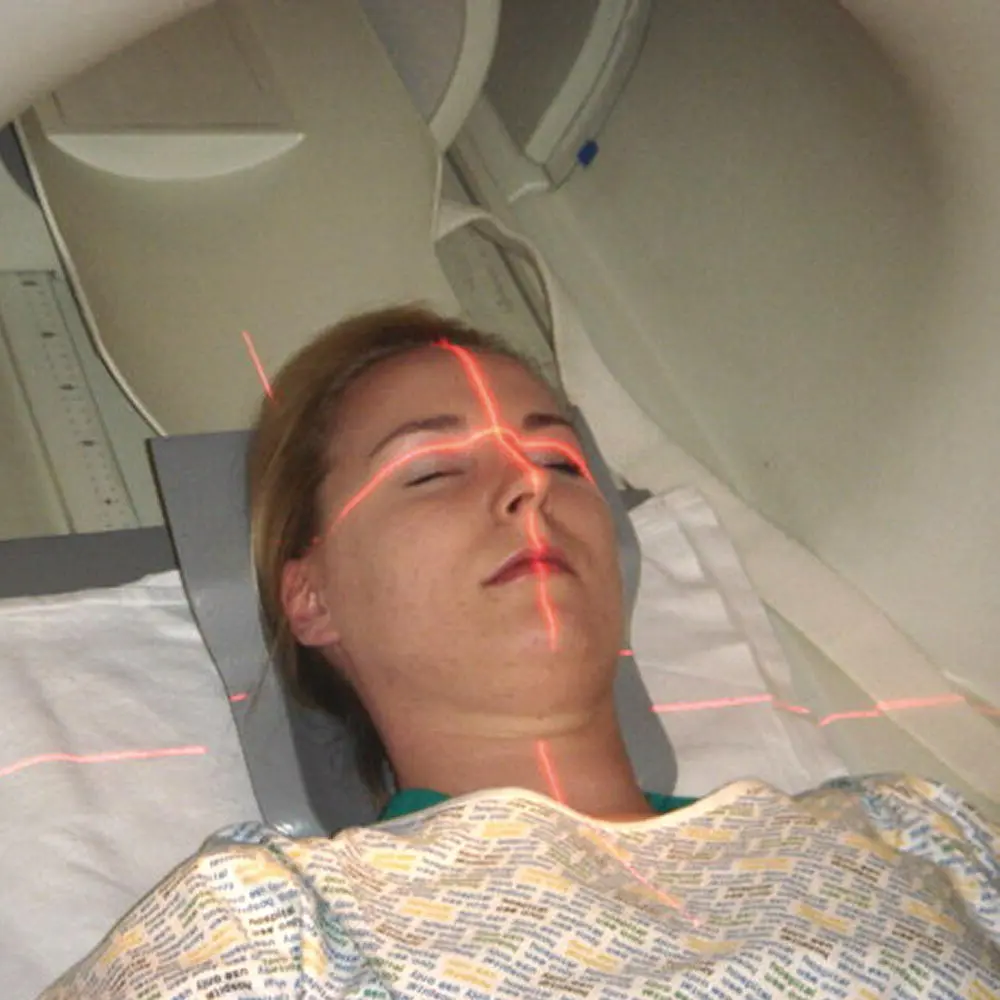
Figure 1.2 Positioning of the alignment lights.
SLICE PRESCRIPTION
This section describes the anatomical landmarks for slice prescription and angulation of imaging planes used in each examination area. All protocols begin with a three‐plane localizer from which slices are prescribed. Imaging coordinates are provided for at least one plane in the three‐plane localizer acquisition.
SUGGESTED PROTOCOL
This is intended as a guideline only. Almost every centre uses different protocols depending on the type of system and radiological preference. However, this section can be helpful for those practitioners scanning without a radiologist, or where the examination is so rare that perhaps neither the radiologist nor the practitioner knows how to proceed. The protocol description is mainly limited to scan plane, weighting, pulse sequence(s) and why it is used. For details of suggested protocol parameters see Table 2.1.
It must be stressed that all the protocols listed are only a reflection of the authors’ practice and research. However, the protocols provided in this section are considered to be the most commonly used. In most examinations, there is a section reserved for Additional techniques . These are not regarded as routine but may be included in the examination. Of course, some practitioners may regard what we call ‘additional’ as ‘routine’, and vice versa.
PROTOCOL OPTIMIZATION
This section is subdivided into:
Technical issues: This includes a discussion of the relationship of SNR, CNR, spatial resolution and scan time pertaining to each examination. Suggestions on how to optimize these factors are described (see Protocol parameters and trade‐offs). The correct use of pulse sequences and various imaging options are also discussed (see also Pulse sequences).
Artefact problems: This contains a description of the common artefacts encountered and ways in which they can be eliminated or reduced (see also Flow phenomena and artefacts).
PATIENT CONSIDERATIONS
This encompasses the condition of the patient, including symptoms and claustrophobia. Suggestions to overcome these are given (see also Patient care and safety ).
CONTRAST USAGE
The reasons for administering a contrast agent in each examination area are discussed. The use of contrast agents varies widely according to radiological preferences. This section should be used as a guide only (see also Contrast agents ).
SUMMARY
Follow this 10‐point plan for good radiographic practice:
1 Review all cases carefully and select appropriate protocols.
2 Have flexible protocols that can reflect the needs of each individual clinical case.
3 Regularly review your protocols and procedures and benchmark them against current best practice.
4 Have clear diagnostic goals including the minimum accepted protocol necessary to obtain a useful diagnostic/clinical outcome.
5 Regularly review your protocols and procedures.
6 Understand the capabilities of your system.
7 Recognize your limitations and, if necessary, refer to another site rather than risk an incomplete or diagnostically unacceptable procedure.
8 Educate all levels of staff to new procedures and/or system capabilities.
9 Be safety paranoid to ensure your unit does not fall victim to the dreaded MRI incident.
10 Most importantly, enjoy your patients and give them the highest standard of care possible.
TERMS AND ABBREVIATIONS USED IN PART 2
Wherever possible, generic terms have been used to describe protocol parameters, particularly pulse sequences and imaging options. Explanations of these can be found in the various sections of Part 1. To avoid ambiguity, the specific following terms have been used:
Fat suppression: includes all fat suppression techniques such as fat saturation (FAT SAT), spectrally selective inversion recovery (SPIR) and Dixon methods.
Gradient moment nulling (GMN): gradient moment rephasing (GMR) and flow compensation (FC).
Oversampling: no phase wrap, anti‐aliasing and anti‐foldover
Rectangular FOV: rectangular or asymmetric FOV
Respiratory compensation (RC): phase reordering and respiratory triggering techniques
Abbreviations are used throughout the book for simplification purposes. A summary of these can be found in Table 1.1. In addition, Table 1.2summarizes the slice prescription parameters for each examination in Part 2and a comparison of acronyms used by certain vendors to describe pulse sequences and imaging options is given in Table 3.1(see Pulse sequences in Part 1).
Table 1.1 Abbreviations used in this book.
| A | Anterior |
| AC | Number of acquisitions |
| ACL | Anterior cruciate ligament |
| ACPC | Anterior–posterior commissure axis |
| ACR | American College of Radiologists |
| ADC | Apparent diffusion coefficient |
| ADEM | Acute disseminating encephalomyelitis |
| AIDS | Autoimmune deficiency syndrome |
| ASIS | Anterior superior iliac spine |
| ASTM | American Society for Testing and Materials |
| AVM | Arteriovenous malformation |
| AVN | Avascular necrosis |
| BFFE | Balanced fast field echo |
| BGRE | Balanced gradient echo |
| BOLD | Blood oxygenation level dependent |
| CDH | Congenital dislocation of the hips |
| CE‐MRA | Contrast enhanced magnetic resonance angiography |
| CNR | Contrast to noise ratio |
| CNS | Central nervous system |
| COPD | Chronic obstructive pulmonary disease |
| CSE | Conventional spin echo |
| CSF | Cerebral spinal fluid |
| CSI | Chemical shift imaging |
| CSR | Chemical shift ratio |
| CT | Computer tomography |
| CVA | Cerebral vascular accident |
| DE Prep | Driven equilibrium magnetization preparation |
| DTI | Diffusion tensor imaging |
| DWI | Diffusion weighted imaging |
| EAM | External auditory meatus |
| ECG | Electrocardiogram |
| EKG | Electrocardiogram (US spelling) |
| EPI | Echo planar imaging |
| ETL | Echo train length |
| FA | Fractional anisotropy |
| FAT SAT | Fat saturation |
| FC | Flow compensation |
| FDA | Food and Drug Administration |
| FFE | Fast field echo |
| FID | Free induction decay |
| FIESTA | Free induction echo stimulated acquisition |
| FISP | Free induction steady precession |
| FLAIR | Fluid attenuated inversion recovery |
| FLASH | Fast low angled shot |
| fMRI | Functional magnetic resonance imaging |
| FOV | Field of view |
| FSE | Fast spin echo |
| Gd | Gadolinium |
| GFE | Gradient field echo |
| GMN | Gradient moment nulling |
| GMR | Gradient moment rephasing |
| GRASS | Gradient recalled acquisition in the steady state |
| GRE | Gradient echo |
| GRE‐EPI | Gradient echo – echo planar imaging |
| HASTE | Half acquisition single‐shot turbo spin echo |
| HIE | Hypoxic ischemic event |
| I | Inferior |
| IAM | Internal auditory meatus |
| ICP | Intracranial pressure |
| IM | Intramuscular |
| IR | Inversion recovery |
| IR‐FSE | Inversion recovery – fast spin echo |
| IR prep | Inversion recovery magnetization preparation |
| IV | Intravenous |
| IVC | Inferior vena cava |
| L | Left |
| MDA | Medical Devices Agency |
| MIP | Maximum intensity projection |
| MOTSA | Multiple overlapping thin slab acquisition |
| MP RAGE | Magnetization prepared rapid acquisition gradient echo |
| MR | Magnetic resonance |
| MRA | Magnetic resonance angiography |
| MRCP | Magnetic resonance cholangiopancreatography |
| MRE | Magnetic resonance enterography |
| MRI | Magnetic resonance imaging |
| MRS | Magnetic resonance spectroscopy |
| MS | Multiple sclerosis |
| MSK | Musculoskeletal |
| MT | Magnetization transfer |
| MVS | Multi‐voxel spectroscopy |
| NEX | Number of excitations |
| NSA | Number of signal averages |
| NSF | Nephrogenic systemic fibrosis |
| P | Posterior |
| PC | Phase contrast |
| PC‐MRA | Phase contrast magnetic resonance angiography |
| PD | Proton density |
| Pe | Peripheral |
| PEAR | Phase encoding artefact reduction |
| PET | Proton emission tomography |
| ppm | Parts per million |
| PRESS | Point resolved spectroscopy |
| PSIF | Reverse FISP |
| R | Right |
| RC | Respiratory compensation |
| REST | Regional saturation technique |
| RF | Radio frequency |
| ROI | Region of interest |
| RR | R to R interval |
| S | Superior |
| SAR | Specific absorption rate |
| SAT | Saturation |
| SE‐EPI | Spin echo – echo planar imaging |
| SNR | Signal to noise ratio |
| SPAMM | Spatial modulation of magnetization |
| SPGR | Spoiled gradient recalled acquisition in the steady state |
| SPIR | Spectrally selective inversion recovery |
| SS | Single shot |
| SS‐EPI | Single‐shot echo planar imaging |
| SSFP | Steady‐state free precession |
| SS‐FSE | Single‐shot fast spin echo |
| SS‐GRE‐EPI | Single‐shot gradient echo EPI |
| STEAM | Stimulated echo acquisition mode |
| STIR | Short TAU inversion recovery |
| SVS | Single‐voxel spectroscopy |
| SW | Susceptibility weighted |
| TE | Time to echo |
| TF | Turbo factor |
| TFE | Turbo field echo |
| TI | Time to inversion |
| TIA | Transient ischaemic attack |
| TLE | Temporal lobe epilepsy |
| TMJ | Temporomandibular joint |
| TOF | Time of flight |
| TOF‐MRA | Time of flight magnetic resonance angiography |
| TR | Time to repeat |
| True FISP | Siemens version of balanced gradient echo |
| TSE | Turbo spin echo |
| VENC | Velocity encoding |
| VNR | Velocity to noise ratio |
| VOI | Volume of interest |
| VQ | Ventilation perfusion scan |
Table 1.2 Summary of slice prescription criteria.
Читать дальшеИнтервал:
Закладка:
Похожие книги на «Handbook of MRI Technique»
Представляем Вашему вниманию похожие книги на «Handbook of MRI Technique» списком для выбора. Мы отобрали схожую по названию и смыслу литературу в надежде предоставить читателям больше вариантов отыскать новые, интересные, ещё непрочитанные произведения.


Обсуждение, отзывы о книге «Handbook of MRI Technique» и просто собственные мнения читателей. Оставьте ваши комментарии, напишите, что Вы думаете о произведении, его смысле или главных героях. Укажите что конкретно понравилось, а что нет, и почему Вы так считаете.