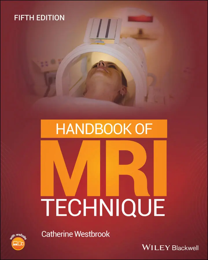
Catherine Westbrook - Handbook of MRI Technique
Здесь есть возможность читать онлайн «Catherine Westbrook - Handbook of MRI Technique» — ознакомительный отрывок электронной книги совершенно бесплатно, а после прочтения отрывка купить полную версию. В некоторых случаях можно слушать аудио, скачать через торрент в формате fb2 и присутствует краткое содержание. Жанр: unrecognised, на английском языке. Описание произведения, (предисловие) а так же отзывы посетителей доступны на портале библиотеки ЛибКат.
- Название:Handbook of MRI Technique
- Автор:
- Жанр:
- Год:неизвестен
- ISBN:нет данных
- Рейтинг книги:3 / 5. Голосов: 1
-
Избранное:Добавить в избранное
- Отзывы:
-
Ваша оценка:
Handbook of MRI Technique: краткое содержание, описание и аннотация
Предлагаем к чтению аннотацию, описание, краткое содержание или предисловие (зависит от того, что написал сам автор книги «Handbook of MRI Technique»). Если вы не нашли необходимую информацию о книге — напишите в комментариях, мы постараемся отыскать её.
FIFTH EDITION Handbook of MRI Technique.
Handbook of MRI Technique
Handbook of MRI Technique — читать онлайн ознакомительный отрывок
Ниже представлен текст книги, разбитый по страницам. Система сохранения места последней прочитанной страницы, позволяет с удобством читать онлайн бесплатно книгу «Handbook of MRI Technique», без необходимости каждый раз заново искать на чём Вы остановились. Поставьте закладку, и сможете в любой момент перейти на страницу, на которой закончили чтение.
Интервал:
Закладка:
| Area | Plane | Angle | Prescription | Coverage | |
|---|---|---|---|---|---|
| Brain | Sagittal | parallel to falx cerebri | lateral borders of each temporal lobe | foramen magnum to vertex and occiput to frontal lobe | |
| Axial | parallel to falx cerebri and ACPC line | foramen magnum to superior surface of the brain | occipital lobe to frontal lobe and both temporal lobes | ||
| Coronal | perpendicular to ACPC line | cerebellum to frontal lobe | foramen magnum to vertex and both temporal lobes | ||
| Temporal lobes | Sagittal | parallel to falx cerebri | lateral borders of each temporal lobe | foramen magnum to vertex and occiput to frontal lobe | |
| Axial | parallel to temporal lobes and falx cerebri | inferior aspect of temporal lobes to superior border of body of corpus callosum | occipital lobe to frontal lobe and both temporal lobes | ||
| Coronal | perpendicular to axial slices and parallel to falx cerebri | posterior cerebellum to anterior border of genu of corpus callosum | foramen magnum and vertex and both temporal lobes | ||
| IAMs and posterior fossa | Sagittal | parallel to falx cerebri | through IAMs on both sides | foramen magnum to superior body of corpus callosum and both temporal lobes | |
| Axial | perpendicular to falx cerebri and parallel to both IAMs | through the IAMs and posterior fossa | occipital lobe to frontal lobe and both temporal lobes | ||
| Coronal | parallel to falx cerebri and a line between both IAMs | posterior border of cerebellum to clivus | foramen magnum to vertex and both temporal lobes | ||
| Pituitary fossa | Sagittal | parallel to falx cerebri | through the pituitary fossa | inferior edge of sphenoid sinus to the superior portion of the lateral ventricles | |
| Axial | parallel to falx cerebri | floor of the pituitary fossa to circle of Willis | occipital lobe to frontal lobe and both temporal lobes | ||
| Coronal | parallel to falx cerebri | posterior to anterior clinoids | inferior border of sphenoid sinus to superior portion of lateral ventricles and both temporal lobes | ||
| Orbits | Sagittal | parallel to rectus muscles and parallel to optic nerve (single orbit) | left to right lateral walls of bony orbit or entire brain (optic neuritis) | foramen magnum to vertex and from occipital to the frontal lobe | |
| Axial | perpendicular to falx cerebri (in true plane or orientated to optic nerve) | inferior margin to above superior margin of orbits | lens of eye, globe, optic nerves and chiasm | ||
| Coronal | perpendicular to optic nerve | optic chiasm to lens of the orbit | left and right lateral walls of the orbit | ||
| Paranasal sinuses | Sagittal | perpendicular to hard palate | through paranasal sinuses | foramen magnum to vertex | |
| Axial | perpendicular to nasal septum | inferior border of maxillary sinuses to superior edge of frontal sinuses | sphenoid sinus, tip of nose and lateral borders of all paranasal sinuses | ||
| Coronal | perpendicular to hard palate | posterior portion of sphenoid sinus to tip of nose | inferior margin of maxillary sinuses to superior border of frontal sinuses | ||
| Pharynx | Sagittal | parallel to cervical spine | left to right lateral walls of pharynx | skull base to thyroid cartilage | |
| Axial | perpendicular to cervical spine | thyroid cartilage to base of skull | soft tissues of neck | ||
| Coronal | parallel to cervical spine | posterior border of cervical cord to anterior surface of neck | skull base to sternoclavicular joints | ||
| Larynx | Sagittal | parallel to cervical spine | left to right skin surfaces of the neck | superior border of hard palate to sternoclavicular joints | |
| Axial | parallel to vocal cords | through laryngeal cartilages and vocal cords | both lateral skin surfaces of the neck | ||
| Coronal | perpendicular to vocal cords | posterior surface of trachea to anterior surface of neck | superior border hard palate to sternoclavicular joints | ||
| Thyroid/parathyroid | Axial | perpendicular to cervical spine | through thyroid | both lateral skin surfaces of neck | |
| Coronal | parallel to cervical spine | through thyroid | mandible to arch of the aorta | ||
| Salivary glands | Sagittal | parallel to cervical spine | left to right skin surfaces of the neck | base of the skull to hyoid bone | |
| Axial | perpendicular to cervical spine | from superior aspect of EAM to angle of jaw or through submandibular glands | all skin surfaces of neck | ||
| Coronal | perpendicular to hard palate and nasal septum | vertebral bodies to superior alveolar process | cervical lymph node chain and skull base | ||
| TMJs | Sagittal | perpendicular to mandibular condyles and parallel to long axis of mandibular condyle | through each TMJ | both TMJs | |
| Axial | orthogonal | through both TMJs | both TMJs | ||
| Coronal | parallel to mandibular condyles | through both TMJs | both TMJs | ||
| Cervical spine | Sagittal | parallel to long axis of spinal cord | from left to right lateral borders of vertebral bodies | base of skull to T2 | |
| Axial | perpendicular to spinal cord and either parallel to disc space or perpendicular to lesion | lamina below to lamina above disc | bony cervical spine and surrounding soft tissue | ||
| Coronal | parallel to long axis of spinal cord | posterior aspect of spinous processes to anterior border of vertebral bodies | base of skull to T2 and left to right borders of neck | ||
| Thoracic spine | Sagittal | parallel to the long axis of the spinal cord | from left to right lateral borders of vertebral bodies | C7 to conus | |
| Axial | perpendicular to spinal cord and either parallel to disc space or perpendicular to lesion | lamina below to lamina above disc | bony thoracic spine and surrounding soft tissue | ||
| Coronal | long axis of spinal cord | posterior aspect of spinous processes to anterior border of vertebral bodies | C7 to conus | ||
| Lumbar spine | Sagittal | parallel to spinal canal | left to right lateral borders of vertebral bodies | conus to sacrum | |
| Axial | parallel to each disc space | lamina below to lamina above disc | exit foramina from T12 to S1 | ||
| Coronal | parallel to long axis of spinal canal | posterior aspect of spinous processes to anterior border of vertebral bodies | conus to sacrum | ||
| Whole spine | Sagittal | parallel to long axis of spinal cord | left to right lateral borders of vertebral bodies | base of skull to sacrum | |
| Axial | parallel to spinal cord | base of skull to sacrum or ROI | bony spine and surrounding soft tissue | ||
| Lungs and mediastinum | Coronal | orthogonal | posterior chest muscles to sternum | apices to lung bases | |
| Axial | orthogonal | apices to lung bases | entire chest to skin surfaces | ||
| Heart and great vessels | Coronal | orthogonal | posterior chest muscles to sternum | apices to lung bases | |
| Axial | orthogonal | inferior border of heart to superior aspect of arch of aorta | entire chest to skin surfaces | ||
| LA view | parallel to intraventricular septum | through left ventricle | entire chest cavity | ||
| Four chamber | through apex of left ventricle and mitral valve | through left ventricle | entire chest cavity | ||
| SA view | parallel to mitral valve | through left ventricle | entire chest cavity | ||
| Thymus | Axial | orthogonal | through thymus | entire chest to skin surfaces | |
| Breast | Sagittal | orthogonal | sternum to axilla | superior axillary tail to nipple and pectoralis muscle and chest wall | |
| Axial | orthogonal | superior axillary tail to inferior margins of breast(s) | superior axillary tail to nipple and pectoralis muscle and chest wall | ||
| Axilla | Sagittal | orthogonal | from sternoclavicular joint to humerus | both axillae or single axilla | |
| Axial | orthogonal | through both axillae and supraclavicular fossae | both axillae | ||
| Coronal | orthogonal | posterior chest muscles to sternum | both axillae | ||
| Brachial plexus | Sagittal | perpendicular to long axis of symptomatic brachial plexus | spinal cord to medial aspect of humerus | C3 to aortic arch | |
| Axial | perpendicular to long axis of C4 to C7 | arch of aorta to C3 | both shoulders and anterior neck | ||
| Coronal | parallel to long axis of C4 to C7 | posterior aspect of cervical cord to sternoclavicular joints | C3 to aortic arch | ||
| Liver/biliary system | Coronal | orthogonal | posterior abdominal peritoneum to anterior abdominal wall | pubis symphysis to diaphragm | |
| Axial | orthogonal | diaphragm to inferior margin of liver | whole abdomen to skin surfaces | ||
| Kidneys/adrenals | Coronal | orthogonal | posterior abdominal peritoneum to anterior abdominal wall | pubis symphysis to gastric ventricle | |
| Axial | orthogonal | inferior margin of kidneys to superior aspect of adrenals | whole abdomen to skin surfaces | ||
| Pancreas | Coronal | orthogonal | posterior abdominal peritoneum to anterior abdominal wall | pubis symphysis to gastric ventricle | |
| Axial | orthogonal | through pancreas | whole abdomen to skin surfaces | ||
| Bowel | Coronal | orthogonal | from posterior abdominal peritoneum to anterior abdominal wall | pubis symphysis to the gastric ventricle | |
| Axial | orthogonal | gastric ventricle to pubis symphysis | whole abdomen to skin surfaces | ||
| Prostate | Sagittal | perpendicular to prostatic/rectal junction | left to right margins of prostate and seminal vesicles | pubis symphysis to iliac crests | |
| Axial | orthogonal or perpendicular to prostatic/rectal junction | pelvic floor to above seminal vesicles | prostate gland and surrounding structures | ||
| Coronal | parallel to prostatic/rectal junction | posterior margin of prostate to symphysis pubis | pubis symphysis iliac crests | ||
| Rectum and testes | Coronal | orthogonal | coccyx to anterior border of pubis symphysis | pubis symphysis to iliac crests | |
| Axial | orthogonal | pelvic floor to iliac crests or through ROI | buttocks and rectum and to skin surfaces | ||
| Ovaries and cervix | Coronal | orthogonal | coccyx to anterior border of pubis symphysis | pubis symphysis iliac crests | |
| Sagittal | orthogonal | left to right pelvic side walls | pubis symphysis to the iliac crests | ||
| Axial | orthogonal | pelvic floor to iliac crests or through ROI | whole pelvis to skin surfaces | ||
| Shoulder | Coronal | parallel to supraspinatus muscle tendon | infraspinatus posteriorly to supraspinatus anteriorly | superior edge of acromion to inferior aspect of subscapularis muscle, deltoid muscle and distal third of supraspinatus muscle | |
| Sagittal | parallel to supraspinatus tendon | medial to glenoid cavity to bicipital groove | distal portion of joint capsule to superior border of acromion | ||
| Axial | orthogonal | from superior acromioclavicular joint (including the supraspinatus muscle) to inferior margin of glenoid | bicipital groove to distal supraspinatus muscle | ||
| Humerus | Coronal | parallel to long axis of humerus and aligned with glenohumeral joint or humeral epicondyles | glenoid to proximal radius and ulna | whole humerus to skin surfaces | |
| Sagittal | parallel to long axis of humerus and aligned with glenohumeral joint or humeral epicondyles | glenoid to proximal radius and ulna | whole humerus to skin surfaces | ||
| Axial | perpendicular to long axis of the humerus | to include lesions seen on coronal or sagittal images | whole humerus or through ROI | ||
| Elbow | Coronal | parallel to a line joining humeral epicondyles | posterior to anterior skin surfaces | whole elbow joint to skin surfaces | |
| Sagittal | perpendicular to a line joining humeral epicondyles | medial to lateral borders of elbow | whole elbow joint to skin surfaces | ||
| Axial | perpendicular to long axis of humerus and forearm | distal humerus to proximal radius and ulna | whole elbow joint to skin surfaces | ||
| Forearm | Coronal | parallel to humeral epicondyles or to distal radio‐ulnar joint | posterior to anterior margins of forearm | whole of forearm from wrist to elbow | |
| Sagittal | perpendicular to humeral epicondyles or to distal radio‐ulnar joint | posterior to anterior margins of forearm | whole of forearm from wrist to elbow | ||
| Axial | perpendicular to coronal slices | well above and below lesions seen in sagittal and coronal planes | whole forearm to skin surfaces | ||
| Wrist and hand | Coronal | parallel to proximal row of carpus | left to right skin surfaces of wrist | inferior border of carpal bones to distal portion of forearm | |
| Sagittal | perpendicular to coronal plane | left to right skin surfaces of wrist | inferior border of carpal bones to distal portion of forearm | ||
| Axial | parallel to proximal row of carpal bones | through ROI | distal radioulnar joint to include triangular fibrocartilage | ||
| Hips | Coronal | angled to compensate for positional rotation of pelvis, demonstrating femoral heads equally on each side | posterior to anterior margins of musculature of hip | junction of ilium and superior acetabulum to below lesser trochanter | |
| Sagittal | perpendicular to superior surface of femoral head | lateral aspect of greater trochanter through articular portions of acetabulum | proximal margin of femoral shaft (below the lesser trochanter) to greater sciatic notch | ||
| Axial | parallel to superior surface of both femoral heads | above articular portion of acetabulum to superior edge of lesser trochanter | junction of ilium and superior acetabulum to below lesser trochanter | ||
| Femur | Coronal | parallel to long axis of femur | anterior to posterior skin surfaces of thigh | entire length of femur | |
| Sagittal | parallel to the long axis of the femur | from the left to right skin surfaces of the thigh | entire length of femur | ||
| Axial | perpendicular to long axis of the femur | prescribed to extend from well below, to well above lesions seen on coronal or sagittal images | entire thigh to skin surfaces | ||
| Knee | Coronal | parallel to posterior surfaces of femoral condyles | femoral condyles to anterior margin of patella | superior edge of patella to inferior edge of tibial tuberosity | |
| Sagittal | parallel with ACL | lateral to medial collateral ligaments | superior edge of patella to below tibial tuberosity | ||
| Axial | perpendicular to posterior surfaces of femoral condyles | superior surface of patella to tibial tuberosity | entire knee to skin surfaces | ||
| Tibia and fibula | Coronal | parallel to interosseous ligament | posterior to anterior skin surfaces of calf | whole of tibia and fibula to skin surfaces | |
| Sagittal | perpendicular to the interosseous ligament | left to right skin margins of calf | whole of tibia and fibula to skin surfaces | ||
| Axial | perpendicular to long axis of the tibia | well above and below lesions seen in sagittal and coronal planes | whole calf to skin surfaces | ||
| Ankle | Coronal | parallel to transmalleolar line | Achilles tendon to base of proximal metatarsals | inferior border of calcaneum to distal portion of tibia | |
| Sagittal | parallel to mortise axially, to distal tibia coronally | from the lateral to medial aspects of the ankle | distal tibia to the sole of the foot and the tarsometatarsal joints | ||
| Axial | perpendicular to long axis of distal tibia | superior margin of tibiofibular margin to bottom of calcaneum and base of fifth metatarsal | entire ankle joint to skin surfaces | ||
| Foot | Coronal | proximally parallel to bases of the first to fourth metatarsals | metatarsophalangeal joints to tarsometatarsal joints | whole foot to skin surfaces | |
| Sagittal | perpendicular to plane joining base of first to fourth metatarsals | lateral to medial aspects of foot | sole of foot to distal tibia | ||
| Axial | perpendicular to metatarsals | metatarophalangeal joints to tarsometatarsal joints | whole foot to skin surfaces |
CONCLUSION
Интервал:
Закладка:
Похожие книги на «Handbook of MRI Technique»
Представляем Вашему вниманию похожие книги на «Handbook of MRI Technique» списком для выбора. Мы отобрали схожую по названию и смыслу литературу в надежде предоставить читателям больше вариантов отыскать новые, интересные, ещё непрочитанные произведения.


Обсуждение, отзывы о книге «Handbook of MRI Technique» и просто собственные мнения читателей. Оставьте ваши комментарии, напишите, что Вы думаете о произведении, его смысле или главных героях. Укажите что конкретно понравилось, а что нет, и почему Вы так считаете.