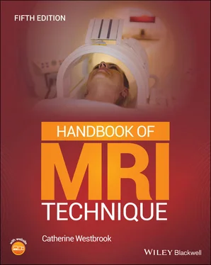
Catherine Westbrook - Handbook of MRI Technique
Здесь есть возможность читать онлайн «Catherine Westbrook - Handbook of MRI Technique» — ознакомительный отрывок электронной книги совершенно бесплатно, а после прочтения отрывка купить полную версию. В некоторых случаях можно слушать аудио, скачать через торрент в формате fb2 и присутствует краткое содержание. Жанр: unrecognised, на английском языке. Описание произведения, (предисловие) а так же отзывы посетителей доступны на портале библиотеки ЛибКат.
- Название:Handbook of MRI Technique
- Автор:
- Жанр:
- Год:неизвестен
- ISBN:нет данных
- Рейтинг книги:3 / 5. Голосов: 1
-
Избранное:Добавить в избранное
- Отзывы:
-
Ваша оценка:
- 60
- 1
- 2
- 3
- 4
- 5
Handbook of MRI Technique: краткое содержание, описание и аннотация
Предлагаем к чтению аннотацию, описание, краткое содержание или предисловие (зависит от того, что написал сам автор книги «Handbook of MRI Technique»). Если вы не нашли необходимую информацию о книге — напишите в комментариях, мы постараемся отыскать её.
FIFTH EDITION Handbook of MRI Technique.
Handbook of MRI Technique
Handbook of MRI Technique — читать онлайн ознакомительный отрывок
Ниже представлен текст книги, разбитый по страницам. Система сохранения места последней прочитанной страницы, позволяет с удобством читать онлайн бесплатно книгу «Handbook of MRI Technique», без необходимости каждый раз заново искать на чём Вы остановились. Поставьте закладку, и сможете в любой момент перейти на страницу, на которой закончили чтение.
Интервал:
Закладка:
8 Chapter 9Figure 9.1 Sagittal view of the spine showing vertebral levels.Figure 9.2 The components of the cervical spine and spinal cord.Figure 9.3 Sagittal localizer showing slice prescription for axial imaging o...Figure 9.4 Sagittal localizer showing slice prescription for axial/oblique s...Figure 9.5 Sagittal FSE/TSE T2‐weighted midline image through the cervical c...Figure 9.6 Axial FSE/TSE T2‐weighted image of the cervical cord demonstratin...Figure 9.7 Axial balanced GRE T2*‐weighted image through the cervical cord....Figure 9.8 Sagittal localizer showing slice prescription for axial imaging o...Figure 9.9 Sagittal FSE/TSE T2‐weighted image of the thoracic spine.Figure 9.10 Axial/oblique FSE/TSE T2‐weighted image through the thoracic cor...Figure 9.11 Sagittal localizer showing slice prescription for axial imaging ...Figure 9.12 Sagittal localizer showing slice prescription for axial imaging ...Figure 9.13 Sagittal FSE/TSE T1‐weighted image demonstrating an acute fractu...Figure 9.14 Axial/oblique FSE/TSE T2‐weighted image of the lumbar spine demo...Figure 9.15 Sagittal STIR of the lumbar spine.Figure 9.16 Sagittal FSE/TSE T1‐weighted image (left) and T2‐weighted image ...Figure 9.17 Sagittal/oblique balanced GRE T2*‐weighted image through the cer...
9 Chapter 10Figure 10.1 Anterior view of the components of the chest cavity.Figure 10.2 Coronal localizer of the chest showing prescription of axial sli...Figure 10.3 Axial CSE T1‐weighted gated image of the chest.Figure 10.4 Axial FSE/TSE T2‐weighted image with radial k ‐space demonstratin...Figure 10.5 Axial FSE/TSE T1‐weighted image of the chest with phase A to P....Figure 10.6 Axial FSE/TSE T1‐weighted image of the chest with phase L to R....Figure 10.7 The great vessels and chambers of the heart.Figure 10.8 The cardiac circulation.Figure 10.9 Coronal localizer through the chest cavity demonstrating slice p...Figure 10.10 Axial localizer through the heart showing slice angulation for ...Figure 10.11 Two‐chamber long‐axis view.Figure 10.12 Long axis view with slice angulation for the four‐chamber view....Figure 10.13 Four‐chamber view.Figure 10.14 Long axis view with slice angulation and boundaries for the sho...Figure 10.15 Short‐axis view.Figure 10.16 Axial images showing bright‐blood imaging (above) and black‐blo...Figure 10.17 Coronal fast spoiled GRE T1‐weighted image acquired after contr...Figure 10.18 Coronary artery imaging after contrast enhancement.Figure 10.19 Sagittal section through the breast.Figure 10.20 Bilateral multi‐channel breast coil.Figure 10.21 Axial spoiled GRE T1‐weighted image of the breasts.Figure 10.22 Axial FSE/TSE T2‐weighted image of the breasts with fat suppres...Figure 10.23 Axial 3D spoiled GRE T1‐weighted image of the breasts pre‐contr...Figure 10.24 Axial 3D spoiled GRE T1‐weighted image of the breasts post‐cont...Figure 10.25 Sagittal 3D spoiled GRE T1‐weighted image of the breasts post‐c...Figure 10.26 MIP post‐processed image of the breasts.Figure 10.27 Sagittal STIR of a breast implant.Figure 10.28 Axial FSE/TSE T1‐weighted unilateral image of a breast.Figure 10.29 The components of the brachial plexus.Figure 10.30 Coronal CSE T1‐weighted image of a normal brachial plexus.Figure 10.31 Coronal 3D STIR image of a normal brachial plexus.
10 Chapter 11Figure 11.1 The components of the liver and biliary system.Figure 11.2 Coronal localizer through the abdomen demonstrating slice prescr...Figure 11.3 Axial spoiled GRE T1‐weighted out of phase image through the liv...Figure 11.4 Axial spoiled GRE T1‐weighted in phase image through the liver....Figure 11.5 Axial 3D FSE/TSE T2‐weighted fat suppressed image through the li...Figure 11.6 Axial SS‐FSE/TSE T2‐weighted image through the liver.Figure 11.7 Axial spoiled GRE T1‐weighted image with fat suppression (Dixon)...Figure 11.8 Coronal SS‐FSE/TSE image of the gallbladder (MRCP). A very long ...Figure 11.9 The urinary system and its vascular supply.Figure 11.10 Coronal localizer through the abdomen demonstrating slice presc...Figure 11.11 Axial spoiled GRE T1‐weighted image with fat suppression (Dixon...Figure 11.12 Axial spoiled GRE T1‐weighted image with fat suppression (Dixon...Figure 11.13 Coronal localizer through the abdomen demonstrating slice presc...Figure 11.14 MRU static‐fluid, coronal 3D FSE T2 highlighting both of the ur...Figure 11.15 MRU excretory, coronal spoiled GRE T1‐weighted with Gd showing ...Figure 11.16 The pancreas and related structures.Figure 11.17 Coronal localizer through the abdomen demonstrating slice presc...Figure 11.18 Axial spoiled GRE T1‐weighted image with fat suppression (Dixon...Figure 11.19 Axial FSE/TSE T2‐weighted image of the pancreas.Figure 11.20 Axial SS‐FSE/TSE T2‐weighted image of the pancreas during free ...Figure 11.21 MRCP of the pancreatic duct.Figure 11.22 Coronal FSE/TSE T2‐weighted with fat suppression through the sm...Figure 11.23 Coronal spoiled GRE T1‐weighted image with fat suppression (Dix...Figure 11.24 Coronal spoiled GRE T1‐weighted with contrast enhancement showi...Figure 11.25 Coronal spoiled GRE T1‐weighted image of the renal arteries dur...
11 Chapter 12Figure 12.1 Sagittal section through the male pelvis showing midline structu...Figure 12.2 Coronal localizer through the pelvis to show slice prescription ...Figure 12.3 Sagittal FSE/TSE T2‐weighted image of the prostate.Figure 12.4 Axial FSE/TSE T2‐weighted image of the prostate.Figure 12.5 Coronal FSE/TSE T2‐weighted image of the prostate.Figure 12.6 Axial FSE/TSE T1‐weighted image of the normal male pelvis.Figure 12.7 Axial DWI of the prostate using an EPI acquisition.Figure 12.8 Axial spoiled GRE T1‐weighted image of the prostate.Figure 12.9 Axial FSE/TSE T2‐weighted image of the scrotum acquired with a s...Figure 12.10 Sagittal section through the female pelvis showing midline stru... Figure 12.11 Sagittal FSE/TSE T2‐weighted image through the female pelvis sh...Figure 12.12 Sagittal FSE/TSE T2‐weighted image demonstrating a large cervic...Figure 12.13 Axial spoiled GRE T1‐weighted fat suppressed (Dixon) image with...
12 Chapter 13Figure 13.1 Anterior view of the right shoulder showing bony structures and ...Figure 13.2 Coronal localizer showing slice prescription boundaries for axia...Figure 13.3 Axial localizer of the shoulder showing the angle of the suprasp...Figure 13.4 Axial localizer showing slice prescription boundaries and angula...Figure 13.5 Coronal localizer showing slice prescription boundaries for sagi... Figure 13.6 Coronal/oblique FSE/TSE T2‐weighted image of the shoulder.Figure 13.7 Coronal/oblique FSE/TSE PD‐weighted image with fat suppression....Figure 13.8 Sagittal/oblique FSE/TSE PD‐weighted image with fat suppression ...Figure 13.9 Coronal/oblique FSE/TSE T1‐weighted MR arthrogram of the shoulde...Figure 13.10 Axial FSE/TSE T1‐weighted MR arthrogram of the shoulder.Figure 13.11 Anterior view of the right humerus.Figure 13.12 Coronal STIR with radial k‐space showing a complete tear of the...Figure 13.13 Coronal FSE/TSE T1‐weighted image of the humerus.Figure 13.14 Axial FSE/TSE PD‐weighted image of the humerus with fat suppres...Figure 13.15 Anterior view of the right elbow showing the bony components.Figure 13.16 Sagittal view of the right elbow showing ligaments on the later...Figure 13.17 Coronal localizer showing slice prescription and angulation for...Figure 13.18 Coronal localizer showing slice prescription and angulation for...Figure 13.19 Coronal FSE/TSE T1‐weighted image of the elbow.Figure 13.20 Coronal FSE/TSE PD‐weighted image of the elbow with fat suppres... Figure 13.21 Sagittal FSE/TSE PD‐weighted image of the elbow with fat suppre...Figure 13.22 Axial FSE/TSE PD‐weighted image of the elbow.Figure 13.23 Anterior view of the right radius and ulna. Figure 13.24 Coronal FSE/TSE T1‐weighted image of the forearm.Figure 13.25 Bony structures of the wrist.Figure 13.26 Axial localizer of the wrist showing slice prescription boundar...Figure 13.27 Coronal localizer showing slice prescription for sagittal imagi...Figure 13.28 Coronal localizer showing slice prescription for axial imaging ... Figure 13.29 Axial FSE/TSE T2‐weighted image through the carpal tunnel.Figure 13.30 Axial FSE/TSE T1‐weighted image through the carpal tunnel.Figure 13.31 Coronal FSE/TSE PD‐weighted image of the hand with fat suppress...Figure 13.32 Axial FSE/TSE PD‐weighted image of the fingers with fat suppres...
Читать дальшеИнтервал:
Закладка:
Похожие книги на «Handbook of MRI Technique»
Представляем Вашему вниманию похожие книги на «Handbook of MRI Technique» списком для выбора. Мы отобрали схожую по названию и смыслу литературу в надежде предоставить читателям больше вариантов отыскать новые, интересные, ещё непрочитанные произведения.
Обсуждение, отзывы о книге «Handbook of MRI Technique» и просто собственные мнения читателей. Оставьте ваши комментарии, напишите, что Вы думаете о произведении, его смысле или главных героях. Укажите что конкретно понравилось, а что нет, и почему Вы так считаете.


