Daniel Buser - 30 Years of Guided Bone Regeneration
Здесь есть возможность читать онлайн «Daniel Buser - 30 Years of Guided Bone Regeneration» — ознакомительный отрывок электронной книги совершенно бесплатно, а после прочтения отрывка купить полную версию. В некоторых случаях можно слушать аудио, скачать через торрент в формате fb2 и присутствует краткое содержание. Жанр: unrecognised, на английском языке. Описание произведения, (предисловие) а так же отзывы посетителей доступны на портале библиотеки ЛибКат.
- Название:30 Years of Guided Bone Regeneration
- Автор:
- Жанр:
- Год:неизвестен
- ISBN:нет данных
- Рейтинг книги:3 / 5. Голосов: 1
-
Избранное:Добавить в избранное
- Отзывы:
-
Ваша оценка:
30 Years of Guided Bone Regeneration: краткое содержание, описание и аннотация
Предлагаем к чтению аннотацию, описание, краткое содержание или предисловие (зависит от того, что написал сам автор книги «30 Years of Guided Bone Regeneration»). Если вы не нашли необходимую информацию о книге — напишите в комментариях, мы постараемся отыскать её.
30 Years of Guided Bone Regeneration — читать онлайн ознакомительный отрывок
Ниже представлен текст книги, разбитый по страницам. Система сохранения места последней прочитанной страницы, позволяет с удобством читать онлайн бесплатно книгу «30 Years of Guided Bone Regeneration», без необходимости каждый раз заново искать на чём Вы остановились. Поставьте закладку, и сможете в любой момент перейти на страницу, на которой закончили чтение.
Интервал:
Закладка:
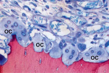
Fig 2-8Light micrograph showing osteoclasts (OC) in Howship lacunae formed in the alveolar bone (fuchsin and toluidine blue stain).
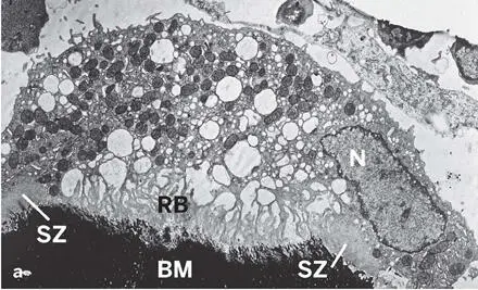
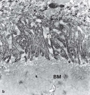
Fig 2-9 (a) Transmission electron micrograph showing an osteoclast with the ruffled border (RB), the sealing zone (SZ), numerous mitochondria, vesicles, and vacuoles, but only a single nuclear profile (N). The ruffled border represents the site of bone matrix (BM) dissolution and degradation (undecalcified ultrathin section). (b) Enlarged transmission electron micrograph of the ruffled border (RB) and bone matrix (BM) (decalcified ultrathin section).
Macrophages in bone (osteal macrophages), another type of cells of hematopoietic origin, are also called osteomacs . These resident cells play substantial roles in bone biology, with key functions in regulating bone formation and remodeling. Furthermore, they are among the first cells that come in contact with implanted biomaterials used for GBR, and they can differentiate toward classic M1 or M2 macrophages or subsequently fuse into osteoclasts or other multinucleated giant cells. 5
Bone matrix formation and mineralization
The osteoblast synthesizes a mixture of macromolecules that are secreted into the extracellular milieu to form the bone matrix—the osteoid or prebone—which consists of water, mineral, collagens, and noncollagenous macromolecules, the latter usually called noncollagenous proteins . The biochemical composition of bone has previously been reviewed, 22 – 27 and collagens play structural and morphogenic roles. 28 In mineralized tissues, they interact with various noncollagenous proteins and provide a scaffold for the accommodation of the mineral crystals. 29 The noncollagenous proteins of bone can roughly be classified into glycoproteins, proteoglycans, plasma-derived proteins, growth factors, and other macromolecules. In addition to its structural function, the bone matrix harbors molecules that play roles in biomineralization and matrix-cell interactions, and it also serves as a reservoir for growth factors and cytokines that may be released while osteoclasts resorb the bone matrix.
At a certain distance from the osteoblast, at the mineralization front, the osteoid converts into mineralized bone. The mineralization of woven bone is initiated by matrix vesicles. In contrast, matrix vesicles are rarely seen in the osteoid of mineralizing lamellar bone. However, the first mineral to appear among the collagen fibrils may be found at small discrete foci that are distributed within the osteoid and accumulate at the mineralization front (Fig 2-10). The colocalization of noncollagenous bone proteins such as osteopontin (Fig 2-11) and bone sialoprotein with these small mineralization foci and with amorphous gray or reticular patches of mineralized bone indicates the association of these proteins with the mineralization process. Bone acidic glycoprotein 75 and osteocalcin, on the other hand, show a diffuse distribution pattern throughout the mineralized bone matrix. 22
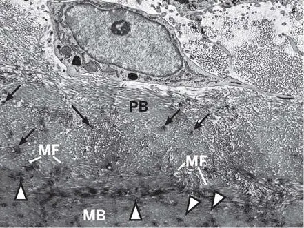
Fig 2-10Transmission electron micrograph illustrating an osteoblast, the osteoid or prebone (PB), the mineralization front (MF), and the mineralized bone matrix (MB). Mineralization foci (arrows) and “gray patches” (arrowheads) are visible in the osteoid and in the mineralized bone matrix, respectively.
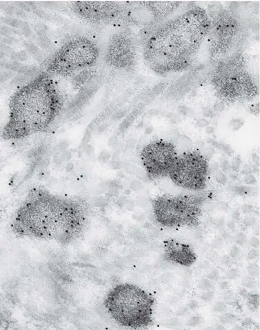
Fig 2-11High-resolution immunocytochemistry showing an association between gold particle labeling for osteopontin and mineralization foci in the osteoid.
Bone Modeling and Remodeling
Structural aspects
Throughout life, the skeleton undergoes continuous physiologic remodeling, which serves the purpose of repair and mechanical adaptation. The terms modeling and remodeling often cause confusion. Modeling indicates a change in shape, whereas remodeling refers to tissue replacement or substitution without a change in architecture. Cortical bone remodeling may be distinguished from trabecular bone remodeling.
Cortical bone
The fundamental structural unit of cortical bone remodeling is the osteon (see Fig 2-2). Primary osteons are formed during appositional growth, whereas secondary osteons are the result of matrix substitution. The remodeling sequence is illustrated in transverse sections in Fig 2-12. First, a resorption canal is formed by osteoclasts. Later, osteoblasts appear and start refilling the canal with concentric lamellae. In compact human bone, completed secondary osteons have an outer diameter of 200 to 250 µm, with the central vascular canal—the Haversian canal—measuring 50 to 80 µm. 30 As a coherent cylindrical structure, secondary osteons rarely measure more than 2 to 3 mm in length. At intervals of 0.5 to 1.0 mm, they are interconnected by transverse vascular channels called Volkmann canals . Longitudinal sections of newly formed osteons have shown that bone resorption and deposition are coupled in time and space and occur in discrete remodeling sites called bone metabolizing units (BMUs). At the advancing front of a resorption canal, osteoclasts are assembled in a cutting cone (Fig 2-13). While the osteoclasts advance longitudinally, they widen the resorption canal up to its final diameter. The tip of a vessel loop follows immediately behind the osteoclasts. This loop lies in the center of the canal and is surrounded by perivascular cells thought to include osteoblast precursor cells. In the reversal phase (ie, between bone resorption and bone matrix deposition), the wall of the canal is lined by mononuclear cells. Further in the back of the cutting cone, osteoblasts appear and deposit the lamellar bone matrix, which will later mineralize. Depending on the species, completion of the osteon requires 2 to 4 months. These measurements and calculations are based on sequential fluorochrome labeling (Fig 2-14) and also allow an accurate determination of the resorption rate of the osteoclast in longitudinal sections, which amounts to 50 to 60 µm per day in dogs. 31 – 33

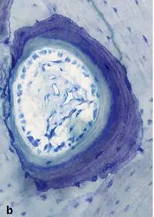
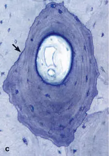
Fig 2-12Cortical bone remodeling in the humerus of an adult man. (a) Resorption canal with osteoclasts (arrows) . (b) Concentric lamellae deposited by osteoblasts. (c) Completed secondary osteon, bound by a cement line (arrow) (transverse sections through evolving secondary osteons; toluidine blue surface stain).
Читать дальшеИнтервал:
Закладка:
Похожие книги на «30 Years of Guided Bone Regeneration»
Представляем Вашему вниманию похожие книги на «30 Years of Guided Bone Regeneration» списком для выбора. Мы отобрали схожую по названию и смыслу литературу в надежде предоставить читателям больше вариантов отыскать новые, интересные, ещё непрочитанные произведения.


Обсуждение, отзывы о книге «30 Years of Guided Bone Regeneration» и просто собственные мнения читателей. Оставьте ваши комментарии, напишите, что Вы думаете о произведении, его смысле или главных героях. Укажите что конкретно понравилось, а что нет, и почему Вы так считаете.