Adrian Becker - Orthodontic Treatment of Impacted Teeth
Здесь есть возможность читать онлайн «Adrian Becker - Orthodontic Treatment of Impacted Teeth» — ознакомительный отрывок электронной книги совершенно бесплатно, а после прочтения отрывка купить полную версию. В некоторых случаях можно слушать аудио, скачать через торрент в формате fb2 и присутствует краткое содержание. Жанр: unrecognised, на английском языке. Описание произведения, (предисловие) а так же отзывы посетителей доступны на портале библиотеки ЛибКат.
- Название:Orthodontic Treatment of Impacted Teeth
- Автор:
- Жанр:
- Год:неизвестен
- ISBN:нет данных
- Рейтинг книги:4 / 5. Голосов: 1
-
Избранное:Добавить в избранное
- Отзывы:
-
Ваша оценка:
Orthodontic Treatment of Impacted Teeth: краткое содержание, описание и аннотация
Предлагаем к чтению аннотацию, описание, краткое содержание или предисловие (зависит от того, что написал сам автор книги «Orthodontic Treatment of Impacted Teeth»). Если вы не нашли необходимую информацию о книге — напишите в комментариях, мы постараемся отыскать её.
Provides protocols for common cases as well as complex and rare presentations Contains individual chapters on the specific aspects of the diagnosis and treatment of impaction in each of the different types of teeth Covers prevalence, etiology, diagnosis, attitudes to treatment, treatment timing, treatment methods, and prognosis Features more than 1,000 high-quality color images and illustrations
remains essential reading for all specialist orthodontists, academic researchers and instructors, oral and maxillofacial surgeons, and advanced students in orthodontics.
Orthodontic Treatment of Impacted Teeth — читать онлайн ознакомительный отрывок
Ниже представлен текст книги, разбитый по страницам. Система сохранения места последней прочитанной страницы, позволяет с удобством читать онлайн бесплатно книгу «Orthodontic Treatment of Impacted Teeth», без необходимости каждый раз заново искать на чём Вы остановились. Поставьте закладку, и сможете в любой момент перейти на страницу, на которой закончили чтение.
Интервал:
Закладка:
The failure of an impacted tooth to erupt will inevitably disturb the eruption patterns of the adjacent teeth, which, as a result, will then assume abnormal relationships to one another. Such relationships are usually characterized by tipping and space loss. The overall result of this is that this situation creates a secondary physical impediment to the eruption of the impacted tooth.
Infra‐occlusion
In Chapter 11we shall demonstrate in greater detail that infra‐occluded permanent teeth are sometimes ankylosed to the surrounding bone and will consequently not respond to orthodontic traction. For the purposes of this present chapter, it should be briefly noted that the ankylosed area of the root is often minimal and may easily be detached by deliberate, but gentle, luxation of the tooth. This will normally be carried out using an elevator or extraction forceps and is done in such a way as to release the rigid and inflexible connection of the bony union. The aim is neither to remove the tooth from its socket nor even to tear the periodontal fibres (which is inevitable). The purpose is to bring the tooth to a greater than normal degree of mobility.
A frequent and undesired consequence of this procedure is a re‐healing and re‐attachment of the tooth to its former ankylotic connection. Accordingly, this approach can only be successful if the traction force is applied to the tooth immediately upon luxation and maintained continuously active. The re‐healing of the bone will be modified by a localized microcosm of distraction osteogenesis [17, 18] created by the traction process. It follows that if the traction force is allowed to decay to ineffectiveness (between patients’ visits for adjustments), re‐ankylosis will result and the tooth will stop moving. Accordingly, in order to be effective, the traction must be of sufficient magnitude and range to cause distraction and to remain active between one visit for adjustment and the next.
The principles of the surgical exposure of impacted teeth
In general, there are two basic approaches to the surgical exposure of impacted teeth: the open eruption technique and the closed eruption technique [19]. A description and comparison of each of these approaches is set out below.
The open eruption technique
Historically, this was the first method used to expose impacted teeth. The tooth was exposed and remained open to the oral environment [20]. Following the removal of the soft tissue and of the bone that covered it, the newly exposed tooth was surrounded by freshly trimmed thick mucosa of the palate or the raw cut edge of the labial/buccal oral mucosa.
The open eruption technique may be performed in several ways but, essentially, these fall into two categories: the window technique and the apically repositioned flap technique.
The window technique involves the surgical excision of a circular section of the overlying mucosa and the thin bony covering. This method is illustrated in the advertising fliers that are distributed widely by companies to market soft tissue laser units. The method has the advantage that it is the simplest, most conservative and most direct method of exposing a tooth, which is palpable immediately under the oral mucosa. It may often be performed using only surface anaesthetic spray or gel. An attachment is immediately bonded to the tooth, enabling orthodontically encouraged eruption to proceed and facilitating complete alignment within a very short time. While this method obviously represents a significant advantage in the treatment of a young patient, the long‐term outcome of the procedure may be characterized by loose oral mucosa on the labial side of the tooth. This is not of attached gustatory epithelium, but rather a mobile, thin, oral mucosa, which does not function well as gingival margin tissue. This situation has been widely documented in the periodontal literature.
It is therefore clear that, in the majority of cases of labially displaced teeth (due to their relative height), this entire surgical procedure is usually inappropriate and contraindicated in relation to the mobile area of the oral mucosa, above the level of attached gingiva. However, if the patient has a very wide band of attached gingiva ( Figure 5.4) and a labially impacted tooth is situated well down in this band, a simple removal of the tissue overlying the crown could satisfactorily still leave 1–2 mm of bound epithelial attachment inferior to the free, movable, oral mucosal lining of the sulcus [7].
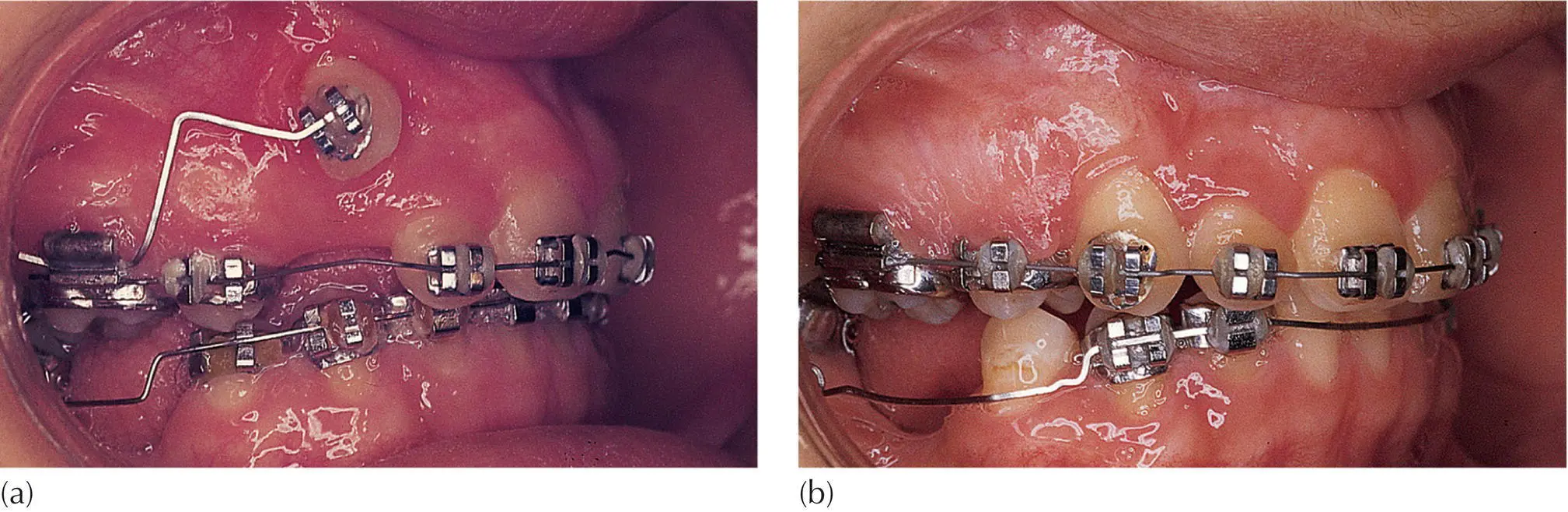
Fig. 5.4 (a) A high buccal canine exposed by circular incision in the very wide band of attached gingiva in this patient. (b) The tooth has been brought down together with attached gingiva on its labial side.
Courtesy of Dr G. Engel.
By contrast, the palatal mucosa is very thick and securely bound down to the underlying bone. In consequence, following its eruption into the palate of a palatally impacted tooth, no parallel precautions need be taken in order to ensure a good attachment for its final periodontal status [5–7].
When the window technique is used on the palatal side, the cut edges of the wound need to be substantially trimmed back and the dental follicle usually needs to be removed in its entirety. The aim of this is to prevent re‐closure of the very considerable thickness of palatal soft tissue over the exposed tooth. Additionally, where there is a deeply buried palatal canine, the patency of the exposure will need to be maintained with the aid of a surgical pack.
The apically repositioned flap technique for performing open exposure is with an apically repositioned flap on the labial/buccal side. This method is designed to improve the periodontal outcome by ensuring, in the final instance, that attached gingiva covers the labial aspect of the erupted tooth. This is achieved by raising a labial flap, taken from the crest of the ridge, and relocating it higher up on the crown of the newly exposed tooth. The method is a recognized and accepted procedure in periodontics and was first described by Vanarsdall and Corn [21, 22] in the context of surgical and orthodontic treatment of unerupted labially displaced teeth. According to these researchers, in the absence of the deciduous canine, a muco‐gingival flap, which incorporates attached gingiva, is raised from the crest of the ridge ( Figure 5.1). If a deciduous canine is present, the flap will be designed to include the entire area of buccal gingiva that invests it and the deciduous tooth itself will be extracted. In either case, the canine is exposed by detaching a flap from the underlying hard tissue, some way up into the vestibulum. The flap is then sutured to the labial side of the exposed crown of the permanent canine and will overlie its cervical area and cover the denuded periosteum. The remainder of the crown will be exposed. Subsequent eruption of the tooth will be accompanied by the healing of the gingival tissue. When the tooth takes up its final position in the arch, it will be found to be invested with a good width of attached gingiva.
The apically repositioned flap method of exposure is best suited to labially/buccally impacted teeth, which are situated above the band of attached gingiva and are not displaced mesially or distally from their place in the dental arch. However, if the case presents with more than a minor degree of mesial or distal displacement, a raised and full‐thickness, soft tissue flap will result in unacceptable denuding of the alveolar bone covering the adjacent tooth and will contraindicate the use of this surgical modality. In order to overcome this, a partial‐thickness surgical flap may be raised, which will then leave the donor area invested with a connective tissue cover [23] to heal over by epithelial proliferation.
Читать дальшеИнтервал:
Закладка:
Похожие книги на «Orthodontic Treatment of Impacted Teeth»
Представляем Вашему вниманию похожие книги на «Orthodontic Treatment of Impacted Teeth» списком для выбора. Мы отобрали схожую по названию и смыслу литературу в надежде предоставить читателям больше вариантов отыскать новые, интересные, ещё непрочитанные произведения.


Обсуждение, отзывы о книге «Orthodontic Treatment of Impacted Teeth» и просто собственные мнения читателей. Оставьте ваши комментарии, напишите, что Вы думаете о произведении, его смысле или главных героях. Укажите что конкретно понравилось, а что нет, и почему Вы так считаете.