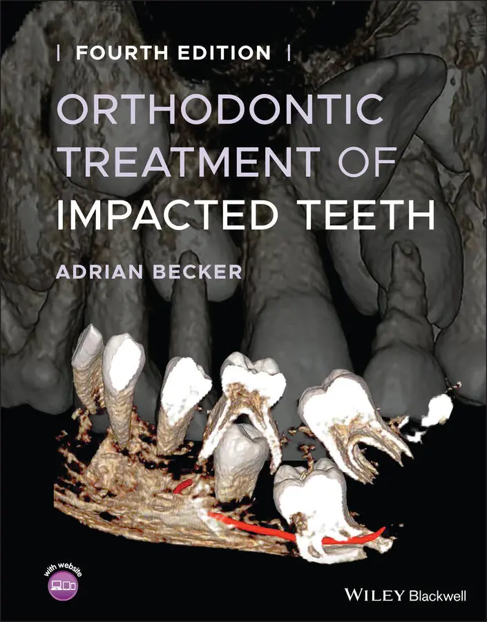
Adrian Becker - Orthodontic Treatment of Impacted Teeth
Здесь есть возможность читать онлайн «Adrian Becker - Orthodontic Treatment of Impacted Teeth» — ознакомительный отрывок электронной книги совершенно бесплатно, а после прочтения отрывка купить полную версию. В некоторых случаях можно слушать аудио, скачать через торрент в формате fb2 и присутствует краткое содержание. Жанр: unrecognised, на английском языке. Описание произведения, (предисловие) а так же отзывы посетителей доступны на портале библиотеки ЛибКат.
- Название:Orthodontic Treatment of Impacted Teeth
- Автор:
- Жанр:
- Год:неизвестен
- ISBN:нет данных
- Рейтинг книги:4 / 5. Голосов: 1
-
Избранное:Добавить в избранное
- Отзывы:
-
Ваша оценка:
Orthodontic Treatment of Impacted Teeth: краткое содержание, описание и аннотация
Предлагаем к чтению аннотацию, описание, краткое содержание или предисловие (зависит от того, что написал сам автор книги «Orthodontic Treatment of Impacted Teeth»). Если вы не нашли необходимую информацию о книге — напишите в комментариях, мы постараемся отыскать её.
Provides protocols for common cases as well as complex and rare presentations Contains individual chapters on the specific aspects of the diagnosis and treatment of impaction in each of the different types of teeth Covers prevalence, etiology, diagnosis, attitudes to treatment, treatment timing, treatment methods, and prognosis Features more than 1,000 high-quality color images and illustrations
remains essential reading for all specialist orthodontists, academic researchers and instructors, oral and maxillofacial surgeons, and advanced students in orthodontics.
Orthodontic Treatment of Impacted Teeth — читать онлайн ознакомительный отрывок
Ниже представлен текст книги, разбитый по страницам. Система сохранения места последней прочитанной страницы, позволяет с удобством читать онлайн бесплатно книгу «Orthodontic Treatment of Impacted Teeth», без необходимости каждый раз заново искать на чём Вы остановились. Поставьте закладку, и сможете в любой момент перейти на страницу, на которой закончили чтение.
Интервал:
Закладка:
2 2. Hunter SB. The radiographic assessment of the unerupted maxillary canine. Br Dent J 1981; 150: 151–155.
3 3. Mason RA. A Guide to Dental Radiography, 2nd ed. Bristol: Wright PSG, 1982.
4 4. Ong A. An alternative technique to the vertex/true occlusal view. Am J Orthod Dentofac Orthop 1994; 106: 621–626.
5 5. Clark CA. A method of ascertaining the relative position of unerupted teeth by means of film radiographs. Proc Roy Soc Med (Sec. Odont) 1910; 3: 87–90.
6 6. Armstrong C, Johnston C, Burden D, Stevenson M. Localizing ectopic maxillary canines – horizontal or vertical parallax? Eur J Orthod 2003; 25: 585–589.
7 7. Jacobs SG. Localisation of the unerupted maxillary canine. Aust Orthod J 1986; 9: 313–316.
8 8. Jacobs SG. Exercises in the localisation of unerupted teeth. Aust Orthod J 1987; 10: 33–35, 58–60.
9 9. Nohadani N, Pohl Y, Ruf S. Displaced premolars in panoramic radiography—fact or fallacy? Angle Orthod 2008, 78: 309–316.
10 10. Chaushu S, Chaushu G, Becker A. The use of panoramic radiographs to localize maxillary palatal canines. Oral Surg Oral Med Oral Pathol Oral Radiol Endod 1999; 88: 511–516.
11 11. Chaushu S, Chaushu G, Becker A. Reliability of a method for the localization of displaced maxillary canines using a single panoramic radiograph. Clin Orthod Res 1999; 2: 194–199.
12 12. Wolf JE, Mattila K. Localization of impacted maxillary canines by panoramic tomography. Dentomaxillofac Radiol 1979; 8: 85–91.
13 13. Fox NA, Fletcher GA, Horner K. Localising maxillary canines using dental panoramic tomography. Br Dent J 1995; 179: 416–420.
14 14. Kohavi D, Zilberman Y, Becker A. Periodontal status following the alignment of buccally ectopic maxillary canine teeth. Am J Orthod 1984; 85: 78–82.
15 15. Becker A, Kohavi D, Zilberman Y. Periodontal status following the alignment of palatally impacted canine teeth. Am J Orthod 1983; 84: 332–336.
16 16. Kohavi D, Becker A, Zilberman Y. Surgical exposure, orthodontic movement and final tooth position as factors in periodontal breakdown of treated palatally impacted canines. Am J Orthod 1984; 85: 72–77.
17 17. Becker A, Chaushu S. Long‐term follow‐up of severely resorbed maxillary incisors following resolution of etiologically‐associated canine impaction. Am J Orthod Dentofacial Orthop 2005; 127: 650–654.
18 18. Ericson S, Kurol J. CT diagnosis of ectopically erupting maxillary canines – a case report. Eur J Orthod 1988; 10: 115–120.
19 19. Ericson S, Kurol J. Resorption of maxillary lateral incisors caused by ectopic eruption of canines. Am J Orthod Dentofacial Orthop 1988; 94: 503–513.
20 20. Ericson S, Kurol J. Radiographic examination of ectopically erupting maxillary canines. Am J Orthod Dentofacial Orthop 1987; 91: 483–492.
21 21. Odegaard J. The treatment of a Class I malocclusion with two horizontally impacted maxillary canines. Am J Orthod Dentofacial Orthop 1997; 111: 357–365.
22 22. Becker A. Comment about making outcome of treatment more predictable. Am J Orthod Dentofacial Orthop 1997; 112: 17A–19A.
23 23. Ericson S, Kurol PJ. Resorption of incisors after ectopic eruption of maxillary canines: a CT study. Angle Orthod 2000; 70: 415–423.
24 24. Bodner L, Bar Ziv J, Becker A. Image accuracy of plain film radiography and computerized tomography in assessing morphological abnormality of impacted teeth. Am J Orthod Dentofacial Orthop 2001; 120: 623–628.
25 25. Dula K, Mini R, van der Stelt PF et al. Hypothetical mortality risk associated with spiral computed tomography of the maxilla and mandible. Eur J Oral Sci 1996; 104: 503–510.
26 26. Dula K, Mini R, van der Stelt PF, Buser D. The radiographic assessment of implant patients: decision‐making criteria. Int J Oral Maxillofac Implants 2001; 16: 80–89.
27 27. Scarfe WC, Azevedo B, Toghyani S, Farman AG. Cone beam computed tomographic imaging in orthodontics. Aust Dent J 2017; 62 (1 Suppl): 33–50.
28 28. Health Protection Agency. Ionising radiation exposure of the UK population: review. Ref: HPA‐RPD‐001. Chilton: Radiation Protection Division, 2005.
29 29. Barish RJ. In‐flight radiation exposure during pregnancy. Obstet Gynecol 2004; 103: 1326–1330.
30 30. Alara. Code of Federal Regulations (10 CFR 20.2003). United States Nuclear Regulatory Commission.
5 Surgical Exposure of Impacted Teeth
Adrian Becker
A brief history of surgery in relation to the treatment of impacted teeth
Aims of surgery for impacted teeth
Surgical intervention without orthodontic treatment
The surgical elimination of pathology
The principles of the surgical exposure of impacted teeth
Partial and full‐flap closure on the palatal side
A conservative attitude to the dental follicle
Pathological pressure necrosis
Quality‐of‐life issues following surgical exposure
Cooperation between surgeon and orthodontist
The team approach to attachment bonding
A brief history of surgery in relation to the treatment of impacted teeth
Prior to the 1950s, few orthodontists were prepared to adapt their skills and their ingenuity to the task of resolving the impaction of maxillary canines and incisors, many preferring to refer these patients to the oral surgeon. The decision regarding the method of treatment of a particular impacted tooth was usually made by the oral and maxillofacial surgeon (OMFS). It was OMFSs who considered the options, chose the one they felt was appropriate and stage‐managed the treatment process.
Surgeons would raise a flap, expose the tooth widely and only then make the decision whether to save the tooth or extract it. If, in their opinion, the impacted tooth could be brought into the dental arch, it would be left open to the oral environment with or without a surgical pack. If, in their judgement, this was unlikely to happen, they would extract the tooth on the spot and then write a note to that effect to the orthodontist. As can be imagined, many potentially retrievable, impacted teeth were thereby condemned to extraction.
The development of the role of the orthodontist in the rescue of impacted teeth was due to the realization that surgical treatment was just not enough. Whereas the elimination of the cause of the impaction and the provision of optimal space (by orthodontic means) did indeed provide a favourable environment to encourage autonomous eruption, it was clear that this alone was far from being universally successful. This led to the second realization: that orthodontic treatment alone was also not enough. It was acknowledged that, in order to achieve a more affirmative and quality result, with greater predictability, surgically afforded access would be required, together with the application of active and positive forces of traction/extrusion directly to the tooth.
From the early 1970s in the Hebrew University‐Hadassah School of Dental Medicine in Jerusalem, Israel, orthodontists joined forces with the OMFS at the chairside and in the operating theatre, to adapt and cement preformed canine orthodontic bands during the surgical procedure itself. This had been the procedure prior to the era of acid‐etching enamel and direct bonding of brackets. As a result, many more of these teeth were reclaimed and, in time, took their rightful place in the dental arch. However, in order to place a band, the entire crown needed to be dissected free of its dental follicle and clear of adjacent bleeding surfaces. This demanded radical surgery and efficient isolation of the tooth during the cementation process. Not every surgeon was willing to cooperate, thereby making the orthodontist much more selective in the choice of surgeon, particularly for difficult cases [1, 2].
Читать дальшеИнтервал:
Закладка:
Похожие книги на «Orthodontic Treatment of Impacted Teeth»
Представляем Вашему вниманию похожие книги на «Orthodontic Treatment of Impacted Teeth» списком для выбора. Мы отобрали схожую по названию и смыслу литературу в надежде предоставить читателям больше вариантов отыскать новые, интересные, ещё непрочитанные произведения.


Обсуждение, отзывы о книге «Orthodontic Treatment of Impacted Teeth» и просто собственные мнения читателей. Оставьте ваши комментарии, напишите, что Вы думаете о произведении, его смысле или главных героях. Укажите что конкретно понравилось, а что нет, и почему Вы так считаете.