Adrian Becker - Orthodontic Treatment of Impacted Teeth
Здесь есть возможность читать онлайн «Adrian Becker - Orthodontic Treatment of Impacted Teeth» — ознакомительный отрывок электронной книги совершенно бесплатно, а после прочтения отрывка купить полную версию. В некоторых случаях можно слушать аудио, скачать через торрент в формате fb2 и присутствует краткое содержание. Жанр: unrecognised, на английском языке. Описание произведения, (предисловие) а так же отзывы посетителей доступны на портале библиотеки ЛибКат.
- Название:Orthodontic Treatment of Impacted Teeth
- Автор:
- Жанр:
- Год:неизвестен
- ISBN:нет данных
- Рейтинг книги:4 / 5. Голосов: 1
-
Избранное:Добавить в избранное
- Отзывы:
-
Ваша оценка:
Orthodontic Treatment of Impacted Teeth: краткое содержание, описание и аннотация
Предлагаем к чтению аннотацию, описание, краткое содержание или предисловие (зависит от того, что написал сам автор книги «Orthodontic Treatment of Impacted Teeth»). Если вы не нашли необходимую информацию о книге — напишите в комментариях, мы постараемся отыскать её.
Provides protocols for common cases as well as complex and rare presentations Contains individual chapters on the specific aspects of the diagnosis and treatment of impaction in each of the different types of teeth Covers prevalence, etiology, diagnosis, attitudes to treatment, treatment timing, treatment methods, and prognosis Features more than 1,000 high-quality color images and illustrations
remains essential reading for all specialist orthodontists, academic researchers and instructors, oral and maxillofacial surgeons, and advanced students in orthodontics.
Orthodontic Treatment of Impacted Teeth — читать онлайн ознакомительный отрывок
Ниже представлен текст книги, разбитый по страницам. Система сохранения места последней прочитанной страницы, позволяет с удобством читать онлайн бесплатно книгу «Orthodontic Treatment of Impacted Teeth», без необходимости каждый раз заново искать на чём Вы остановились. Поставьте закладку, и сможете в любой момент перейти на страницу, на которой закончили чтение.
Интервал:
Закладка:
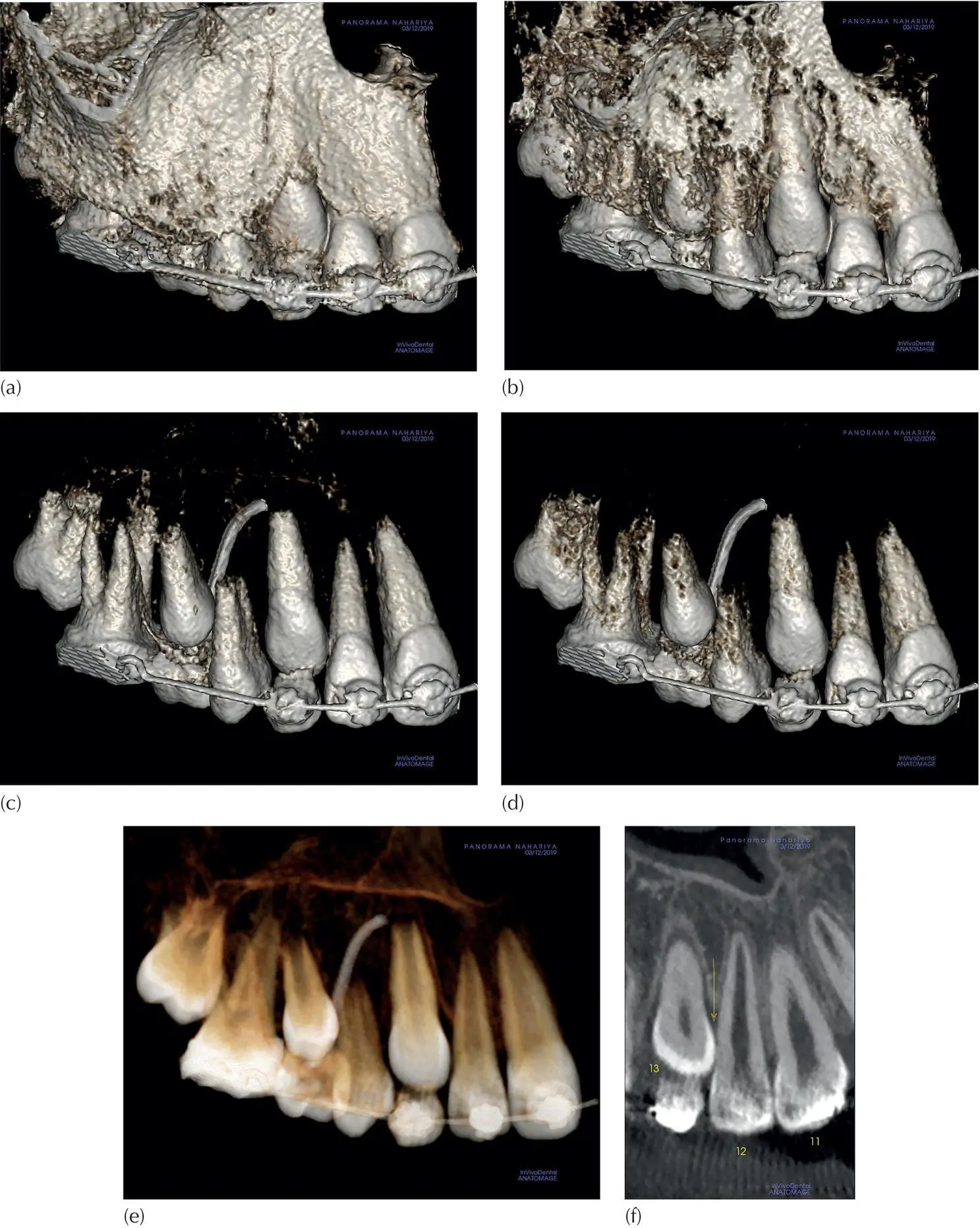
Fig. 4.13 Bone peeling in 3D. (a–d) Progressive bone peeling and how it may mislead by altering the teeth volume and interproximal contact. (e) Volume rendering in the 3D transparent mode. (f) Longitudinal slice cropped from the multi‐planar reconstruction screen ( Figure 4.14) showing the deepest point of interproximal contact (see text).
The multi‐planar reconstruction (MPR) screen presentation in Figure 4.14is a typical example. In order to define the exact mesio‐distal contact area between the lateral incisor and canine on the right side, the sagittal ( Figure 4.14b) and coronal ( Figure 4.14c) planes are tilted until the long axis of the incisor is brought exactly vertical. Once the tooth is vertically positioned, it may be rotated on its axis. The yellow line with arrows at both ends is a custom section tool, which has the ability to rotate around an axis marked at its centre. It is placed on the axial ( Figure 4.14a) view with its centre at the point at which it meets the tooth axis, on which rotation may be made. The window in Figure 4.14(d) displays the cut produced by the rotation tool. By rotating the tool, the tooth outline may be depicted at any point on the 360° circle. The deepest point of contact is illustrated in the window in Figure 4.14(d) and enlarged separately in Figure 4.13(f).
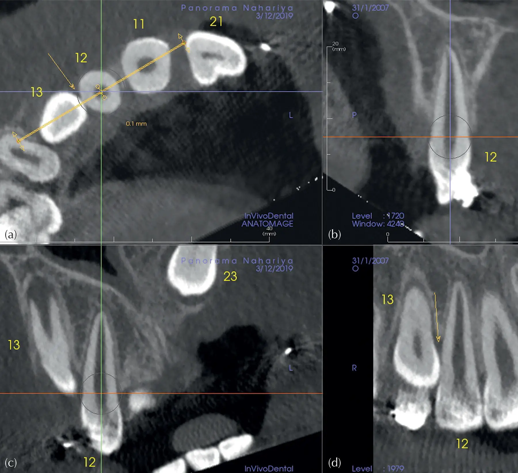
Fig. 4.14 A view of the multi‐planar reconstruction screen for Case 1, as presented in InVivoDental software (see text).
Peeling the 3D opaque bony mode and exploiting the 3D transparent mode offer many advantages in appreciating the inter‐relations of the teeth and the surrounding structures. Understanding of the process by which they are produced and their reducing effect on tooth volume are factors that often need to be taken into account.
In light of the foregoing discussion, it will not come as a surprise to learn that peeling of bone in the body of the mandible will result in much loss of teeth volume, particularly the thick bone of the buccal side of the mandible. In practice, because the densities of the teeth and the surrounding compact bone are of a similar order, it is largely impossible to peel this thick buccal bone without excessively reducing tooth volume and it becomes necessary to combine clipping and, in some cases, sculpting to achieve the desired results (as demonstrated in Case 2).
Case 2: Peeling, clipping and sculpting
In this case the clinical aim was to find aetiological evidence for the failure to erupt of the first mandibular molar on the right side. In order to enhance the 3D view, a combination of mild bone peeling, clipping and sculpting away part of the lingual cortical bone was executed (Animation 4.1 on this book’s website). Two additional animations (Animation 4.2 and Animation 4.3) will enhance the perception of the case. The latter animation shows how, by tilting the chin upwards (in the SW) and thereby placing the molar in a vertical position, animation becomes more informative (orthodontic treatment by Dr Ronen Zoizner).
The lesser density of the bone in the upper jaw makes the situation there much more favourable for obtaining good‐quality imaging of the impacted teeth, while maintaining tooth volume.
It is important to note that the 3D transparent view tends to deceptively show tooth volumes to be smaller than they are and, as such, should be treated with caution.
Automatic tooth segmentation
This procedure, based on artificial intelligence (AI), is already in use ( Figure 4.15). At the time of writing it is a third‐party service, which will undoubtedly be added to the CBCT software by the manufacturers. This is an excellent feature because it saves time and effort, but should only be used to obtain a general impression. Presently it is not accurate enough in relation to tooth volume. In the not too distant future, however, it is expected to improve, as with most applications that depend on AI. Tooth contact or minor resorption will need to be confirmed, preferably in the MPR screen.
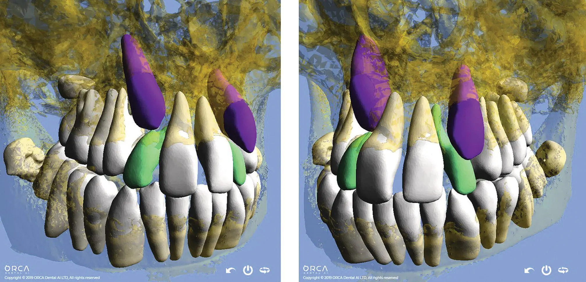
Fig. 4.15 Automatic segmentation, artificial intelligence (AI) driven. The software algorithm automatically segments the teeth and bone for each jaw separately and the inferior alveolar canal. A viewer that can run on a PC or a smartphone will allow each entity to be coloured, rotated in all axes and moved separately, as well as being removed and much more.
Courtesy of ORCA Dental AI.
Multi‐planar reconstruction
This is the basic screen in all CT software and displays the three anatomical planes: axial, coronal and sagittal. The initial display is dictated by the orientation in space in which the patient was positioned during the scan. The operator may then tilt the volume in all three axes and reach any desired position in space, i.e. the position of the patient's head may be virtually altered after the scan was taken. An initial overall impression of the situation may be gained by exploring the scanned volume, using the 3D volume‐rendering module. At the outset, however, in order to discover if there are any relevant and unexpected details in the general area, a careful examination of each of the three planes for incidental findings is essential.
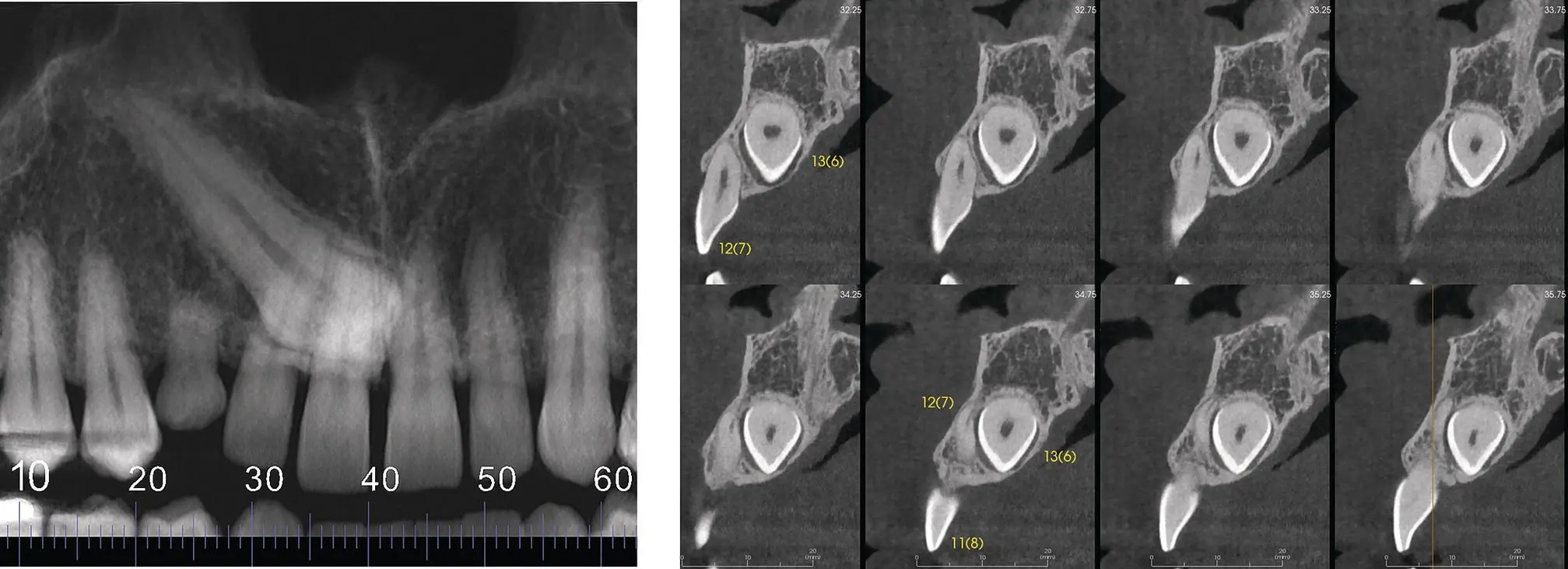
Fig. 4.16 Diagnosing resorption, cross‐sections. The lateral incisor #12(7) is tipped mesially. Cross‐sections are vertical cuts. Therefore in this case the cross‐sections cannot reveal the resorption to its full extent.
From this point on, the focus of the investigation is directed specifically to the impacted tooth/teeth. Many orthodontists will rely on the bucco‐lingual information, gleaned from a series of cross‐sectional cuts that are perpendicular to the curved panoramic cut. It is a common misconception that all the information in the bucco‐lingual plane should lie in these cross‐sections, which may often be 1 mm thick with 1 mm spacing from each other. In fact, this concept is a clear recipe for negligently failing to identify important information, because the angle of these sections cannot reveal all the relevant data (Case 3, Figures 4.16– 4.18).
The MPR screen contains all the information and it is a very important tool. The first step to be taken is a thorough three‐plane scrolling of the ROI, with slice thickness set to a minimum (the voxel size), in the absence of sharpening filters. The ability to tilt one or more planes is an advantage in achieving the appropriate and more diagnostic cutting/slicing angle for visualization. The action that adds the ultimate level of diagnosis is viewing each involved tooth separately. It is necessary to tilt each tooth to a vertical position and to examine it while rotating it through 360° and covering every possible angle ( Figures 4.14and 4.17). Additional scrolling through the axials along the vertical tooth will complete the process. Similarly, if the impacted tooth is located at an almost horizontal angle, it is tilted so that its long axis is exactly oriented in the horizontal and sagittal posture. Using the same rotating tool, on this occasion the tool should be placed on the tooth axis in the coronal plane window ( Figure 4.19c). After rotating the tool through 360°, additional scrolling through coronals along the tooth will complete the diagnostic information.
Читать дальшеИнтервал:
Закладка:
Похожие книги на «Orthodontic Treatment of Impacted Teeth»
Представляем Вашему вниманию похожие книги на «Orthodontic Treatment of Impacted Teeth» списком для выбора. Мы отобрали схожую по названию и смыслу литературу в надежде предоставить читателям больше вариантов отыскать новые, интересные, ещё непрочитанные произведения.


Обсуждение, отзывы о книге «Orthodontic Treatment of Impacted Teeth» и просто собственные мнения читателей. Оставьте ваши комментарии, напишите, что Вы думаете о произведении, его смысле или главных героях. Укажите что конкретно понравилось, а что нет, и почему Вы так считаете.