Adrian Becker - Orthodontic Treatment of Impacted Teeth
Здесь есть возможность читать онлайн «Adrian Becker - Orthodontic Treatment of Impacted Teeth» — ознакомительный отрывок электронной книги совершенно бесплатно, а после прочтения отрывка купить полную версию. В некоторых случаях можно слушать аудио, скачать через торрент в формате fb2 и присутствует краткое содержание. Жанр: unrecognised, на английском языке. Описание произведения, (предисловие) а так же отзывы посетителей доступны на портале библиотеки ЛибКат.
- Название:Orthodontic Treatment of Impacted Teeth
- Автор:
- Жанр:
- Год:неизвестен
- ISBN:нет данных
- Рейтинг книги:4 / 5. Голосов: 1
-
Избранное:Добавить в избранное
- Отзывы:
-
Ваша оценка:
Orthodontic Treatment of Impacted Teeth: краткое содержание, описание и аннотация
Предлагаем к чтению аннотацию, описание, краткое содержание или предисловие (зависит от того, что написал сам автор книги «Orthodontic Treatment of Impacted Teeth»). Если вы не нашли необходимую информацию о книге — напишите в комментариях, мы постараемся отыскать её.
Provides protocols for common cases as well as complex and rare presentations Contains individual chapters on the specific aspects of the diagnosis and treatment of impaction in each of the different types of teeth Covers prevalence, etiology, diagnosis, attitudes to treatment, treatment timing, treatment methods, and prognosis Features more than 1,000 high-quality color images and illustrations
remains essential reading for all specialist orthodontists, academic researchers and instructors, oral and maxillofacial surgeons, and advanced students in orthodontics.
Orthodontic Treatment of Impacted Teeth — читать онлайн ознакомительный отрывок
Ниже представлен текст книги, разбитый по страницам. Система сохранения места последней прочитанной страницы, позволяет с удобством читать онлайн бесплатно книгу «Orthodontic Treatment of Impacted Teeth», без необходимости каждый раз заново искать на чём Вы остановились. Поставьте закладку, и сможете в любой момент перейти на страницу, на которой закончили чтение.
Интервал:
Закладка:
Case 3: Diagnosing Resorption
The left‐hand image in Figure 4.16represents the anterior portion of a reconstructed panoramic view, depicting a typical, palatally impacted and strongly tipped canine. At the same time, the root of the lateral incisor is tipped mesially. The right‐hand image in Figure 4.16shows a row of eight serial cross‐sectional cuts across the root of the lateral incisor, presenting a suspicion of root resorption, due to the proximity of the canine crown. Because the cross‐sectional cuts are always vertical on a reconstructed panoramic view, the tipped root of the lateral incisor cannot be sectioned to reveal the resorption to its full extent. The MPR screen ( Figure 4.17) is the place to look for the extent of the resorption. The coronal ( Figure 4.17c) and sagittal ( Figure 4.17b) planes are tilted to bring the lateral incisor long axis to a perfect vertical posture. The rotating tool is placed on the tooth axis in the axial ( Figure 4.17a) plane. The tool is then rotated 360°, thus depicting its outline at every possible angle. The window in Figure 4.17d is recording the resorption in the disto‐palatal aspect. The tool continues on its way around the tooth axis and in Figure 4.18a resorption in the palatal aspect is recorded, indicating the breadth of the resorption lesion. There is, indeed, no substitute for this diagnostic ability in the aspect of the tooth long axis (orthodontic treatment by Dr Ronen Zoizner).
Case 4: Multi‐planar reconstruction for an incisor that is almost horizontal
The clinical aim was to learn why the right central incisor had refused to erupt, and to establish its exact location and anatomy and assess its proximity to other teeth and structures. The right central incisor was horizontally and sagitally re‐aligned in the axial ( Figure 4.19a) and sagittal ( Figure 4.19b) planes and the centre of the rotating tool was placed on its long axis in the coronal window ( Figure 4.19c). Rotating the tool has revealed the tooth anatomy and interproximal contact areas. Figure 4.19(d) represents the cut produced by the rotating tool. In this case it demonstrates the point of contact with the lateral incisor (Animation 4.4) (orthodontic treatment by Dr Morris Strauss).
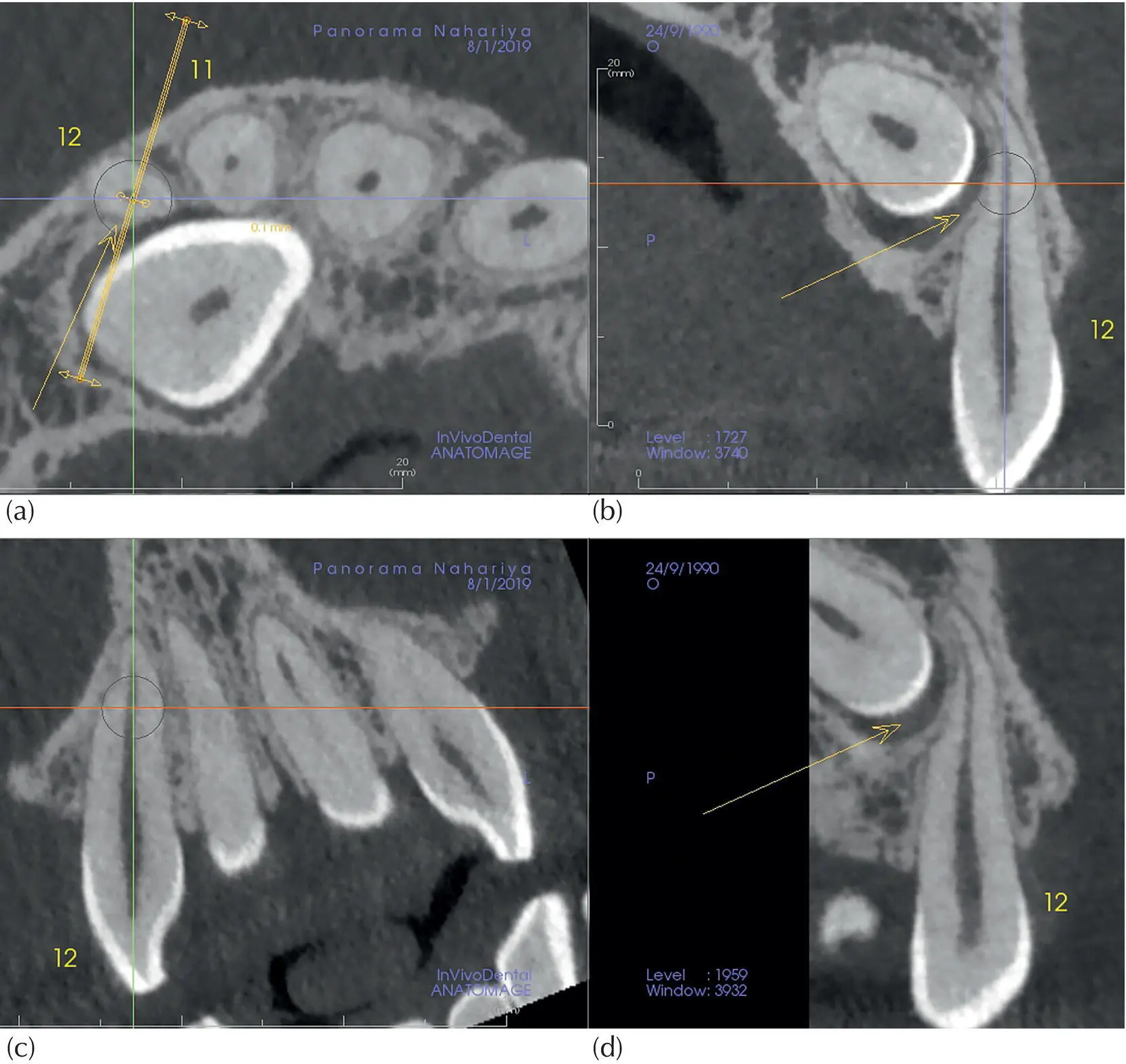
Fig. 4.17 Diagnosing resorption with multi‐planar reconstruction. The long axis of the lateral incisor from Figure 4.16has been tilted to a vertical position in the coronal (c) and sagittal (b) planes. The rotating tool is placed on the tooth axis in the axial (a) plane. Window (d) shows the resorption in the disto‐palatal aspect.
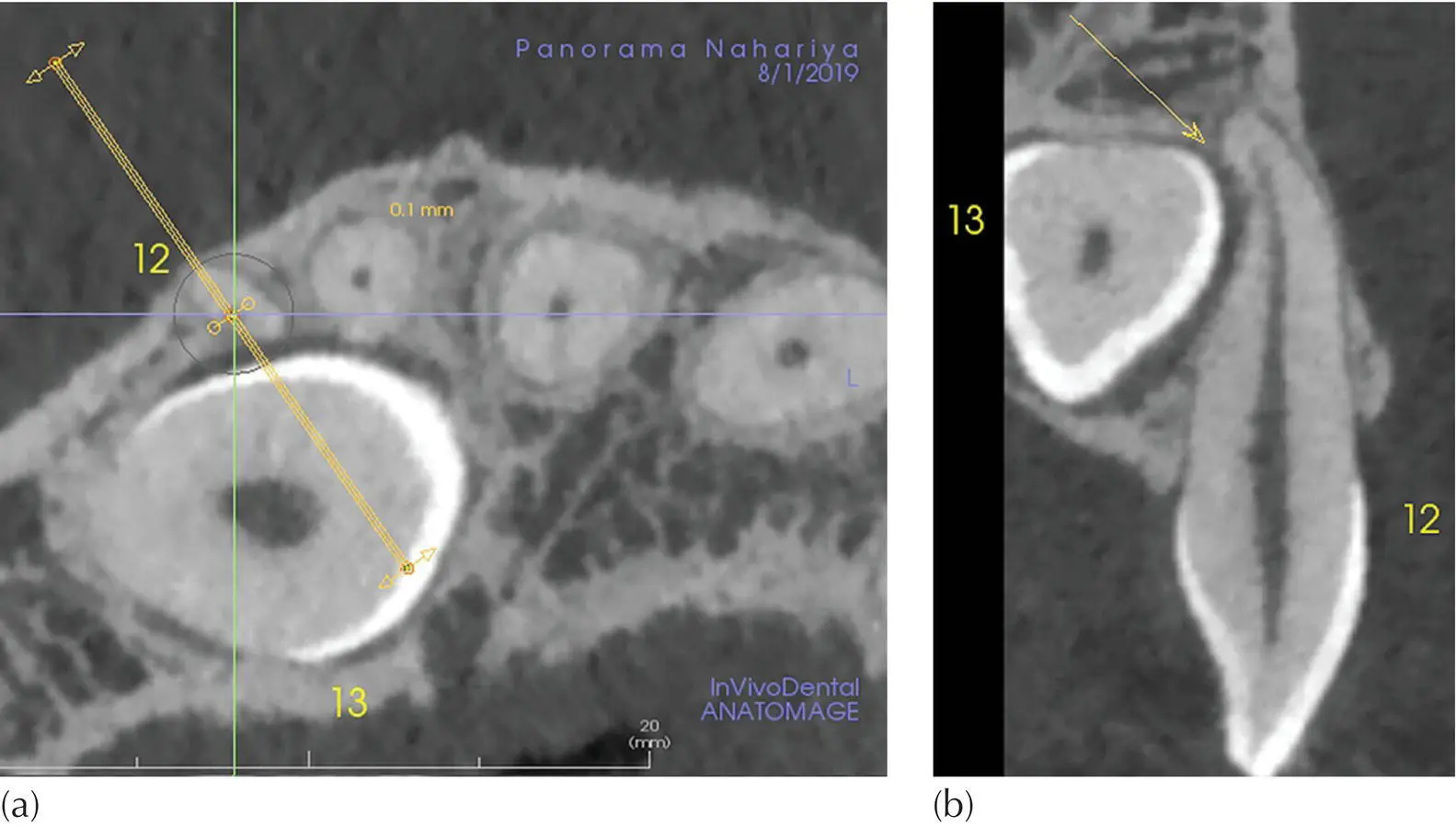
Fig. 4.18 Diagnosing resorption with multi‐planar reconstruction (MPR). The axial (a) and the special tool window (b) are cropped from the MPR screen. The appearance in (b) reveals the resorption in the palatal aspect.
Inferior dental canal marking
Tracing the inferior alveolar and marking it in red is a helpful feature. Once marked, it is embedded in the volume and will show as a red dot or line in any cut that crosses its path. This method is used principally in planning the placement of dental implants. However, for much the same reason, it has value in respect of the inter‐relations between the inferior dental (ID) canal and a severely and deeply infra‐occluded/impacted molar or premolar, whose roots are developing in close proximity to it ( Figure 4.20and Case 5).
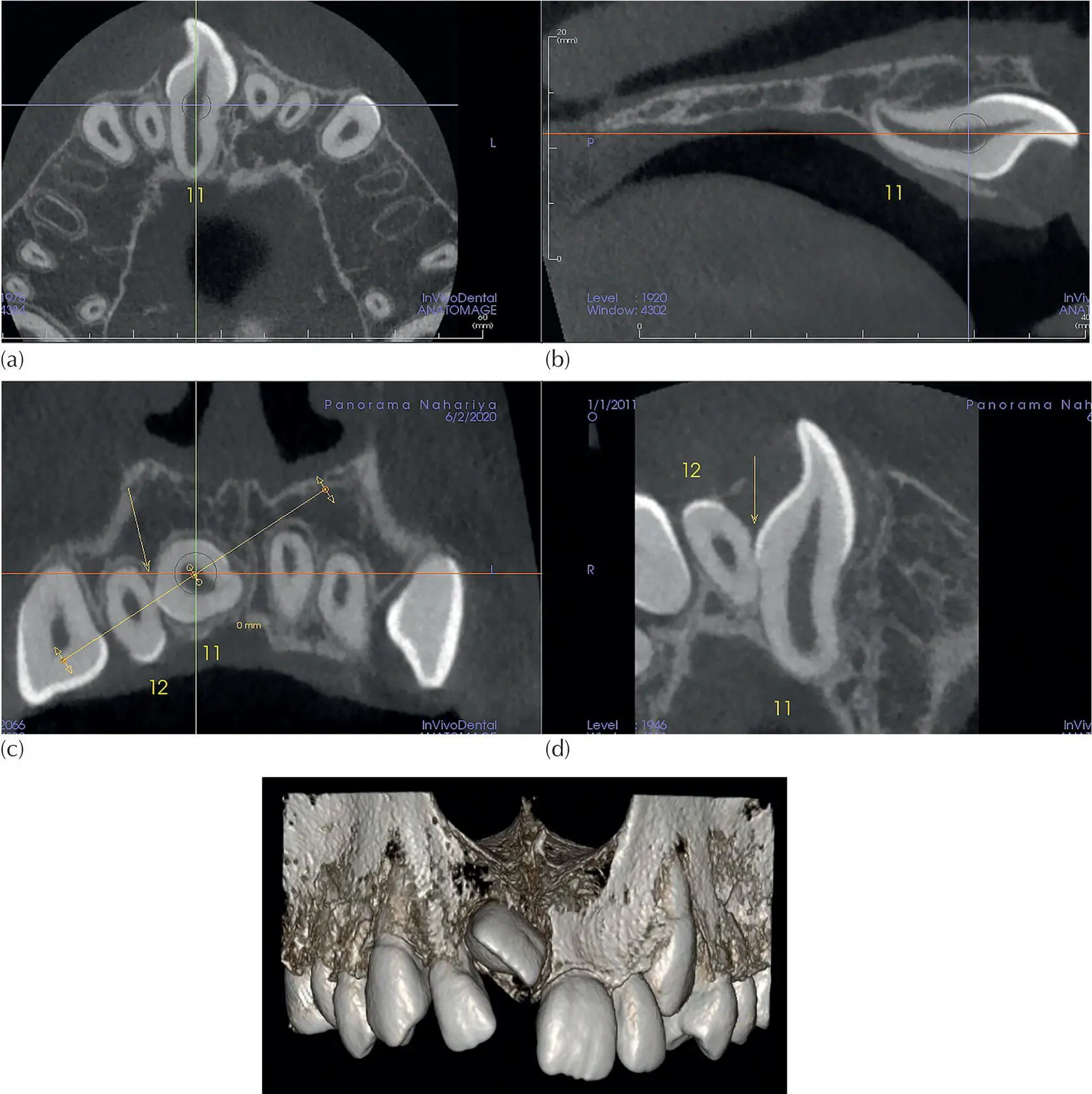
Fig. 4.19 Multi‐planar reconstruction for an incisor that is almost horizontal. The right central incisor was horizontally and sagitally re‐aligned in the axial (a) and sagittal (b) planes and the centre of the rotating tool is placed on its long axis in the coronal window (c). Window (d) represents the cut produced by the rotating tool, in which the contact point with the lateral incisor is demonstrated.
Case 5: Inter‐relations between the inferior dental canal and the first molar
The clinical aim was to find aetiological evidence for the failure to erupt of the first mandibular molar on the left side. The first impression from looking into the 3D bony view ( Figure 4.20a) was that the ID canal was embraced by both distal and mesial roots (Animation 4.5). Such a condition is usually caused by some obstruction in the way of the eruption path of the tooth, as a result of which the roots continue to develop and, in this case, to embrace the canal. The obstruction can be caused by a physical blockage or by ankylosis preventing the natural eruption of the tooth. While exploring the 3D view, a resorption lesion was observed in the mesial root next to the furcation. Figure 4.20(b) shows clipping of the buccal side all the way to the furcation, leaving the volume with only the lingual side of #36 (19), thereby enhancing the lesion area view (Animation 4.6). The 3D module is a wonderful tool for gaining a general impression, but the investigation is actually carried out on the MPR screen. Figure 4.21is a cropped MPR screen where the arrows indicate the ICRR lesion. A scroll through all three planes of the MPR screen can be viewed in Animation 4.7.
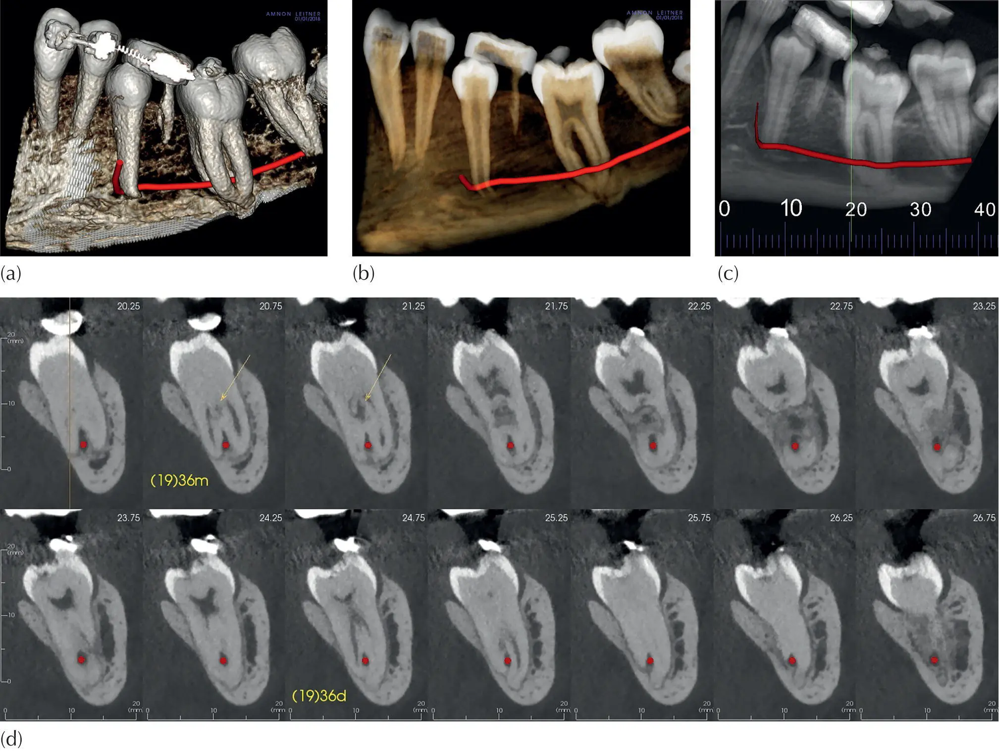
Fig. 4.20 First mandibular molar embracing inferior dental canal. (a) 3D bony view using bone peeling, sculpting and clipping. (b) 3D transparent view, buccal side clipped up to molar furcation, leaving only the lingual side for visualization. (c) Panoramic view with defining cross‐sectional grid. (d) A series of cross‐sectional slices showing the neurovascular bundle on each slice.
It is important to note that the majority of ICRR lesions originate at the cemento‐enamel junction (CEJ). When searching for aetiological evidence for an impacted tooth eruption failure, it is important to check the CEJ carefully, while rotating the tooth 360° as explained in Case 3, for an early‐stage ICRR.
With the earlier introduction of spiral CT and subsequently of CBCT, much debate was generated in relation to the justification for their use in orthodontics in general, and their value in the diagnosis and treatment planning of impacted teeth in particular. For planar radiography to provide a comparable level of positional information, a number of different views of the impacted tooth would need to be taken and the accumulated level of radiation that these would generate is of the same order as that emitted by the new CBCT machines.
The many studies that have been undertaken, for patients with an impacted tooth, to compare the advantages of using CBCT over plain 2D radiography have not provided the conclusive evidence that one would expect. It is clearly an indisputable fact that 3D imaging has, at least in theory, provided an infinite number of possible angles from which to view the tooth, as compared with a 2D radiological representation. So why was there no conclusive result? Could it be that the range and depth of CBCT post‐processing techniques that were performed were not as sophisticated as they should have been? Or perhaps only a minimum/standard orthodontic service was offered by the imaging technicians for the individuals comprising the patient samples. It is our opinion that the technicians in many of the radiological institutes in most of the Westernized countries we have visited or with whom we have had professional communication have not succeeded in mastering the complexities involved in the sophisticated interpretation of the CBCT imaging modality.
Читать дальшеИнтервал:
Закладка:
Похожие книги на «Orthodontic Treatment of Impacted Teeth»
Представляем Вашему вниманию похожие книги на «Orthodontic Treatment of Impacted Teeth» списком для выбора. Мы отобрали схожую по названию и смыслу литературу в надежде предоставить читателям больше вариантов отыскать новые, интересные, ещё непрочитанные произведения.


Обсуждение, отзывы о книге «Orthodontic Treatment of Impacted Teeth» и просто собственные мнения читателей. Оставьте ваши комментарии, напишите, что Вы думаете о произведении, его смысле или главных героях. Укажите что конкретно понравилось, а что нет, и почему Вы так считаете.