Veterinary Surgical Oncology
Здесь есть возможность читать онлайн «Veterinary Surgical Oncology» — ознакомительный отрывок электронной книги совершенно бесплатно, а после прочтения отрывка купить полную версию. В некоторых случаях можно слушать аудио, скачать через торрент в формате fb2 и присутствует краткое содержание. Жанр: unrecognised, на английском языке. Описание произведения, (предисловие) а так же отзывы посетителей доступны на портале библиотеки ЛибКат.
- Название:Veterinary Surgical Oncology
- Автор:
- Жанр:
- Год:неизвестен
- ISBN:нет данных
- Рейтинг книги:5 / 5. Голосов: 1
-
Избранное:Добавить в избранное
- Отзывы:
-
Ваша оценка:
Veterinary Surgical Oncology: краткое содержание, описание и аннотация
Предлагаем к чтению аннотацию, описание, краткое содержание или предисловие (зависит от того, что написал сам автор книги «Veterinary Surgical Oncology»). Если вы не нашли необходимую информацию о книге — напишите в комментариях, мы постараемся отыскать её.
The new edition of the most comprehensive resource on surgical oncology, covering both basic and advanced surgical oncology procedures in small animals Veterinary Surgical Oncology
Veterinary Surgical Oncology
Veterinary Surgical Oncology, Second Edition
Veterinary Surgical Oncology — читать онлайн ознакомительный отрывок
Ниже представлен текст книги, разбитый по страницам. Система сохранения места последней прочитанной страницы, позволяет с удобством читать онлайн бесплатно книгу «Veterinary Surgical Oncology», без необходимости каждый раз заново искать на чём Вы остановились. Поставьте закладку, и сможете в любой момент перейти на страницу, на которой закончили чтение.
Интервал:
Закладка:
Fine needle aspiration of internal organs can also be performed and may be helpful in guiding diagnostic and treatment choices. Image guidance should be utilized when obtaining FNAs of masses within a body cavity. Aspirates of lung and other thoracic organs can be performed safely in most cases. In one study, fine needle aspiration of lung masses had a sensitivity of 77% and a specificity of 100% (DeBerry et al. 2002). The aspiration of cranial mediastinal masses is beneficial, as thymomas can be diagnosed by cytology (Rae et al. 1989; Atwater et al. 1994; Lana et al. 2006). Cytologic diagnosis of thymoma requires the presence of a population of unequivocal malignant epithelial cells. The presence of mast cells is also common in thymoma and often supports the diagnosis (Atwater et al. 1994). Flow cytometry is another diagnostic tool that will differentiate thymoma from lymphoma using an FNA sample. Thymomas will contain both CD4+ and CD8+ lymphocytes, whereas lymphoma would typically contain a clonal expansion of one lymphocyte type (Lana et al. 2006).
Fine needle aspiration of hepatic and splenic neoplasia has been described in several studies (Osborne et al. 1974; Hanson et al. 2001; Roth 2001; Wang et al. 2004). Successful diagnosis of hepatic neoplasia with fine needle aspiration is variable. A study has reported diagnostic rates for liver cytology of multiple pathologies (including neoplasia) as high as 80% (Roth 2001); however, another study demonstrated less success with diagnostic rates of 14% in dogs and 33% in cats for fine needle aspiration of hepatic neoplasia (Wang et al. 2004). In cases of suspected splenic hemangiosarcoma, fine needle aspiration is generally not recommended, as an accurate diagnosis is unlikely due to the abundance of blood‐filled cavities. Additionally, complications may include severe bleeding from the aspiration site. Fine needle aspiration of splenic neoplasia such as lymphoma and mast cell tumors is often diagnostic (Hanson et al. 2001).
Other tumors in which fine needle aspiration has been utilized to obtain diagnostic information include gastrointestinal tumors and bony tumors. The accuracy of fine needle aspiration in the diagnosis of gastrointestinal neoplasia is often dependent on the type of neoplasia present. For instance, fine needle aspiration of gastrointestinal lymphoma tends to have a higher sensitivity than aspiration of gastrointestinal carcinoma/adenocarcinoma or leiomyoma/leiomyosarcoma (Bonfanti et al. 2006). The specificity of the diagnosis is similar among these neoplastic diseases with fine needle aspiration (Bonfanti et al. 2006). In one study, ultrasound‐guided fine needle aspiration of osteosarcoma lesions was found to have a sensitivity of 97% and specificity of 100% for the diagnosis of a sarcoma (Britt et al. 2007). Another study found that cytology after fine needle aspiration agreed with incisional and excisional biopsies of bony lesions in 71% of cases (Berzina et al. 2008). In a more recent study, histology of a bone lesion was superior to cytology (Sabattini et al. 2017). Histology of a biopsy had a sensitivity of 72%, specificity of 100%, and accuracy of 82%, whereas cytology had a sensitivity of 83%, specificity of 80%, and accuracy of 83% (Sabattini et al. 2017).
As with any procedure, FNAs are not without risk. In certain cases, bleeding or fluid leakage can be problematic, especially within a closed body cavity where it cannot be easily controlled. Tumor seeding and implantation along the needle tract is a rare occurrence, but in certain tumors has been reported more frequently. Localized tumor implantation following ultrasound‐guided FNA of transitional cell carcinoma of the bladder has been reported (Nyland et al. 2002) and should be a consideration when deciding on methods for diagnosing bladder masses. Fine needle aspiration of mast cell tumors brings the risk to cause degranulation, and clinicians should be prepared to treat untoward systemic effects following aspiration of a suspicious or known mast cell tumor. Despite the risks associated with needle aspiration, it remains an effective, inexpensive, and valuable tool in the preoperative planning process.
Biopsy
Clinicians often use the term “biopsy” as a nonspecific description of obtaining a tissue sample for histopathologic interpretation. Because of this, two major categories of biopsy have been designated: pretreatment biopsy (tissue obtained before treatment initiation) or posttreatment biopsy (tissue obtained at the time of definitive tumor resection). All biopsy procedures, whether pretreatment or posttreatment, should be carefully planned with several factors in mind. These factors include known patient comorbidities, anatomic location of the mass, differential diagnoses, biopsy technique, eventual definitive treatment, and any neoadjuvant/adjuvant therapies that may need to be incorporated.
Pretreatment Biopsy
Needle Core Biopsy
This technique is commonly used for soft tissue, visceral, and thoracic masses (Osborne et al. 1974; Atwater et al. 1994; deRycke et al. 1999). Image guidance is recommended when using this technique in closed body cavities. Most patients require sedation and local anesthesia but may not need general anesthesia.
Instrumentation includes a needle core biopsy instrument (automated or manual) ( Figure 1.3), #11 scalpel blade, local anesthetic, and a 22 g hypodermic needle. To perform the procedure, the area surrounding the mass is clipped free of fur and prepared with aseptic technique. If intact skin is to be penetrated and the animal is not anesthetized, the skin overlying the area to be penetrated is anesthetized with lidocaine or bupivacaine. A 1–2 mm stab incision is made over the mass to allow for placement of the needle core biopsy instrument. The instrument is oriented properly and fired, and the instrument is withdrawn. The 22 g needle can be used to gently remove the biopsy from the trough of the needle core instrument. This identical procedure is performed for masses within a body cavity; however, it is necessary to use image guidance (most commonly ultrasound) for proper placement of the instrument within the desired tissue. Imaging can be used to determine the depth of penetration and to safely avoid nearby vital structures.
Punch Biopsy
This technique is most effective for cutaneous lesions as well as intraoperatively for biopsies of masses within organs such as the liver, spleen, and kidney. Subcutaneous lesions can be biopsied using this method, but it is best to incise the skin overlying the mass and then obtain the sample using the biopsy instrument.
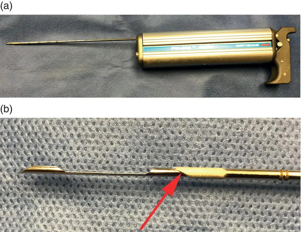
Figure 1.3 (a) Automated needle core biopsy instrument. (b) The tip of the needle has an indentation, which is filled with the tumor tissue when inserted. There is a sleeve with a cutting edge (red arrow), which cuts the piece of tissue in the indentation of the needle.
Instrumentation includes a punch biopsy instrument ( Figure 1.4), which typically comes in sizes of 2, 4, 6, and 8 mm; #11 scalpel blade; local anesthetic; Metzenbaum scissors; forceps; and suture. The area containing the mass is clipped free of fur and prepared with aseptic technique. If intact skin will be penetrated and the animal is not anesthetized, the skin overlying the lesion is anesthetized with lidocaine or bupivacaine. For cutaneous masses, an incision is not necessary. For subcutaneous masses, make an incision in the skin over the mass and dissect tissues overlying the mass if present to allow for the procurement of a better sample. The skin incision should be large enough for the punch biopsy instrument to be placed and allow it to be twisted without engaging skin. Twist the punch biopsy instrument until the device is embedded into the mass to the hub. The punch biopsy instrument is then withdrawn from the mass to expose the tissue sample. Gently grasp the sample with forceps, utilize Metzenbaum scissors to sever the deep aspect of the sample from the rest of the tissue, and remove the sample. A single suture is generally sufficient to close the incision. The same procedure can be performed on visceral organs.
Читать дальшеИнтервал:
Закладка:
Похожие книги на «Veterinary Surgical Oncology»
Представляем Вашему вниманию похожие книги на «Veterinary Surgical Oncology» списком для выбора. Мы отобрали схожую по названию и смыслу литературу в надежде предоставить читателям больше вариантов отыскать новые, интересные, ещё непрочитанные произведения.


Обсуждение, отзывы о книге «Veterinary Surgical Oncology» и просто собственные мнения читателей. Оставьте ваши комментарии, напишите, что Вы думаете о произведении, его смысле или главных героях. Укажите что конкретно понравилось, а что нет, и почему Вы так считаете.