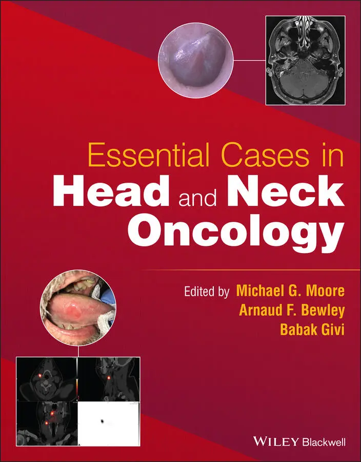
Essential Cases in Head and Neck Oncology
Здесь есть возможность читать онлайн «Essential Cases in Head and Neck Oncology» — ознакомительный отрывок электронной книги совершенно бесплатно, а после прочтения отрывка купить полную версию. В некоторых случаях можно слушать аудио, скачать через торрент в формате fb2 и присутствует краткое содержание. Жанр: unrecognised, на английском языке. Описание произведения, (предисловие) а так же отзывы посетителей доступны на портале библиотеки ЛибКат.
- Название:Essential Cases in Head and Neck Oncology
- Автор:
- Жанр:
- Год:неизвестен
- ISBN:нет данных
- Рейтинг книги:5 / 5. Голосов: 1
-
Избранное:Добавить в избранное
- Отзывы:
-
Ваша оценка:
Essential Cases in Head and Neck Oncology: краткое содержание, описание и аннотация
Предлагаем к чтению аннотацию, описание, краткое содержание или предисловие (зависит от того, что написал сам автор книги «Essential Cases in Head and Neck Oncology»). Если вы не нашли необходимую информацию о книге — напишите в комментариях, мы постараемся отыскать её.
Essential Cases in Head and Neck Oncology This textbook also includes:
Covers the full spectrum of head and neck surgeries, including reconstructive procedures Discusses ethics related to cancer treatments, medical research, and other care issues Promotes multidisciplinary critical thinking, clinical problem-solving, communication, and collaboration Helps medical students and trainees evaluate their learning and contextualize their knowledge Features high-quality images and succinct explanatory text throughout
is an indispensable study aid for trainee clinicians, residents, and fellows studying for board certification and other exams, and an excellent reference guide for oncologists, otolaryngologists, surgeons, and other practitioners working in medical oncology, radiation oncology, and oromaxillofacial surgery.
Essential Cases in Head and Neck Oncology — читать онлайн ознакомительный отрывок
Ниже представлен текст книги, разбитый по страницам. Система сохранения места последней прочитанной страницы, позволяет с удобством читать онлайн бесплатно книгу «Essential Cases in Head and Neck Oncology», без необходимости каждый раз заново искать на чём Вы остановились. Поставьте закладку, и сможете в любой момент перейти на страницу, на которой закончили чтение.
Интервал:
Закладка:
Table of Contents
1 Cover
2 Title Page Essential Cases in Head and Neck Oncology Edited by MICHAEL G. MOORE, MD, FACS Arilla Spence DeVault ProfessorVice Chair of Academic AffairsDepartment of Otolaryngology‐Head and Neck SurgeryIndiana University School of MedicineMedical Director, IUH Joe & Shelly Schwarz Cancer Center Indianapolis, IN, USA ARNAUD F. BEWLEY, MD Associate ProfessorDepartment of Otolaryngology‐Head and Neck Surgery Chief of Head and Neck SurgeryUniversity of California, Davis Sacramento, CA, USA BABAK GIVI, MD, FACS Associate Professor, Section Chief Patient Safety‐Quality Improvement Officer Department of Otolaryngology‐Head and Neck SurgeryNYU Langone Health New York, NY, USA Endorsed by: American Head and Neck Society Los Angeles, CA, USA
3 Copyright Page
4 List of Authors
5 SECTION 1: Oral Cavity CASE 1 CASE 2 CASE 3 CASE 4 CASE 5 CASE 6 Multiple Choice Questions Reference Suggested Readings
6 SECTION 2: Oropharynx CASE 7 CASE 8 CASE 9 CASE 10 CASE 11 Multiple Choice Questions References Suggested Reading
7 SECTION 3: Nasopharynx CASE 12 CASE 13 CASE 14 CASE 15 Multiple Choice Questions Suggested Reading
8 SECTION 4: Laryngeal Cancer CASE 16 CASE 17 CASE 18 CASE 19 CASE 20 CASE 21 Multiple Choice Questions Suggested Reading
9 SECTION 5: Hypopharynx CASE 22 CASE 23 CASE 24 Multiple Choice Questions Suggested Reading
10 SECTION 6: Thyroid CASE 25 CASE 26 CASE 27 CASE 28 CASE 29 CASE 30 Multiple Choice Questions Suggested Reading
11 SECTION 7: Parathyroid CASE 31 CASE 32 CASE 33 Multiple Choice Questions Suggested Reading
12 SECTION 8: Paraganglioma CASE 34 CASE 35 CASE 36 CASE 37 Suggested Reading
13 SECTION 9: Neck CASE 38 CASE 39 CASE 40 CASE 41 CASE 42 Multiple Choice Questions References Suggested Reading
14 SECTION 10: Trachea CASE 43 CASE 44 CASE 45 Multiple Choice Questions Suggested Reading
15 SECTION 11: Skull Base CASE 46 CASE 47 CASE 48 Multiple Choice Questions Suggested Reading
16 SECTION 12: Cutaneous Malignancies CASE 49 CASE 50 CASE 51 CASE 52 CASE 53 CASE 54 Multiple Choice Questions Suggested Reading
17 SECTION 13: Salivary CASE 55 CASE 56 CASE 57 CASE 58 CASE 59 CASE 60 Suggested Reading
18 SECTION 14: Reconstruction CASE 61 CASE 62 CASE 63 CASE 64 CASE 65 Multiple Choice Questions Suggested Reading
19 SECTION 15: Ethics CASE 66 CASE 67 CASE 68 CASE 69 Multiple Choice Questions Suggested Reading
20 Index
21 End User License Agreement
List of Tables
1 Chapter 3 TABLE 14.1 T staging of nasopharyngeal cancer per AJCC 8.TABLE 14.2 N staging of nasopharyngeal cancer per AJCC 8.TABLE 14.3 Group staging of nasopharyngeal cancer per AJCC 8.
2 Chapter 5Table 24.1 Pharyngeal reconstructive techniques.
3 Chapter 7TABLE 32.1 Multiple endocrine neoplasia: summary of associated neoplasms.
4 Chapter 10TABLE 43.1 Tracheal neoplasms.TABLE 45.1 Cotton–Myer classification of subglottic stenosis. a
5 Chapter 11TABLE 46.1 T‐staging: nasal cavity and ethomoid sinus.TABLE 46.2 Kadish staging of olfactory neuroblastoma.TABLE 46.3 Hyams grading system for esthesioneuroblastomas.
List of Illustrations
1 Chapter 1 FIGURE 1.1 This photo demonstrates the patient's right lateral tongue ulcera... FIGURE 1.2 This image shows the patient's fused CT‐ lymphoscintigraphy image... FIGURE 2.1 This photo demonstrates the ulcerative mucosal lesion of the righ... FIGURE 2.2 These axial images are from the patient's CT (a, b) as well as th... FIGURE 3.1 Axial (a) and coronal (b) cut of the primary lesion. Note there i... FIGURE 3.2 Axial cut of the neck portion of the CT demonstrating the patholo... FIGURE 3.3 Intraoperative photo of the right oral cavity defect after surgic... FIGURE 4.1 This intraoral photograph shows the lesion of the patient's left ... FIGURE 4.2 CT scan shows an extensive and destructive process of the left ma... FIGURE 5.1 The patient’s lower lip mass. The tumor extendsfrom the right Co... FIGURE 5.2 These representative axial cuts for the patient's neck CT with IV... FIGURE 5.3 This intraoperative photo shows the patient's total lower lip def... FIGURE 5.4 Bernard-Webster bilateral advancement flap reconstruction. FIGURE 6.1 This transoral photograph shows mild submucosal fullness of the l... FIGURE 6.2 An axial T1‐weighted MRI image without contrast (a) and a coronal... FIGURE 6.3 These intraoperative images show the surgical approach transorall... FIGURE 6.4 This intraoperative photo shows the palate defect after reconstru...
2 Chapter 2 FIGURE 7.1 This is a fused axial image of a PET/CT scan at the level of the ... FIGURE 8.1 These axial images of the fused PET/CT (left) and CT of the neck ... FIGURE 9.1 This image from a transnasal fiberoptic laryngoscopy shows no obv... FIGURE 9.2 A contrast‐enhanced CT of the neck demonstrates a solitary, enlar... FIGURE 9.3 A PET/CT demonstrates no distant metastatic sites but shows focal... FIGURE 9.4 Histologic images of a poorly differentiated squamous cell carcin... FIGURE 10.1 AxialAxial CT image. FIGURE 10.2 18FDG‐PET/CT axial view demonstrating an intense right tonsil tu... FIGURE 10.3 This intraoperative photo demonstrates an open approach to the o... FIGURE 11.1 A CT of the neck was performed with intravenous contrast. There ... FIGURE 11.2 This is an axial cut of the patient's PET/CT. There is slight so... FIGURE 11.3 This intraoperative photo shows the resection bed following a ro...
3 Chapter 3 FIGURE 12.1 This axial cut of a T1‐weighted MRI of the skull base demonstrat... FIGURE 13.1 (a) and (b) These photos from the patient's fiberoptic nasophary... FIGURE 13.2 This figure shows enlargement of the adenoid tissue and lingual ... FIGURE 13.3 This is a representative fused axial image from the patient's PE... FIGURE 14.1 (a) and (b) On these representative contrast‐enhanced axial cuts... FIGURE 14.2 There is no obvious mucosal‐based primary tumor, but there is a ... FIGURE 14.3 Operative nasopharyngolaryngoscopy indicates a slight fullness a...FIGURE 15.1 Endophytic ulcerative lesion on the right lateral aspect of the ...FIGURE 15.2 Right nasopharyngeal mass on CT scan without bony invasion.
4 Chapter 4FIGURE 16.1 CT scan with contrast demonstrated exophytic mass of the right A...FIGURE 16.2 Direct laryngoscopy demonstrating an exophytic mass arising from...FIGURE 17.1 Fiberoptic scope examination indicating an ulcerative mass of th...FIGURE 17.2 CT scan of the neck with contrast demonstrates a glottic tumor w...FIGURE 17.3 Laryngectomy and bilateral neck dissection specimen.FIGURE 18.1 Fiberoptic nasolaryngoscopy demonstrating right glottic tumor.FIGURE 18.2 CT scan of the neck indicating an enhancing mass involving the r...FIGURE 18.3 Intraoperative photograph demonstrating the radial forearm free ...FIGURE 19.1 Intraoperative photo demonstrating an exophytic lesion of the ri...FIGURE 19.2 Intraoperative photo demonstrating the surgical field following ...FIGURE 20.1 Flexible nasolaryngoscopic view of the glottis demonstrating the...FIGURE 20.2 Axial cuts of the CT scan of the neck with contrast indicates a ...FIGURE 20.3 Coronal and sagittal views of the mass demonstrate high‐grade ob...FIGURE 21.1 Flexible nasolaryngoscopy demonstrating the presence of a submuc...FIGURE 21.2 A CT scan of the neck with contrast indicates the presence of a ...FIGURE 21.3 High‐power H&E stain of a laryngeal chondrosarcoma (left) shows ...FIGURE 21.4 This schematic indicates the resection of this patient's tumor....
5 Chapter 5FIGURE 22.1 Intraoperative photo before transoral robotic removal of a poste...FIGURE 22.2 Intraoperative photo after transoral robotic removal of a poster...FIGURE 23.1 Transnasal flexible endoscopy indicates the presence of a mass e...FIGURE 23.2 In these axial cuts of a contrast‐enhanced CT of the neck, there...FIGURE 23.3 FDG PET/CT indicates uptake involving the left pyriform sinus ma...FIGURE 24.1 CT scan of the neck with contrast indicates a mass of the left p...FIGURE 24.2 Barium swallow after total laryngopharyngectomy with free flap r...
Читать дальшеИнтервал:
Закладка:
Похожие книги на «Essential Cases in Head and Neck Oncology»
Представляем Вашему вниманию похожие книги на «Essential Cases in Head and Neck Oncology» списком для выбора. Мы отобрали схожую по названию и смыслу литературу в надежде предоставить читателям больше вариантов отыскать новые, интересные, ещё непрочитанные произведения.


Обсуждение, отзывы о книге «Essential Cases in Head and Neck Oncology» и просто собственные мнения читателей. Оставьте ваши комментарии, напишите, что Вы думаете о произведении, его смысле или главных героях. Укажите что конкретно понравилось, а что нет, и почему Вы так считаете.