Essential Cases in Head and Neck Oncology
Здесь есть возможность читать онлайн «Essential Cases in Head and Neck Oncology» — ознакомительный отрывок электронной книги совершенно бесплатно, а после прочтения отрывка купить полную версию. В некоторых случаях можно слушать аудио, скачать через торрент в формате fb2 и присутствует краткое содержание. Жанр: unrecognised, на английском языке. Описание произведения, (предисловие) а так же отзывы посетителей доступны на портале библиотеки ЛибКат.
- Название:Essential Cases in Head and Neck Oncology
- Автор:
- Жанр:
- Год:неизвестен
- ISBN:нет данных
- Рейтинг книги:5 / 5. Голосов: 1
-
Избранное:Добавить в избранное
- Отзывы:
-
Ваша оценка:
Essential Cases in Head and Neck Oncology: краткое содержание, описание и аннотация
Предлагаем к чтению аннотацию, описание, краткое содержание или предисловие (зависит от того, что написал сам автор книги «Essential Cases in Head and Neck Oncology»). Если вы не нашли необходимую информацию о книге — напишите в комментариях, мы постараемся отыскать её.
Essential Cases in Head and Neck Oncology This textbook also includes:
Covers the full spectrum of head and neck surgeries, including reconstructive procedures Discusses ethics related to cancer treatments, medical research, and other care issues Promotes multidisciplinary critical thinking, clinical problem-solving, communication, and collaboration Helps medical students and trainees evaluate their learning and contextualize their knowledge Features high-quality images and succinct explanatory text throughout
is an indispensable study aid for trainee clinicians, residents, and fellows studying for board certification and other exams, and an excellent reference guide for oncologists, otolaryngologists, surgeons, and other practitioners working in medical oncology, radiation oncology, and oromaxillofacial surgery.
Essential Cases in Head and Neck Oncology — читать онлайн ознакомительный отрывок
Ниже представлен текст книги, разбитый по страницам. Система сохранения места последней прочитанной страницы, позволяет с удобством читать онлайн бесплатно книгу «Essential Cases in Head and Neck Oncology», без необходимости каждый раз заново искать на чём Вы остановились. Поставьте закладку, и сможете в любой момент перейти на страницу, на которой закончили чтение.
Интервал:
Закладка:
Answer:Since the disease is stage III, consideration of adjuvant treatment is warranted. Radiotherapy should be considered after discussion of the case at a multidisciplinary tumor board. The benefit of radiotherapy is not as clear in N1 disease; however, limited data exist that shows tumors with a depth of invasion of greater than 4 mm are at increased risk of regional failure without adjuvant therapy. Since there is no evidence of primary site positive margins or extranodal extension, there is no indication for adjuvant chemotherapy.
Question: The patient completes a course of adjuvant radiotherapy. What is your recommended regimen for follow‐up and clinical surveillance?
Answer:Based on National Comprehensive Cancer Network ( NCCN) guidelines, baseline imaging at 12 weeks after completion of adjuvant treatment should be obtained, followed by physical examination every 1–3 months in the first year post‐treatment and then 4–6 months in the second year. In years 3–5, a physical exam every 4–8 months is recommended and annually after 5 years. Annual thyroid‐stimulating hormone ( TSH) testing is recommended since the neck has received radiotherapy. Dental, nutrition, and ongoing depression evaluation are also recommended.
Key Points
Oral tongue SCC is the most common malignancy of the oral cavity. The most important risk factors are tobacco, alcohol, poor dentition, diets low in fruits and vegetables, and Fanconi anemia.
The risk of occult metastases in early stage oral cavity cancers is usually upward of 20%. Level I clinical trial evidence exists in the survival benefit of elective neck dissection in early stage tongue cancer and clinically negative cervical nodes when the depth of invasion is >3 mm. Currently, imaging techniques are not sensitive enough to identify occult metastases, and a negative CT or PET scan does not rule out microscopic metastases.
Sentinel node biopsy in oral cavity cancers has been studied and shown to be reliable enough to identify the majority of occult metastases. Sentinel node sensitivity is reported as 86% with a negative predictive value of 95% based on the European Organization for Research and Treatment of Cancer.
Recommended primary treatment of oral cavity cancers is primarily surgical. Wide local excision with a 1 cm margin and lymph node dissection (selective node dissection in clinically negative neck) is the current recommendation.
Depth of invasion is an important prognostic factor. A depth of invasion of more than 3 mm is associated with an increased risk of lymph node metastases.
The current indications for adjuvant radiotherapy are (i) close or positive margins, (ii) nodal involvement, (iii) perineural invasion, and (iv) advanced stage tumor (T3–4).
Concurrent chemotherapy with platinum‐based agents is only recommended in positive margins or extranodal extension.
CASE 2
Babak Givi
History of Present Illness
A 64‐year‐old woman presents with a nonhealing ulcer of the right mandibular alveolus for the past month after extraction of the second molar. The lesion is not painful and does not bleed. She has a more than 40 pack‐year history of smoking and drinking two alcoholic drinks a day for the past 30 years. She does not report any history of other medical problems or prior malignancy.
Physical Examination
No palpable neck mass is identified. Oral cavity exam shows an ulcerative lesion limited to the occlusal surface of the right mandibular alveolus measuring 2 × 1 cm. The lesion is not tender to touch, does not extend to the buccal mucosa or floor of the mouth (see Figure 2.1). The rest of the exam, including flexible laryngoscopy, is within normal limits.
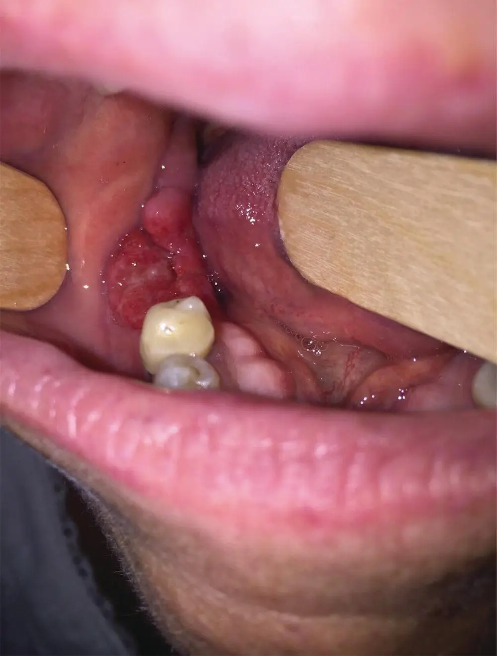
FIGURE 2.1 This photo demonstrates the ulcerative mucosal lesion of the right mandible gingiva.
Management
Question: What would you recommend next?
Answer:Since the lesion has been present for more than 2 weeks and a significant history of tobacco and alcohol abuse exists, tissue diagnosis via incisional or punch biopsy is warranted.
Question: The biopsy is performed and shows invasive, moderately differentiated keratinizing SCC. How would you stage this disease?
Answer:The clinical stage of the disease at this point is T2N0M0, stage II. However, the alveolar lesions can invade the mandible early in the process. Therefore, imaging of the mandible to better delineate osseous involvement is indicated.
Question: How would you determine the involvement of the mandible?
Answer:No imaging modality can offer 100% sensitivity and specificity. CT scanning, especially cone‐beam CT, has been utilized as a modality with high sensitivity. Magnetic resonance imaging (MRI) could be useful in determining the involvement of the bone marrow. In the absence of any single definitive modality, attention to the overall clinical exam and imaging findings could guide the clinician to determine the extent of the disease. For oral cavity lesions with suspected mandibular involvement, most surgeons would start with a CT scan with contrast of the head and neck to evaluate the primary site lesion and regional lymphatics.
A PET/MRI and neck CT with IV contrast are obtained. The CT of the neck shows significant thinning of the lingual cortex of the mandible at the site of the lesion and the absence of the buccal cortex superiorly. The inferior alveolar canal appears intact (see Figure 2.2). The PET/MRI shows intense activity at the primary lesion without clear evidence of lymph node metastases. On MRI, there is an abnormal marrow signal at the location of the lesion, suspicious for marrow invasion. There is no evidence of involvement of the mandibular canal.
Question: Based on imaging findings, what is the clinical stage of the disease?
Answer:Since there is radiographic evidence of osseous invasion, the disease is upstaged to T4aN0M0, stage IVa.
Question: What is the recommended course of treatment?
Answer:Oral cavity SCC is primarily treated with surgery, followed by risk‐adjusted adjuvant treatment. In this case, the recommended treatment is composite resection of the lesion with segmental mandibulectomy, selective neck dissection (levels I–III), and reconstruction with microvascular free tissue transfer, such as fibula free flap, to provide the best functional outcome.
The patient undergoes segmental mandibulectomy and selective neck dissection with fibula free flap reconstruction. The final pathology shows a 2 × 1 cm ulcerative SCC of the gingiva with invasion of the mandible. All margins are free of tumor (>5 mm). There is no perineural invasion. There is a microscopic focus of carcinoma present in 1 out of 32 lymph nodes (level Ib node, 2.3 cm) with no evidence of extracapsular extension.
Question: What is the pathologic stage?
Answer:Based on mandibular invasion and nodal involvement, the pathologic stage is pT4aN1M0, stage IVa.
Question: What adjuvant treatment regimen would you recommend to this patient?
Answer:In spite of negative margins and no perineural invasion, advanced T stage and involvement of regional nodes warrant adjuvant radiotherapy. There is no indication for chemotherapy (positive margins and extranodal extension). Therefore, adjuvant radiotherapy is adequate.
Читать дальшеИнтервал:
Закладка:
Похожие книги на «Essential Cases in Head and Neck Oncology»
Представляем Вашему вниманию похожие книги на «Essential Cases in Head and Neck Oncology» списком для выбора. Мы отобрали схожую по названию и смыслу литературу в надежде предоставить читателям больше вариантов отыскать новые, интересные, ещё непрочитанные произведения.


Обсуждение, отзывы о книге «Essential Cases in Head and Neck Oncology» и просто собственные мнения читателей. Оставьте ваши комментарии, напишите, что Вы думаете о произведении, его смысле или главных героях. Укажите что конкретно понравилось, а что нет, и почему Вы так считаете.