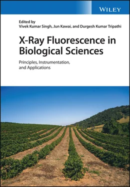
X-Ray Fluorescence in Biological Sciences
Здесь есть возможность читать онлайн «X-Ray Fluorescence in Biological Sciences» — ознакомительный отрывок электронной книги совершенно бесплатно, а после прочтения отрывка купить полную версию. В некоторых случаях можно слушать аудио, скачать через торрент в формате fb2 и присутствует краткое содержание. Жанр: unrecognised, на английском языке. Описание произведения, (предисловие) а так же отзывы посетителей доступны на портале библиотеки ЛибКат.
- Название:X-Ray Fluorescence in Biological Sciences
- Автор:
- Жанр:
- Год:неизвестен
- ISBN:нет данных
- Рейтинг книги:5 / 5. Голосов: 1
-
Избранное:Добавить в избранное
- Отзывы:
-
Ваша оценка:
- 100
- 1
- 2
- 3
- 4
- 5
X-Ray Fluorescence in Biological Sciences: краткое содержание, описание и аннотация
Предлагаем к чтению аннотацию, описание, краткое содержание или предисловие (зависит от того, что написал сам автор книги «X-Ray Fluorescence in Biological Sciences»). Если вы не нашли необходимую информацию о книге — напишите в комментариях, мы постараемся отыскать её.
Discover a comprehensive exploration of X-ray fluorescence in chemical biology and the clinical and plant sciences X-Ray Fluorescence in Biological Sciences: Principles, Instrumentation, and Applications
X-Ray Fluorescence in Biological Sciences: Principles, Instrumentation, and Applications
X-Ray Fluorescence in Biological Sciences — читать онлайн ознакомительный отрывок
Ниже представлен текст книги, разбитый по страницам. Система сохранения места последней прочитанной страницы, позволяет с удобством читать онлайн бесплатно книгу «X-Ray Fluorescence in Biological Sciences», без необходимости каждый раз заново искать на чём Вы остановились. Поставьте закладку, и сможете в любой момент перейти на страницу, на которой закончили чтение.
Интервал:
Закладка:
9 9 Yoneda, H. and Huriuchi, T. (1971). Optical flats for use in X‐ray spectrochemical microanalysis. Rev. Sci. Instrum. 42: 1069.
10 10 Klockenkamper, R. (1996). Total Reflection X‐Ray Fluorescence, Analysis, Chemical Analysis, vol. 140. New York: Wiley.
11 11 Kunimura, S. and Kawai, J. (2007). Portable total reflection X‐ray fluorescence spectrometer for nanogram Cr detection limit. Anal. Chem. 79: 2593–2595.
Note
1 *Retired, Email: nlmisra@yahoo.com
5 Micro X‐Ray Fluorescence and X‐Ray Absorption near Edge Structure Analysis of Heavy Metals in Micro‐organism
Changling Lao1,2,3, Liqiang Luo2, Yating Shen2, and Shuai Zhu2
1 Guilin University of Technology, Guilin, China
2 National Research Center of Geoanalysis, Beijing, China
3 China University of Geosciences (Beijing), Beijing, China
5.1 Introduction
With the ever‐increasing exploitation of mineral resources, heavy metal pollution has led to a serious effect on the ecosystem. Micro‐organisms which participate in nearly all biochemical reactions exist widely in the environment. They play an important role in the dissolution, migration, transformation, and precipitation of heavy metals. High concentrations of heavy metals possess toxic effects on micro‐organisms, including the disruption of cell membranes, alteration of enzyme specificity and protein structure, and the sometimes fatal destruction of DNA [1]. In environments, heavy metals contaminate some micro‐organisms, which are detoxified through biological processes such as adsorption, accumulation, and biotransformation, and further influence the circulation of elements and chemical species. X‐ray fluorescence (XRF) and X‐ray absorption spectroscopy (XAS) develop alongside synchrotron (SR) technology, which can be applied to understand the distribution, migration, and transformation of heavy metals.
5.2 Effects of Heavy Metals on Microbial Growth
The biogeochemical cycle of elements will affect the growth of micro‐organisms. Some metals are essential nutrients for the growth of micro‐organisms (such as Mg, Na, Fe, Co, Cu, Mo, Ni, W, V, and Zn) [2]. Some metal elements can get energy for the metabolism of micro‐organisms through the redox reaction process (such as As(III/V), Fe(II/III), Mn(II/IV), V(IV/V), Se(IV/VI), U(IV/VI)) [3]. There are also some heavy metal ions (such as Ag +, Hg 2+, Cd 2+, Co 2+, CrO 4 2+, Cu 2+, Ni 2+, Pb 2+, and Zn 2+) which possess a great affinity to thiol‐containing groups, and thus can easily replace essential metal ions that are typically present and necessary in many enzymes [4, 5].
The composition of the microflora is governed by their physical and chemical properties [6]. The concentration, chemical speciation, and bioavailability of elements in the soils have obvious effects on microbial growth [7, 8]. At the same time, the production of reactive oxygen species (ROS), including hydrogen peroxide (H 2O 2), superoxide dismutase (O 2 −) and hydroxyl radicals (OH −) may alter the intracellular redox status and ultimately cause cell death when the ROS exceeds the antioxidant capacity of the micro‐organism [9]. Low concentration of AuCl 4 −(<130 μmol/l) could stimulate Rhizopusoryzae to secrete reductive proteins for biological detoxification, but when the concentration of AuCl 4 −was higher than 130 μmol/l, the ultrastructure of the cells would be destroyed, and growth was significantly inhibited.
5.3 Application of μ‐XRF and XAS in Understanding the Cycling of Elements Driven by Micro‐organism
Micro‐organisms are key‐drivers of carbon‐, nitrogen‐, sulfur‐, and metal‐cycling on Earth, and they directly and indirectly drive element cycling [10]. Micro‐organisms can influence the cycling process of heavy metal elements through dissolution, adsorption, transformation, and other processes. XRF and XAS analysis techniques play important roles in revealing the mechanism of heavy metals migration between water, soils, plants, and gases. The toxicity of heavy metals is mainly influenced by its chemical species and mobility.
XANES analysis, used to detect the species of As, can help to understand the redox mechanism of arsenic, the circulation of arsenic between solid and liquid phases, and the release mechanism of arsenic from sediments into water. Under reducing conditions, 74–85% of the arsenic exists as As (III), which has a high ecological risk. After switching to a toxic condition, the native micro‐organism can oxidize As (III) to As (V) and adsorb it to the solid phase, indicating that oxidation conditions can reduce the solubility and toxicity of As [11].
Iron‐bearing minerals have strong adsorption capacity for heavy metals. The interaction between micro‐organisms and iron minerals is of great significance to the cycle of heavy metals in the environment. Arsenate is strongly associated with soil minerals, particularly iron(hydr)oxides [12]. The catalytic oxidation of iron has an important effect on the adsorption and chemical species of arsenate. Analysis of XRD and XANES showed that Shewanella putrefaciens could oxidize Fe (II) to feroxyhyte (δ ′‐FeOOH) and goethite (α‐FeOOH), while Sb(V) was reduced to Sb (III) and adsorbed on the surface of these secondary Fe(III) oxides, which can effectively reduce the toxic effect of Sb in aqueous solutions [13]. The mine water contained high concentrations of iron and other heavy metals, and the formation of iron‐containing biofilms may affect the cycling of heavy metals. 16S rDNA, TEM, XRD, XANES, and scanning‐particle induced X‐ray emission analysis (S‐PIXE) results revealed that under the action of Gallionella sp., the concentration of Fe and As decreased significantly, accompanied by the formation of a yellowish biofilm precipitate. The precipitate was confirmed to be schwertmannite containing Fe, As, and S, and consequently As was deposited in the mineral. These results indicated that Gallionella Sp. could promote the formation of iron‐bearing minerals, which improved the adsorption capacity to arsenic [14].
In the reduction condition, some micro‐organisms may promote the release of arsenic. In anoxic sediments, Geobacter sp. one can use Fe (III) as the electron acceptor to reduce Fe (III) to Fe (II) to dissolve iron‐bearing minerals, accompanied by reduction of As (V) to As (III), so the As originally adsorbed on the surface of iron‐bearing minerals was dissolved and released into the aquatic environment, increasing the ecological risk of As entering the food chain [15].
5.4 Application of μ‐XRF and XAS in Understanding the Transformation of Elements Driven by Micro‐organisms
In the heavy metal enriched environment, the chemical speciation of heavy metal elements is one of the important factors affecting the growth of micro‐organisms. Toxic heavy metal ions can easily replace essential metal ions that are necessary in many enzymes [4, 5]. Micro‐organisms can detoxify through biological processes and survive by changing the chemical speciation, bioavailability, and toxicity of heavy metals.
Redox of toxic elements by micro‐organisms is an important detoxification mechanism. Au (I/III)‐complexes are highly toxic to organisms, while Au nanoparticles (AuNPs) are relatively non‐toxic [16, 17]. Once Au (I/III)‐complexes enter the cells, they easily bind with glutathione and react with oxygen molecules to form oxidized glutathione and hydrogen peroxide, causing high oxidative stress and inhibiting catalase activity [18, 19]. Some micro‐organisms can detoxify through reductive precipitation of Au (III)‐complexes to less toxic Au (0) nanoparticles. Different micro‐organisms have different reduction and detoxification mechanisms for Au (I/III)‐complexes. Micro‐organisms can reduce Au (I/III) to Au (0) through extracellular secretions, reduced proteins on cell membranes, or intracellular reductase ( Figure 5.1) [20]. Delftia acidovorans can survive in a solution containing 100 μmol/l AuCl 3, but was significantly inhibited when they could not secrete secondary metabolites, suggesting that secondary metabolites can help them to reduce Au (III) to Au(0) [21]. The membrane vesicles produced by many gram‐negative bacteria can be used as a protective shield against the toxic environment [22]. When cyanobacteria were exposed to an Au (III) solution, the cyanobacteria would release the membrane vesicles to wrap the cells and prevent the entrance of Au (III) into the cells, while the reduction of Au (III) to metallic Au involves the formation of Au (I)‐sulfide, as confirmed by XANES [23]. Analysis of XANES and μ‐XRF revealed that Cupriavidus metallidurans accumulated Au(III)‐complexes from the solution, and the cellular Au was coupled to the formation of Au(I)‐S species intermediates, finally leading to the formation of Au(0) in both intra‐ and extracellular reductive precipitation, accompanied by a efflux outside the cell [19].
Читать дальшеИнтервал:
Закладка:
Похожие книги на «X-Ray Fluorescence in Biological Sciences»
Представляем Вашему вниманию похожие книги на «X-Ray Fluorescence in Biological Sciences» списком для выбора. Мы отобрали схожую по названию и смыслу литературу в надежде предоставить читателям больше вариантов отыскать новые, интересные, ещё непрочитанные произведения.
Обсуждение, отзывы о книге «X-Ray Fluorescence in Biological Sciences» и просто собственные мнения читателей. Оставьте ваши комментарии, напишите, что Вы думаете о произведении, его смысле или главных героях. Укажите что конкретно понравилось, а что нет, и почему Вы так считаете.


