X-Ray Fluorescence in Biological Sciences
Здесь есть возможность читать онлайн «X-Ray Fluorescence in Biological Sciences» — ознакомительный отрывок электронной книги совершенно бесплатно, а после прочтения отрывка купить полную версию. В некоторых случаях можно слушать аудио, скачать через торрент в формате fb2 и присутствует краткое содержание. Жанр: unrecognised, на английском языке. Описание произведения, (предисловие) а так же отзывы посетителей доступны на портале библиотеки ЛибКат.

- Название:X-Ray Fluorescence in Biological Sciences
- Автор:
- Жанр:
- Год:неизвестен
- ISBN:нет данных
- Рейтинг книги:5 / 5. Голосов: 1
-
Избранное:Добавить в избранное
- Отзывы:
-
Ваша оценка:
X-Ray Fluorescence in Biological Sciences: краткое содержание, описание и аннотация
Предлагаем к чтению аннотацию, описание, краткое содержание или предисловие (зависит от того, что написал сам автор книги «X-Ray Fluorescence in Biological Sciences»). Если вы не нашли необходимую информацию о книге — напишите в комментариях, мы постараемся отыскать её.
Discover a comprehensive exploration of X-ray fluorescence in chemical biology and the clinical and plant sciences X-Ray Fluorescence in Biological Sciences: Principles, Instrumentation, and Applications
X-Ray Fluorescence in Biological Sciences: Principles, Instrumentation, and Applications
X-Ray Fluorescence in Biological Sciences — читать онлайн ознакомительный отрывок
Ниже представлен текст книги, разбитый по страницам. Система сохранения места последней прочитанной страницы, позволяет с удобством читать онлайн бесплатно книгу «X-Ray Fluorescence in Biological Sciences», без необходимости каждый раз заново искать на чём Вы остановились. Поставьте закладку, и сможете в любой момент перейти на страницу, на которой закончили чтение.
Интервал:
Закладка:
Now, if a thin film of sample having a thickness of a few nanometers is placed on the sample support, at the intersection of incident and totally reflected beams, the path of incident beam shall not change much, as the thickness of the sample on the support is insignificant. However, the incident beam shall excite the sample. After the beam is totally reflected from the support, the totally reflected beam shall also pass through the sample and its path shall not change much, once again due to the negligible thickness of the sample. This beam shall also excite the sample. Thus, the sample is excited doubly, one by the incident beam and again by the totally reflected beam, whereas in XRF the sample excitation is done only once by the incident beam which is absorbed in the sample as well as the sample support. This is a simple explanation of TXRF and has been shown in Figure 4.3. Due to this phenomenon (TXRF), the intensity of the characteristic X‐rays coming out of the sample shall be approximately double that of when the X‐rays fall at an angle greater than the critical angle (off TXRF condition). Since the critical angle is dependent on the falling X‐ray beam energy on the support, the beam falling on the sample should be preferably monochromatic. If the beam is not monochromatic, the X‐rays of energy lower than the cutoff energy shall be totally reflected but those above the cutoff energy shall not be totally reflected, and will penetrate deep into the sample support, thus increasing the spectral background. These high energy X‐rays shall not excite the sample efficiently as they shall be far away from the absorption edges of the analytes and thus, are merely a nuisance. Due to this reason, a monochromatic beam of X‐ray is required for the ideal sample excitation. For producing a monochromatic beam, a monochromator ‐ either a synthetic multilayer or a crystal cut from a particular plane ‐ is chosen [10]. Multilayer monochromators have high reflectivity, are rugged, and thus are preferred compared to crystals which have low reflectivity and do not possess much strength. However, some researchers have found that during monochromatization, a large part of the X‐ray beam is cut off and remains unutilized, concluding that it is better to use polychromatic excitation where the increase in spectral background is compensated for by the high flux excitation by the polychromatic beam accompanied by a simple instrumentation. Nonetheless, monochromatic excitation is ideal for TXRF analysis [11].
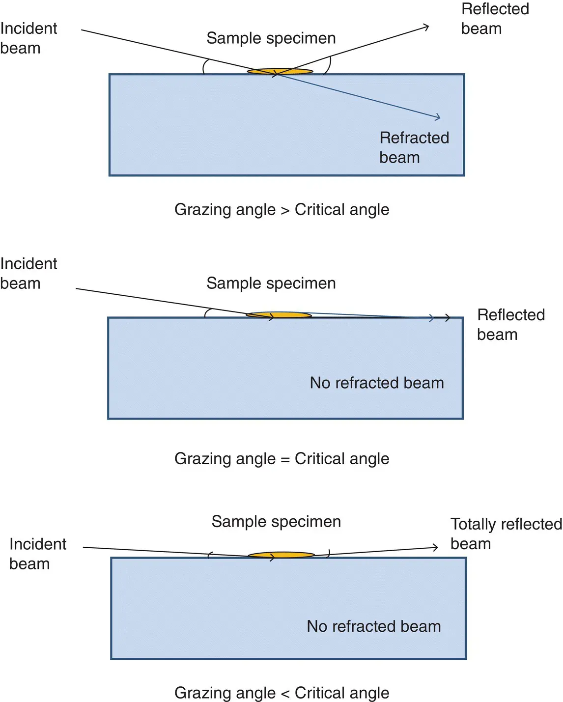
Figure 4.3 A representation of the advantages of TXRF analysis involving efficient sample excitation, X‐ray detection and reduction in spectra background compared to that of EDXRF background.
4.4.3 TXRF Instrumentation for Trace Element Determination
For trace element determination in TXRF, monochromatic X‐rays coming from an X‐ray source, as mentioned above, are passed through a slit so that a parallel beam of X‐rays is obtained and unwanted scattered X‐rays are cutoff. This parallel monochromatic beam is made to fall on a sample support containing a thin specimen of the sample at an angle less than the critical angle, corresponding to the monochromatic beam and sample support being used for sample excitation[5,8,10]. The sample is excited efficiently, as mentioned above, and the emitted X‐rays are detected using a Si(Li) or any other solid state detector. The totally reflected beam is stopped by a beam stopper so that it does not pass further and harm any laboratory personnel in the area. A schematic of the TXRF configuration is shown in Figure 4.4given below.
4.4.4 Sample Preparation for TXRF Analysis
The main steps in TXRF analysis of any sample are:
1 Dissolution of the sample in a suitable medium (mostly acids).
2 Matrix separation, if required.
3 Mixing of the remaining sample solution after matrix separation with a suitable internal standard.
4 Aliquoting a small volume (2–10 μl) of the above solution on the center of a sample support and drying it under an IR lamp to produce a thin film specimen.
5 Loading of the specimen in the TXRF spectrometer to measure its spectra.
6 Processing of the TXRF spectra obtained: identification of the peaks, deconvolution of the interfering peaks, determining the intensity of the X‐ray lines, and conversion of the intensities to the concentration.
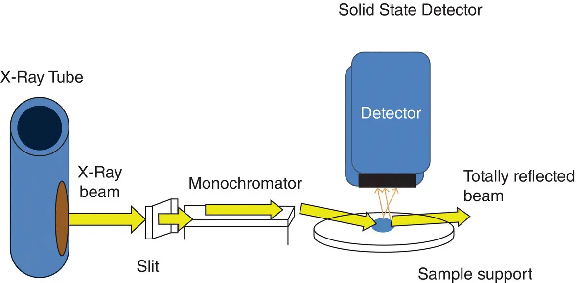
Figure 4.4 A schematic depiction of TXRF instrumentation for trace element analysis.
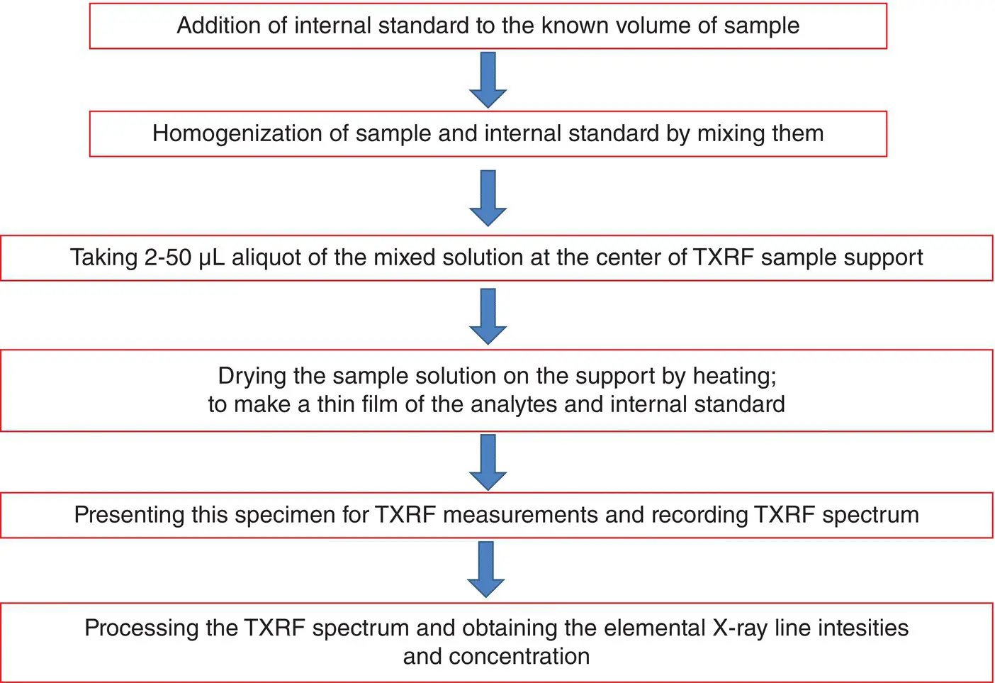
Figure 4.5 Schematic flow chart of sample preparation in TXRF analysis.
The above steps are shown in Figure 4.5. An aliquot of 2–10 μl of a sample and internal standard, mixed with each other thoroughly, is placed on a sample support and dried so that a thin film of the sample is formed. For trace element determinations in large matrix sample, the matrix separation is necessary because if the matrix is not separated, even 2 μl solutions shall form a thick deposit and produce matrix effects. In addition, the spectral background shall be very high. After selective separation of matrix, even a concentrated solution becomes very dilute, having analytes only predominantly in the solution. Normally, solvent extraction or anion exchange resin is used for such extraction. However, for biological samples, since the matrix is organic tissues made of low Z elements, the matrix separation is not required in most cases and the samples can be measured directly without any processing. This is a great advantage of TXRF for biological samples, as the possibility of samples being contaminated, or analyte being lost during complicated sample processing, as in other conventional techniques of analysis, is negligible. The X‐ray beams generally used for sample excitation in TXRF analysis are Rh Kα (20.216 keV), Mo Kα (17.479 keV), W Lα (8.396 keV) and W Lβ (9.671 keV). For low atomic number (Z) element analysis, low energy X‐rays, e.g. Rh Lα (2.698 keV), Mo Lα (2.295 keV), Αl Kα (1.487 keV), Cr Kα (5.415 keV) and Sc Kα (4.090 keV) etc. are preferred. The TXRF spectra are measured by keeping the samples in an ambient air atmosphere. However, for low Z elemental determinations, vacuum atmosphere sample chambers are required to avoid absorption of low Z elemental X‐ray lines in air atmosphere, and reduce the background due to scattering from the atmospheric air component atoms in the sample chamber and X‐ray beam path. A beam path of helium atmosphere between the detector and samples is also useful, especially to avoid interference of Ar Kα with the analytes as Ar is present (about 1%) in atmosphere. Using different sample excitation X‐ray sources and sample atmospheres (air, helium, or vacuum), elements from C to U can be determined by TXRF with different detection limits. Since the fluorescence yield of different elemental X‐ray lines are different, a correction for the sensitivity factors is required. The sensitivity values are defined as the ratio of intensity of a particular X‐ray line obtained from the TXRF spectrum and its concentration (or amount) deposited on the support. It may change with instrumental parameters such as X‐ray tube voltage and current, area of the sample exposed to the exciting X‐rays, as well as the area of the sample seen by the detector's solid angle on the sample. It is very difficult to make the sample area excited constant in each TXRF measurement and hence the sensitivity values of the elemental X‐ray lines measured changes with different specimens, even if the TXRF instrumental parameters are same. However, the change in the sensitivity values, due to the above reasons, is by a fixed factor for all the elements present in a particular sample during one measurement. This factor may change from one specimen to another. However, the ratio of sensitivity values of elemental X‐ray lines with respect to a particular elemental X‐ray line shall remain constant for all the specimens of a sample, if instrumental conditions are same. Due to this reason, a known amount of an internal standard, of an element which is not present in the sample, is added and the elemental X‐ray line`s sensitivities are determined with respect to this elemental X‐ray line intensity. The elemental X‐ray line intensities are normalized with the respective relative sensitivity values and the analyte concentrations are determined using the formula:
Читать дальшеИнтервал:
Закладка:
Похожие книги на «X-Ray Fluorescence in Biological Sciences»
Представляем Вашему вниманию похожие книги на «X-Ray Fluorescence in Biological Sciences» списком для выбора. Мы отобрали схожую по названию и смыслу литературу в надежде предоставить читателям больше вариантов отыскать новые, интересные, ещё непрочитанные произведения.


Обсуждение, отзывы о книге «X-Ray Fluorescence in Biological Sciences» и просто собственные мнения читателей. Оставьте ваши комментарии, напишите, что Вы думаете о произведении, его смысле или главных героях. Укажите что конкретно понравилось, а что нет, и почему Вы так считаете.