X-Ray Fluorescence in Biological Sciences
Здесь есть возможность читать онлайн «X-Ray Fluorescence in Biological Sciences» — ознакомительный отрывок электронной книги совершенно бесплатно, а после прочтения отрывка купить полную версию. В некоторых случаях можно слушать аудио, скачать через торрент в формате fb2 и присутствует краткое содержание. Жанр: unrecognised, на английском языке. Описание произведения, (предисловие) а так же отзывы посетителей доступны на портале библиотеки ЛибКат.
- Название:X-Ray Fluorescence in Biological Sciences
- Автор:
- Жанр:
- Год:неизвестен
- ISBN:нет данных
- Рейтинг книги:5 / 5. Голосов: 1
-
Избранное:Добавить в избранное
- Отзывы:
-
Ваша оценка:
X-Ray Fluorescence in Biological Sciences: краткое содержание, описание и аннотация
Предлагаем к чтению аннотацию, описание, краткое содержание или предисловие (зависит от того, что написал сам автор книги «X-Ray Fluorescence in Biological Sciences»). Если вы не нашли необходимую информацию о книге — напишите в комментариях, мы постараемся отыскать её.
Discover a comprehensive exploration of X-ray fluorescence in chemical biology and the clinical and plant sciences X-Ray Fluorescence in Biological Sciences: Principles, Instrumentation, and Applications
X-Ray Fluorescence in Biological Sciences: Principles, Instrumentation, and Applications
X-Ray Fluorescence in Biological Sciences — читать онлайн ознакомительный отрывок
Ниже представлен текст книги, разбитый по страницам. Система сохранения места последней прочитанной страницы, позволяет с удобством читать онлайн бесплатно книгу «X-Ray Fluorescence in Biological Sciences», без необходимости каждый раз заново искать на чём Вы остановились. Поставьте закладку, и сможете в любой момент перейти на страницу, на которой закончили чтение.
Интервал:
Закладка:
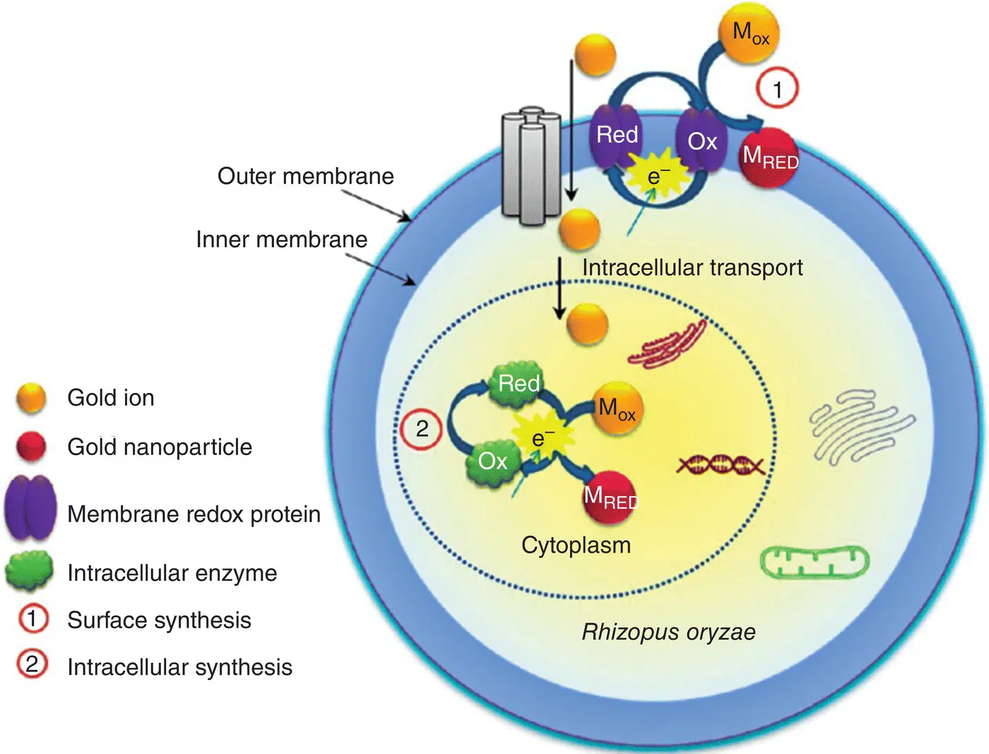
Figure 5.1 Reduction and detoxification mechanism of gold ions by Rhizopous oryzae cells.
Source: Reproduced from Das et al. [20] with the permission from American Chemical Society.
5.5 Application of μ‐XRF and XAS in Understanding the Mechanism of Using Micro‐organisms in Bioremediation
In the environment, heavy metal pollution is a serious problem. However, it is well known that microbial remediation is highly efficient and indispensable in repairing heavy metal pollution. Thus, understanding the mechanism of using micro‐organisms in bioremediation may help to improve remediation efficiency.
The biomineralization of heavy metals inside and outside the micro‐organism cells can precipitate heavy metals, which then reduces their bioavailability. Biomineralization is an important bioremediation method that includes phosphate mineralization, carbonate mineralization, and sulfide mineralization. XAS results showed that Shewanella oneidensis MR‐1 could reduce dissolved U(VI) to UO 2minerals with poor solubility to achieve the purpose of bioremediation. The addition of S, Si, and P elements in the environment could promote the formation of EPS (Extracellular Polymeric Substances), and the proportion of UO 2increases by 55–95%, which improved the efficiency of bioremediation significantly [24]. Uranium oxides were mineralized to secondary minerals by fungus, and the main processes were as follows: (1) When the fungus cells were exposed to UO 3and U 3O 8, the cells secreted large amounts of oxalic acid, which caused UO 3and U 3O 8to dissolve and form (UO 2) 2+complexes; (2) U was adsorbed by cells, and the adsorption capacity could reach 80 mg/g; (3) The dissolved (UO 2) 2+further complexed with P to form UO 2HPO 4and [UO 2(HPO 4) 2] 2−, and finally mineralized to form the uranyl phosphate mineral NH 4(UO 2)(PO 4).3H 2O and H 2(UO 2) 2(PO 4) 2.8H 2O [25].
The chemical species of heavy metals in the rhizosphere microenvironment are one of the important factors that affect the process of plant absorption of heavy metals. Study on the mechanism of interaction between rhizosphere micro‐organisms and heavy metals have important significance for the development of phytoremediation. It has been shown that arbuscular mycorrhizal fungi could enhance the capacity of plants to absorb Cr through mineralized and immobilized Cr in the rhizospheric environment, and the main mechanism listed as follows: (i) When exposed to Cr 6+solution, the EPS secreted by fungal cells increased significantly, and Cr mainly combined with the EPS on the cell surface to form a granular substance; (ii) Cr 6+was reduced to Cr 3+, and mainly accumulated in fungal cells in the form of phosphate, histidine, acetate and K 2CrO 4; (iii) EXAFS results showed that Cr was mainly combined with O, P, and C atoms in fungal cells; (iv) Most of Cr was absorbed by arbuscular mycorrhizal fungi, and only a small amount of Cr entered the plants, which alleviates the toxic effect of Cr on plants; (v) Cr in plant root cells mainly existed in the form of acetate, histidine analogs, phosphate, and K 2CrO 4.
5.6 The Advantage of Using μ‐XRF and XAS to Explore the Interaction Mechanism Between Micro‐organisms and Heavy Metals
XRF and XRS analysis techniques have obvious advantages in the analysis of environmental biological samples, especially in microbial samples. Most analysis methods require pretreatment the sample to create a uniform composition. They then detect the average concentration of elements. Heavy metals in biological samples are usually unevenly distributed. Determination of the distribution characteristics of heavy metal elements in a tissue or cell can help to understand the interaction mechanism between micro‐organisms and heavy metals. Synchrotron radiation‐XRF analysis has spatial resolution, and it can be used to analyze biological samples at the micron or even nano scale, which can be applied to investigate the distribution of heavy metal elements in the samples at the cellular level. With the development of third‐generation synchrotron radiation technology, micro‐beam X‐ray fluorescence spectroscopy (μ‐XRF), combined with micro‐beam X‐ray absorption structure (μ‐XANES) analysis technology make it possible to investigate the distribution characteristics and chemical species in a microbial cell. In addition, XAS can be used for the determination of amorphous materials, including solid, liquid, and even gas samples requiring no pretreatment.
The combination of μ‐XRF, STXM, and XANES methods are used for the in‐situ measurements of the distribution and morphological characteristics of heavy metals in the cells, which is an important means of exploring the microbial detoxification mechanism. High‐energy XRF with a spatial resolution of 150 nm was used to make elemental maps and qualitative chemical analyses of individual Pseudomonas fluorescens of planktonic and attached cells exposed to Cr(VI), and found that: (i) a large amount of the toxic element Cr is enriched in planktonic cells; (ii) notable excesses of Ca and P were found in attached cells, and Cr does not increase significantly in the cell, but is enriched within the 1–3 μm of surface‐adherent outside the cell; (iii) μ‐XANES analysis showed that Cr 6+was reduced to Cr 3+and associated with a phosphoryl functional group, which prevented Cr from entering into attached cells [26]. Researchers used XRF and XANES technology to investigate the distribution and chemical species status of heavy metals in C. metallidurans cells exposed to Au and Pt solutions, and found that gold was unevenly distributed in single cells, while Pt was evenly distributed. C. metallidurans cells accumulated less Pt, while more Au was absorbed in the center of the cell as nanoparticles, indicating that Pt had fewer toxic effects on cells than Au [27]. Reith et al. [19] combined XRF, TEM‐EDXA and XANE with a spot size of 120 × 150 nm to monitor the distribution of Au in C. metallidurans cells after being exposed to HAuCl 4for a different duration, and they found that the Au in the cell increased and then decreased after 144 hours of exposure. XANE results show that cellular Au formed Au(I)‐S compounds and finally produced Au nanoparticles. C. metallidurans reduced Au(III) to intra‐ and extracellular Au(0) and eliminated Au outside the cells [19]. Research on the detoxification mechanism of Penicillium oxalicum SL2 to Cr(VI) showed that when cells secreted cysteine and oxalic acid, Cr(VI) was reduced to chromium oxalate coupled to the formation of Cr(III)‐Cys intermediate complexes. Cr(VI) distributed in the hyphae and cytoplasm, indicating that Cr(VI) could translocate across the cell wall and cytomembrane into the hyphae, and Cr(VI) and Cr(III) had the same distribution characteristics in the hyphae.
XANES can be used to determine the chemical speciation in a specified area of samples that is preserved in its native state without pretreatment, which makes the results more authentic and reliable. Zeng et al. [28] used HPLC‐HG‐AFS and in‐situ XANES technology to analyze the species transformation of As(III) in Trichoderma asperellum , Penicillium janthinellum, and Fusarium oxysporum , and did not find methylated As in the cells of fungi using HPLC–HG–AFS, while MMA or/and DMA were observed using the XANES method. This may be due to the fact that for HPLC–HG–AFS analysis, mixed cellular samples must be acquired in advance via cellular lysis and centrifugation, which may change the chemical species of arsenic during pretreatment process. Thus, XANES can be applied directly to analyze the chemical species of samples in‐situ without pretreatment and the data obtained will be more reliable.
Читать дальшеИнтервал:
Закладка:
Похожие книги на «X-Ray Fluorescence in Biological Sciences»
Представляем Вашему вниманию похожие книги на «X-Ray Fluorescence in Biological Sciences» списком для выбора. Мы отобрали схожую по названию и смыслу литературу в надежде предоставить читателям больше вариантов отыскать новые, интересные, ещё непрочитанные произведения.


Обсуждение, отзывы о книге «X-Ray Fluorescence in Biological Sciences» и просто собственные мнения читателей. Оставьте ваши комментарии, напишите, что Вы думаете о произведении, его смысле или главных героях. Укажите что конкретно понравилось, а что нет, и почему Вы так считаете.